



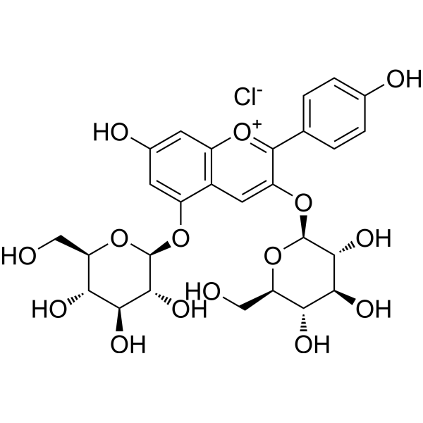
Cyanidin 3,5-diglucoside chloride

Based on 1 Customer Validation
Cyanidin 3,5-diglucoside chloride is an anthocyanin. Cyanidin 3,5-diglucoside chloride inhibits inflammatory cytokines (IL-1α, IL-1β, and IL-6) expression and NO production. Cyanidin 3,5-diglucoside chloride inhibits the phosphorylation of STAT3, IκB, ERK, p38, and AKT. Cyanidin 3,5-diglucoside chloride inhibits high pressure-induced decrease in GLAST. Cyanidin 3,5-diglucoside chloride exerts anti-inflammatory and skin barrier modulating effects. Cyanidin 3,5-diglucoside chloride can be used in retinal research.
For research use only. We do not sell to patients.
- Purity : 99.90%
- CAS No.: 2611-67-8
- Formula: C27H31ClO16
- Molecular Weight:646.98
-
Storage:
4°C, sealed storage, away from moisture and light
* In solvent : -80°C, 6 months; -20°C, 1 month (sealed storage, away from moisture and light)

To place orders, for customer services and technical support, please contact: MedChemExpress USA
Tel: 609-228-6898 E-mail: [email protected] [email protected]


-
 Biological Activity
-
Chemical Information
-
Solvent & Solubility
- Protocols
- Purity & Documentation
- References
-
 Help & FAQs


-
HY-N8408
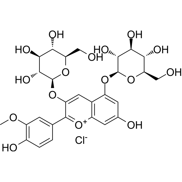
Peonidin 3,5-diglucosideAnthocyanin -
HY-W040045
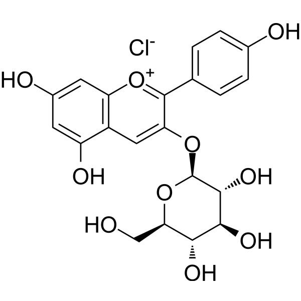
Callistephin chlorideROS/iNOS/TNF-α/COX-2 Inhibitor -
HY-18085
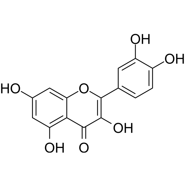
QuercetinFlavonoid Antioxidant -
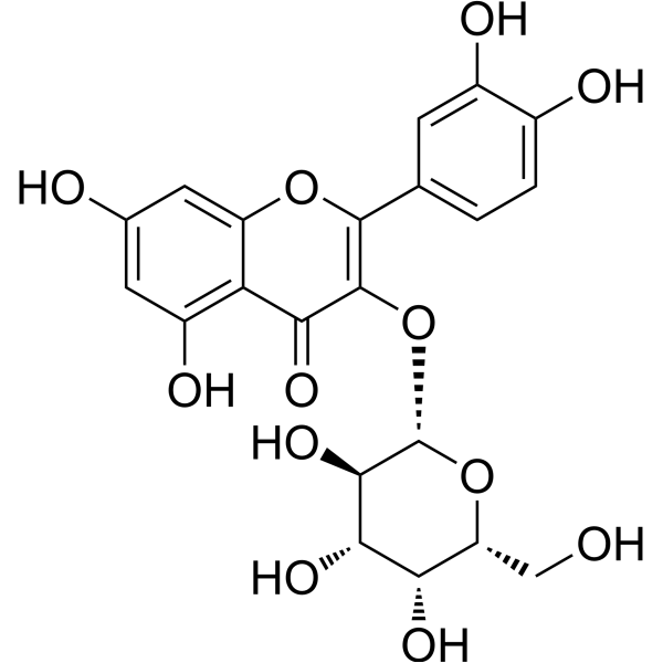
HY-N0452
HyperosideApoptosis Inducer -
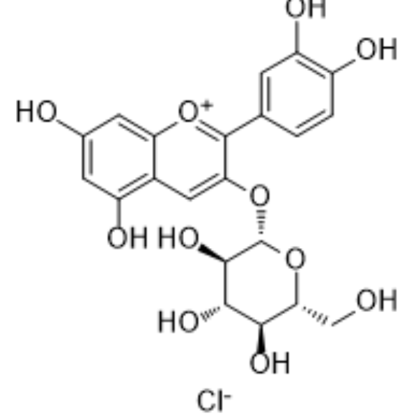
HY-N0640
Kuromanin chlorideBlood glucose regulator -
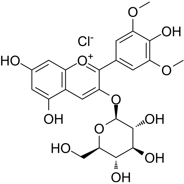
HY-125740
Malvidin-3-glucoside chlorideNF-κB Inhibitor -
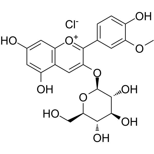
HY-W040127
Peonidin 3-O-glucoside chlorideInsulin Secretagogue -
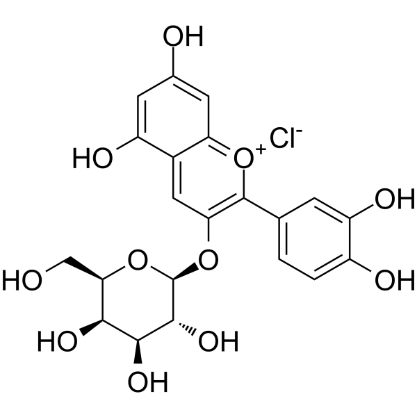
HY-N4142
Cyanidin-3-O-galactoside chlorideAntioxidant Agent -
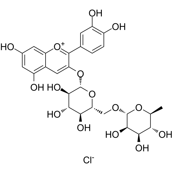
HY-105935
Keracyanin chlorideAntioxidant Agent -
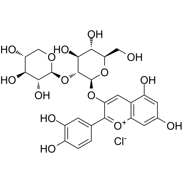
HY-N2533
Cyanidin 3-sambubioside chlorideAnthocyanin Compound -
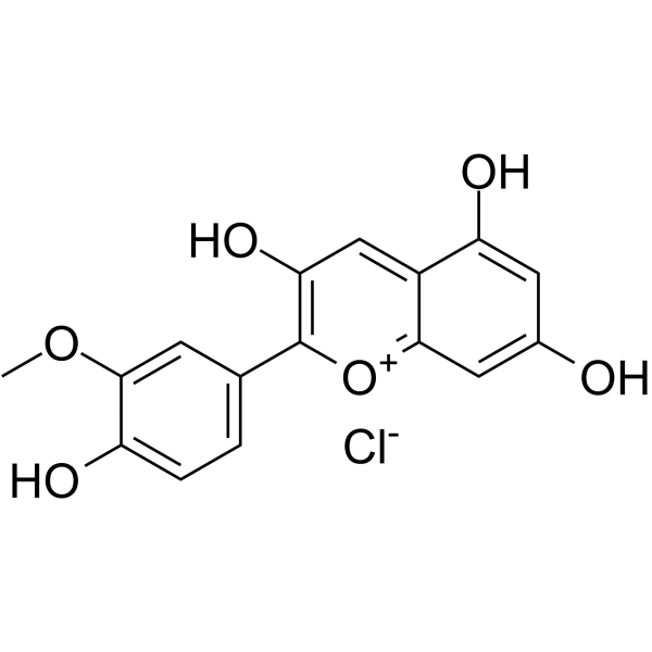
HY-N2459
Peonidin chlorideAnthocyanin Compound -
HY-123894
Pelargonidin 3,5-diglucoside chlorideNatural Pigment


Biological Activity
Description
|
IL-1β |
IL-1α |
IL-6 |
STAT3 |
In Vitro
Cyanidin 3,5-diglucoside chloride (10-50 μM) can significantly increase the survival rate of rat retinal Müller cells induced by hyperbaric pressure, inhibit the decrease in the expression of glutamate-aspartate transporter (GLAST) mRNA and protein in these cells caused by hyperbaric pressure[1].
Cyanidin 3,5-diglucoside chloride (1-10 μg/mL; 0-24 h) exert anti-inflammatory and skin barrier regulatory effects in the TNF-α/IL-17A/IFN-γ-induced HaCaT psoriasis model[2].
MedChemExpress (MCE) has not independently confirmed the accuracy of these methods. They are for reference only. Further protocols information, click here.
-
Cell Line:HaCaT human keratinocytes
-
Concentration:1 μg/mL, 5 μg/mL, 10 μg/mL
-
Incubation Time:2 h
-
Result:Concentration-dependently inhibited the phosphorylation of STAT3, IκB, ERK, p38, and AKT.


Chemical Information
-
CAS No. 2611-67-8
-
Appearance Solid
-
Molecular Weight 646.98
-
Formula C27H31ClO16
-
Color Brown to black
-
SMILES
O[C@@H]1[C@@H](O)[C@H](O)[C@@H](CO)O[C@H]1OC2=CC(O)=CC3=[O+]C(C4=CC=C(O)C(O)=C4)=C(O[C@H]5[C@H](O)[C@@H](O)[C@H](O)[C@@H](CO)O5)C=C23.[Cl-]
-
Structure Classification
-
Initial Source
-
Shipping
Room temperature in continental US; may vary elsewhere.
-
Storage
4°C, sealed storage, away from moisture and light
* In solvent : -80°C, 6 months; -20°C, 1 month (sealed storage, away from moisture and light)


Solvent & Solubility
In Vitro:
H2O : 50 mg/mL (77.28 mM; Need ultrasonic)
Please refer to the solubility information to select the appropriate solvent. Once prepared, please aliquot and store the solution to prevent product inactivation from repeated freeze-thaw cycles.
Storage method and period of stock solution: -80°C, 6 months; -20°C, 1 month (sealed storage, away from moisture and light). When stored at -80°C, please use it within 6 months. When stored at -20°C, please use it within 1 month.
* Note: If you choose water as the stock solution, please dilute it to the working solution, then filter and sterilize it with a 0.22 μm filter before use.
Please refer to the solubility information to select the appropriate solvent. Once prepared, please aliquot and store the solution to prevent product inactivation from repeated freeze-thaw cycles.
Storage method and period of stock solution: -80°C, 6 months; -20°C, 1 month (sealed storage, away from moisture and light). When stored at -80°C, please use it within 6 months. When stored at -20°C, please use it within 1 month.
* Note: If you choose water as the stock solution, please dilute it to the working solution, then filter and sterilize it with a 0.22 μm filter before use.
Concentration (start) × Volume (start) = Concentration (final) × Volume (final)

Protocols
-
Kinase activity and phosphorylation assays
Kinase activity assays measure the ability of kinases to transfer phosphate groups from ATP to specific substrates, while phosphorylation assays detect the presence and levels of phosphorylated proteins. Common methods include radiolabeled ATP incorporation (e. g. ,), ADP release detection via bioluminescence (e. g. ,[3]), enzyme-linked immunosorbent assays (ELISA) for phospho-specific epitopes (e. g. ,[6]), and microtiter-based formats for high-throughput screening (e. g. ,[8]). The ADP-Glo assay quantifies kinase activity by measuring ADP produced during phosphorylation using a luciferase-based system. Radiometric assays involve autoradiography or scintillation counting after incorporation of 32P-labeled ATP into substrate proteins. ELISA-based approaches rely on phospho-specific antibodies to detect activated kinases in cell lysates or purified samples.
-
RNA extraction experimental
By lysing cells, releasing RNA, and removing impurities such as proteins and DNA, high-purity RNA products are finally obtained. The commonly used traditional method is the guanidine isothiocyanate/phenol/chloroform method (Trizol), which is suitable for a variety of animal materials including animal tissues, microorganisms, cultured cells, etc., and most plant materials.
-
Western Blot
Western blotting (WB) is a commonly used experimental method in molecular biology, biochemistry, and immunogenetics for identifying and quantifying target proteins. It combines gel electrophoresis with immunoassay, enabling researchers to analyze protein expression, post-translational modifications, and molecular weight.
-
LPS-Induced Endotoxemia/Systemic Inflammation
Lipopolysaccharide (LPS)-induced endotoxemia is a widely used in vivo model of acute systemic inflammation in which LPS, a Gram-negative bacterial endotoxin, activates innate immune signaling primarily through TLR4, leading to rapid and transient induction of pro-inflammatory cytokines such as TNF-α, IL-6, and IL-1β in circulation and tissues. This cytokine surge is commonly used as a measurable readout of systemic inflammatory activation and immune dysregulation, and is typically assessed within hours after intraperitoneal LPS administration in mouse models of endotoxemia. The model captures key features of systemic inflammatory response syndrome, including cytokine release, immune cell activation, and downstream tissue responses, and has been used to evaluate anti-inflammatory interventions such as cytokine modulation, lipid mediators, and immune cell-targeting therapies.
-
Protocol for Kinase activity and phosphorylation assays
Kinase activity assays measure transfer of phosphate from ATP to a protein or peptide substrate, generating phosphorylated substrate, ADP, or incorporated radiolabeled phosphate as the readout; phosphorylation assays measure site-specific phosphorylation in cells or tissues as a proxy for kinase-pathway activation, inhibition, or substrate regulation. Phosphorylation can be detected by phospho-specific Western blot, immunoprecipitation kinase assay, phospho-immunofluorescence, phospho-flow cytometry, luminescent ADP detection, radiolabeled ATP incorporation, or reporter-based pathway assays, and these readouts can be applied to cancer cells, primary neurons, mouse tumors, organoids, inflammatory macrophages, ferroptosis studies, and mitophagy studies when the kinase target is biologically relevant.
-
Research Protocol for Inflammation-related Diseases
The NLRP3 inflammasome is a cytosolic innate immune signaling platform that integrates priming signals and danger-signal activation to promote caspase-1 activation, maturation of IL-1β and IL-18, and gasdermin D-mediated pyroptotic cell death. The core experimental logic is to determine whether inflammatory disease phenotypes are driven by increased NLRP3 expression, ASC-containing inflammasome assembly, caspase-1 cleavage, GSDMD cleavage, and extracellular release of IL-1β/IL-18 rather than by nonspecific cell injury alone. The pathway is strongly linked to inflammation-related disease phenotypes because monosodium urate crystals activate NALP3/NLRP3 inflammasome signaling in gout-like crystal inflammation, cholesterol crystals activate NLRP3 inflammasomes in atherogenesis models, and DSS-induced intestinal inflammation has been reported to involve NLRP3 inflammasome activity. However, experimental colitis studies also show context-dependent protective effects of NLRP3 inflammasome co
-
Pyroptosis Solutions
Pyroptosis is a lytic inflammatory cell-death pathway executed by gasdermin pores, most classically through inflammasome-mediated activation of caspase-1, cleavage of gasdermin D, membrane pore formation, LDH release, and secretion of IL-1β and IL-18. The canonical pathway is commonly modeled by priming cells with an inflammatory signal such as LPS to induce pro-IL-1β and inflammasome components, followed by an activation signal such as ATP or nigericin to activate NLRP3, ASC speck formation, caspase-1 cleavage, GSDMD cleavage, cytokine release, and pyroptotic membrane rupture. The non-canonical pathway is triggered when cytosolic LPS activates mouse caspase-11 or human caspase-4/5, leading to GSDMD cleavage and pyroptosis, and this can secondarily activate NLRP3-dependent IL-1β release. Pyroptosis is linked to inflammatory injury, infection, cancer, liver disease, ocular disease, placental inflammation, and other disease phenotypes, but unresolved questions include which gasdermin fam


Purity & Documentation
-
Data Sheet (282 KB)
-
SDS (394 KB)
- English - EN (394 KB)
- Français - FR (394 KB)
- Deutsch - DE (394 KB)
- Norwegian - NO (394 KB)
- Español - ES (394 KB)
- Swedish - SV (394 KB)
- Italian - IT (394 KB)
- Korean - KR (394 KB)
- Portuguese - PT (394 KB)
-
Handling Instructions (2659 KB)
References


Complete Stock Solution Preparation Table
Please refer to the solubility information to select the appropriate solvent. Once prepared, please aliquot and store the solution to prevent product inactivation from repeated freeze-thaw cycles.
Storage method and period of stock solution: -80°C, 6 months; -20°C, 1 month (sealed storage, away from moisture and light). When stored at -80°C, please use it within 6 months. When stored at -20°C, please use it within 1 month.
| Optional Solvent | Concentration Solvent Mass | 1 mg | 5 mg | 10 mg | 25 mg |
|---|---|---|---|---|---|
| H2O | 1 mM | 1.5456 mL | 7.7282 mL | 15.4564 mL | 38.6411 mL |
| 5 mM | 0.3091 mL | 1.5456 mL | 3.0913 mL | 7.7282 mL | |
| 10 mM | 0.1546 mL | 0.7728 mL | 1.5456 mL | 3.8641 mL | |
| 15 mM | 0.1030 mL | 0.5152 mL | 1.0304 mL | 2.5761 mL | |
| 20 mM | 0.0773 mL | 0.3864 mL | 0.7728 mL | 1.9321 mL | |
| 25 mM | 0.0618 mL | 0.3091 mL | 0.6183 mL | 1.5456 mL | |
| 30 mM | 0.0515 mL | 0.2576 mL | 0.5152 mL | 1.2880 mL | |
| 40 mM | 0.0386 mL | 0.1932 mL | 0.3864 mL | 0.9660 mL | |
| 50 mM | 0.0309 mL | 0.1546 mL | 0.3091 mL | 0.7728 mL | |
| 60 mM | 0.0258 mL | 0.1288 mL | 0.2576 mL | 0.6440 mL |
* Note: If you choose water as the stock solution, please dilute it to the working solution, then filter and sterilize it with a 0.22 μm filter before use.


Cyanidin 3,5-diglucoside chloride Related Classifications
HY-129138 Related Classifications
 Powered by Bioz