



Manumycin A
Based on 2 publication(s) in Google Scholar
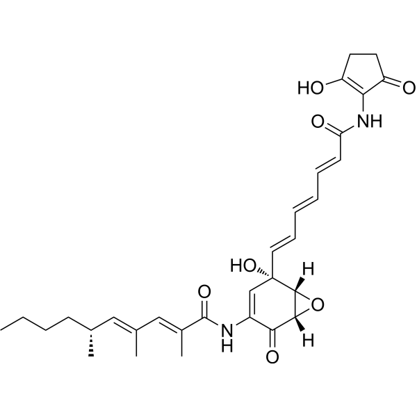
Manumycin A is a polyketide antibiotic and an inhibitor of thioredoxin reductase 1 (TrxR-1). Manumycin A can inhibit the growth of breast cancer cells and exert its anti-tumor activity through LC3. Manumycin A can downregulate the release of pro-inflammatory cytokines in human monocytes stimulated by TNF α, and has potential anti-inflammatory activity. Manumycin A can inhibit the Ras/Raf/ERK1/2 signaling and hnRNP H1 in castration resistant prostate cancer cells to suppress exosome biogenesis and secretion.
For research use only. We do not sell to patients.
- Purity : 98.30%
- CAS No.: 52665-74-4
- Formula: C31H38N2O7
- Molecular Weight:550.64
-
Storage:
-20°C, sealed storage, away from moisture
* In solvent : -80°C, 6 months; -20°C, 1 month (sealed storage, away from moisture)

To place orders, for customer services and technical support, please contact: MedChemExpress USA
Tel: 609-228-6898 E-mail: [email protected] [email protected]


-
 Biological Activity
-
Chemical Information
-
Solvent & Solubility
- Protocols
- Purity & Documentation
- References
-
 Help & FAQs


-
HY-19363
GW4869N-SMase Inhibitor -
HY-10502
TipifarnibFTase Inhibitor -
HY-15872A
FTI-277 hydrochlorideFTase Inhibitor -
HY-100223
CalpeptinCalpain Inhibitor -
HY-15136
LonafarnibFarnesyltransferase Inhibitor -

HY-10071
Y-27632ROCK Inhibitor -
HY-16592
Brefeldin AProtein Transport Inhibitor -

HY-18739
Phorbol 12-myristate 13-acetatePKC Activator -
HY-50876
DaporinadNMPRT Inhibitor -
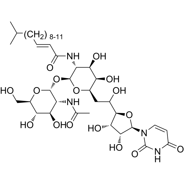
HY-A0098
TunicamycinER Stress Inducer -

HY-13259
MG-132Proteasome Inhibitor -
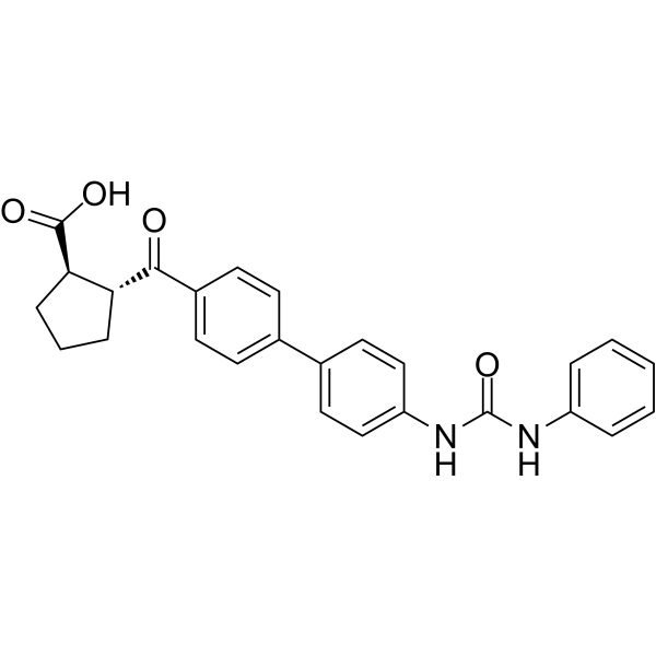
HY-10038
A 922500DGAT-1 Inhibitor
Publications Citing Use of MedChemExpress (MCE) Manumycin A
MoreAll Antibiotic Isoforms
MoreAll Phospholipase Isoforms
More

Biological Activity
Description
IC50 & Target
In Vitro
Manumycin A (0.25-5 μM, 24 h) downregulates the release of pro-inflammatory cytokines in human monocytes stimulated by TNF α, thus possessing potential anti-inflammatory properties[1]. Manumycin A (0-25 μM, 72 h) can enhance its inhibitory effect on lung cancer cells after inhibiting HSF1 mediated heat shock response (HSR)[2]. Manumycin A (5 μM, 72 h) inhibits the growth of triple negative breast cancer cells through LC3 mediated cytoplasmic vacuolation death[3]. Manumycin A (250 nM, 48 h) inhibits exosome biogenesis and secretion by targeting Ras/Raf/ERK1/2 signaling and hnRNP H1 in castration resistant prostate cancer cells[4].
MedChemExpress (MCE) has not independently confirmed the accuracy of these methods. They are for reference only. Further protocols information, click here.
-
Cell Line:Human monocytic leukemia cells THP-1
-
Concentration:0.25-5 μM
-
Incubation Time:24 h
-
Result:Inhibited the production and release of IL-1 β, IL-6, and IL-8.
-
Cell Line:Lung cancer cell lines NCI-H1299, NCI-H23, A549
-
Concentration:0-25 μM
-
Incubation Time:72 h
-
Result:Enhanced growth inhibition of NSCLC cells after inhibiting HSPA1 expression.
-
Cell Line:Triple-negative breast cancer (TNBC) cells (MDA-MB-231, HCC1937, BT-20)
-
Concentration:5 μM
-
Incubation Time:72 h
-
Result:Inhibited the cell viability of different TNBC cell lines.
-
Cell Line:C4-2B cell
-
Concentration:250 nM
-
Incubation Time:48 h
-
Result:Reduced protein levels of extracellular vesicle biogenic markers Alix, Hrs, and Rab27a.
In Vivo
MedChemExpress (MCE) has not independently confirmed the accuracy of these methods. They are for reference only.
-
Animal Model:Female nude mice with breast cancer transplanted with MDAMB-231 cells[3].
-
Dosage:5 mg/kg
-
Administration:Intraperitoneal injection (i.p.); Once every two days; 2 weeks
-
Result:Inhibited tumor growth and increased levels of ER stress markers such as Bip, CHOP, LC3, and p62 proteins, as well as higher molecular weight ubiquitinated proteins.
-
Animal Model:HSALR transgenic mice[5].
-
Dosage:3 μg
-
Administration:Intramuscular injection (i.m.); once
-
Result:Reduced the content of Clcn1 exon 7A introns.


Chemical Information
-
CAS No. 52665-74-4
-
Appearance Solid
-
Molecular Weight 550.64
-
Formula C31H38N2O7
-
Color Yellow to orange
-
SMILES
CCCC[C@@H](C)/C=C(C)/C=C(C)/C(NC1=C[C@](/C=C/C=C/C=C/C(NC2=C(O)CCC2=O)=O)(O)[C@]3([H])O[C@]3([H])C1=O)=O
-
Structure Classification
-
Initial Source
new Streptomyces strain
-
Shipping
Room temperature in continental US; may vary elsewhere.
-
Storage
-20°C, sealed storage, away from moisture
* In solvent : -80°C, 6 months; -20°C, 1 month (sealed storage, away from moisture)


Publications (2)
-
Journal Impact Factor
-
Most Recent
-
Inflamm Res
Lipopolysaccharide (LPS) induces sclerostin secretion by extracellular vesicle via TLR4/miR-92a-3p/PTEN/NF-κB signalling pathway in murine macrophage. [Abstract]2025 Jan 25;74(1):27. PMID: 39862242 -


Solvent & Solubility
In Vitro:
DMSO : 50 mg/mL (90.80 mM; Need ultrasonic; Hygroscopic DMSO has a significant impact on the solubility of product, please use newly opened DMSO)
Please refer to the solubility information to select the appropriate solvent. Once prepared, please aliquot and store the solution to prevent product inactivation from repeated freeze-thaw cycles.
Storage method and period of stock solution: -80°C, 6 months; -20°C, 1 month (sealed storage, away from moisture). When stored at -80°C, please use it within 6 months. When stored at -20°C, please use it within 1 month.
Please refer to the solubility information to select the appropriate solvent. Once prepared, please aliquot and store the solution to prevent product inactivation from repeated freeze-thaw cycles.
Storage method and period of stock solution: -80°C, 6 months; -20°C, 1 month (sealed storage, away from moisture). When stored at -80°C, please use it within 6 months. When stored at -20°C, please use it within 1 month.
Concentration (start) × Volume (start) = Concentration (final) × Volume (final)
In Vivo:
Select the appropriate dissolution method based on your experimental animal and administration route.
- For the following dissolution methods, please ensure to first prepare a clear stock solution using an In Vitro approach and then sequentially add co-solvents:
- To ensure reliable experimental results, the clarified stock solution can be appropriately stored based on storage conditions. As for the working solution for In Vivo experiments, it is recommended to prepare freshly and use it on the same day.
- The percentages shown for the solvents indicate their volumetric ratio in the final prepared solution. If precipitation or phase separation occurs during preparation, heat and/or sonication can be used to aid dissolution.
Add each solvent one by one: 10% DMSO 40% PEG300 5% Tween-80 45% Saline
Solubility: ≥ 1.25 mg/mL (2.27 mM); Clear solution
This protocol yields a clear solution of ≥ 1.25 mg/mL (saturation unknown).
Taking 1 mL working solution as an example, add 100 μL DMSO stock solution (12.5 mg/mL) to 400 μL PEG300, and mix evenly; then add 50 μL Tween-80 and mix evenly; then add 450 μL Saline to adjust the volume to 1 mL.
Preparation of Saline: Dissolve 0.9 g sodium chloride in ddH₂O and dilute to 100 mL to obtain a clear Saline solution.
In Vivo Dissolution Calculator
Please enter the basic information of animal experiments:
-
-
-
-
Recommended: Prepare an additional quantity of animals to account for potential losses during experiments.
Please enter your animal formula composition:
-
%DMSO +
Recommended: Keep the proportion of DMSO in working solution below 2% if your animal is weak.
-
%+
-
+%Tween-80 + +
-
%Saline +
The co-solvents required include: DMSO, . All of co-solvents are available by MedChemExpress (MCE). , Tween 80. All of co-solvents are available by MedChemExpress (MCE).
Working solution concentration: 0.22 mg/mL
Method for preparing stock solution: mg drug dissolved in μL DMSO. Stock solution concentration: mg/mL. * In solvent : -80°C, 6 months; -20°C, 1 month (sealed storage, away from moisture)
1. Take μL DMSO stock solution;
2. Add μL .
μL , mix evenly;
3. Then add μL Tween 80, mix evenly;
4. Then add μL
Please ensure that the stock solution in the first step is dissolved to a clear state, and add co-solvents in sequence. You can use ultrasonic heating (ultrasonic cleaner, recommended frequency 20-40 kHz), vortexing, etc. to assist dissolution.

Protocols
-
LPS-Induced Endotoxemia/Systemic Inflammation
Lipopolysaccharide (LPS)-induced endotoxemia is a widely used in vivo model of acute systemic inflammation in which LPS, a Gram-negative bacterial endotoxin, activates innate immune signaling primarily through TLR4, leading to rapid and transient induction of pro-inflammatory cytokines such as TNF-α, IL-6, and IL-1β in circulation and tissues. This cytokine surge is commonly used as a measurable readout of systemic inflammatory activation and immune dysregulation, and is typically assessed within hours after intraperitoneal LPS administration in mouse models of endotoxemia. The model captures key features of systemic inflammatory response syndrome, including cytokine release, immune cell activation, and downstream tissue responses, and has been used to evaluate anti-inflammatory interventions such as cytokine modulation, lipid mediators, and immune cell-targeting therapies.
-
Cotton Pellet Granuloma
Cotton pellet granuloma is a classical in vivo chronic inflammation model used to evaluate the anti-inflammatory potential of test substances by measuring their ability to inhibit granuloma tissue formation around an implanted foreign body (cotton pellet) in rodents. The method is based on the biological response to a sterile implanted material, which induces proliferative phase inflammation characterized by fibroblast proliferation and collagen-rich granuloma formation, and the final readout reflects the extent of chronic inflammatory tissue growth surrounding the pellet. In multiple preclinical pharmacological evaluations, inhibition of cotton pellet-induced granuloma formation has been used as an indicator of anti-inflammatory activity in both synthetic and natural product screening contexts.
-
Carrageenan-Induced Paw Edema
Carrageenan-induced paw edema is an acute inflammation model in which intraplantar injection of carrageenan induces localized inflammatory swelling characterized by vascular permeability, leukocyte infiltration, and production of inflammatory mediators such as prostaglandins and cytokines, making it widely used to evaluate anti-inflammatory agents in vivo. The resulting paw volume or thickness increase is quantified over time as a direct readout of inflammatory intensity and drug efficacy, typically reflecting cyclooxygenase-mediated prostaglandin-driven edema formation and immune cell recruitment in peripheral tissue[20].
-
Autophagy
Autophagy is a process in which eukaryotic cells use lysosomes to degrade their own cytoplasmic proteins and damaged organelles under the regulation of autophagy related gene (Atg). Microtubule-associated proteins light chain 3 (LC3) is recognized as autophagy marker, which transfers from cytoplasmic LC3 (LC3-I) to membrane type (LC3-II). LC3-II/I ratio could be detected by Western Blot and fluorescence microscopy.
-
Research Protocol for Inflammation-related Diseases
The NLRP3 inflammasome is a cytosolic innate immune signaling platform that integrates priming signals and danger-signal activation to promote caspase-1 activation, maturation of IL-1β and IL-18, and gasdermin D-mediated pyroptotic cell death. The core experimental logic is to determine whether inflammatory disease phenotypes are driven by increased NLRP3 expression, ASC-containing inflammasome assembly, caspase-1 cleavage, GSDMD cleavage, and extracellular release of IL-1β/IL-18 rather than by nonspecific cell injury alone. The pathway is strongly linked to inflammation-related disease phenotypes because monosodium urate crystals activate NALP3/NLRP3 inflammasome signaling in gout-like crystal inflammation, cholesterol crystals activate NLRP3 inflammasomes in atherogenesis models, and DSS-induced intestinal inflammation has been reported to involve NLRP3 inflammasome activity. However, experimental colitis studies also show context-dependent protective effects of NLRP3 inflammasome co
-
Breast Cancer Modeling
Breast cancer is a heterogeneous cancer, and it has been distinguished into four subtypes: luminal A, luminal B, HER2-positive and basal-like. Molecular mutations, epigenetic alterations, hormone exposure and immune microenvironment are related to the progression of breast cancer.
-
Macroautophagy Solutions
Macroautophagy is a conserved lysosome-dependent degradation pathway in which cytoplasmic material is sequestered into double-membrane autophagosomes and delivered to lysosomes for degradation and recycling. The pathway supports cellular homeostasis during nutrient limitation, organelle stress, protein-aggregate accumulation, infection, differentiation, and tissue remodeling by coupling cargo sequestration, autophagosome maturation, lysosomal fusion, and degradation of cargo-derived macromolecules. The core molecular sequence includes initiation by nutrient- and stress-regulated autophagy machinery, autophagosome nucleation, LC3/ATG8-family conjugation to autophagosomal membranes, cargo selection through receptors such as SQSTM1/p62, autophagosome-lysosome fusion, and lysosomal degradation. LC3 was identified as a mammalian homolog of yeast Atg8 that localizes to autophagosomal membranes after processing, and p62/SQSTM1 was shown to connect ubiquitinated cargo with autophagic degradati


Purity & Documentation
-
Data Sheet (280 KB)
-
SDS (393 KB)
- English - EN (393 KB)
- Français - FR (393 KB)
- Deutsch - DE (393 KB)
- Norwegian - NO (393 KB)
- Español - ES (393 KB)
- Swedish - SV (393 KB)
- Italian - IT (393 KB)
- Korean - KR (393 KB)
- Portuguese - PT (393 KB)
-
Handling Instructions (2659 KB)
References
[1]. Cecrdlova E, et al. Manumycin A downregulates release of proinflammatory cytokines from TNF alpha stimulated human monocytes. Immunol Lett. 2016 Jan;169:8-14. [Content Brief]
[2]. Sojka DR, et al. Inhibition of the Heat Shock Protein A (HSPA) Family Potentiates the Anticancer Effects of Manumycin A. Cells. 2021 Jun 7;10(6):1418. [Content Brief]
[4]. Datta A, et al. Manumycin A suppresses exosome biogenesis and secretion via targeted inhibition of Ras/Raf/ERK1/2 signaling and hnRNP H1 in castration-resistant prostate cancer cells. Cancer Lett. 2017 Nov 1;408:73-81. [Content Brief]
[5]. Oana K, et al. Manumycin A corrects aberrant splicing of Clcn1 in myotonic dystrophy type 1 (DM1) mice. Sci Rep. 2013;3:2142. [Content Brief]


Complete Stock Solution Preparation Table
Please refer to the solubility information to select the appropriate solvent. Once prepared, please aliquot and store the solution to prevent product inactivation from repeated freeze-thaw cycles.
Storage method and period of stock solution: -80°C, 6 months; -20°C, 1 month (sealed storage, away from moisture). When stored at -80°C, please use it within 6 months. When stored at -20°C, please use it within 1 month.
| Optional Solvent | Concentration Solvent Mass | 1 mg | 5 mg | 10 mg | 25 mg |
|---|---|---|---|---|---|
| DMSO | 1 mM | 1.8161 mL | 9.0803 mL | 18.1607 mL | 45.4017 mL |
| 5 mM | 0.3632 mL | 1.8161 mL | 3.6321 mL | 9.0803 mL | |
| 10 mM | 0.1816 mL | 0.9080 mL | 1.8161 mL | 4.5402 mL | |
| 15 mM | 0.1211 mL | 0.6054 mL | 1.2107 mL | 3.0268 mL | |
| 20 mM | 0.0908 mL | 0.4540 mL | 0.9080 mL | 2.2701 mL | |
| 25 mM | 0.0726 mL | 0.3632 mL | 0.7264 mL | 1.8161 mL | |
| 30 mM | 0.0605 mL | 0.3027 mL | 0.6054 mL | 1.5134 mL | |
| 40 mM | 0.0454 mL | 0.2270 mL | 0.4540 mL | 1.1350 mL | |
| 50 mM | 0.0363 mL | 0.1816 mL | 0.3632 mL | 0.9080 mL | |
| 60 mM | 0.0303 mL | 0.1513 mL | 0.3027 mL | 0.7567 mL | |
| 80 mM | 0.0227 mL | 0.1135 mL | 0.2270 mL | 0.5675 mL |


Manumycin A Related Classifications
HY-N6796 Related Classifications
 Powered by Bioz