



EF24
Based on 2 publication(s) in Google Scholar
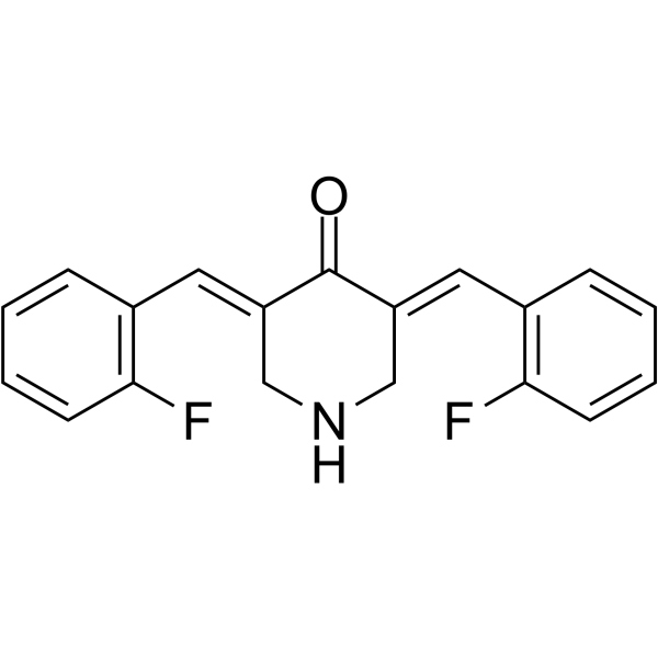
EF24, a curcumin analogue, is an NF-kB inhibitor with great anti-tumor efficacy and oral bioavailability via deactivation of the MAPK/ERK signaling pathway in oral squamous cell carcinoma (OSCC). EF24 is active against melanoma and breast cancer cell lines with GI50 values of 0.7 μM and 0.8 μM, respectively. EF24 induces cell cycle arrest and apoptosis in MDA-MB-231 human breast cancer cells and DU-145 human prostate cancer cells. EF24 increases the levels of activated caspase 3 and 9, and decreases the phosphorylated forms of MEK1 and ERK.
For research use only. We do not sell to patients.
- Purity : 99.34%
- CAS No.: 342808-40-6
- Formula: C19H15F2NO
- Molecular Weight:311.33
-
Storage:Powder -20°C, 3 years , 4°C, 2 years ; In solvent -80°C, 6 months , -20°C, 1 month

To place orders, for customer services and technical support, please contact: MedChemExpress USA
Tel: 609-228-6898 E-mail: [email protected] [email protected]


-
 Biological Activity
-
Chemical Information
-
Solvent & Solubility
- Protocols
- Purity & Documentation
- References
-
 Help & FAQs


-
HY-18085
QuercetinFlavonoid Antioxidant -
HY-17364
TemozolomideDNA Alkylator -

HY-10227
BortezomibProteasome Inhibitor -
HY-N0182
FisetinTNF Receptor Inhibitor -
HY-N0005
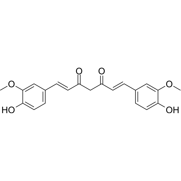
Curcuminp300 Histone Acetylatransferase Inhibitor -
HY-10181
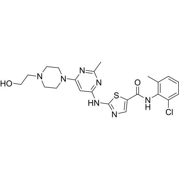
DasatinibSrc/Bcr-Abl Inhibitor -

HY-100579
Ferrostatin-1Ferroptosis Inhibitor -
HY-50846
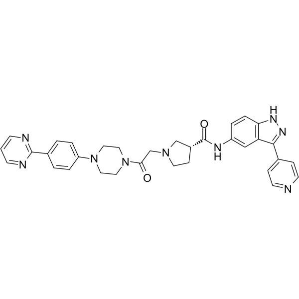
SCH772984ERK1/2 Inhibitor -
HY-15816
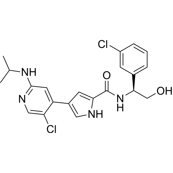
UlixertinibERK1/2 Inhibitor -
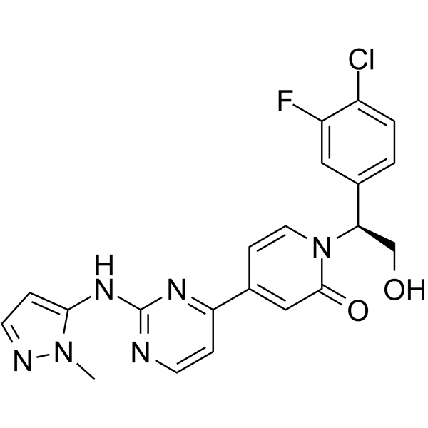
HY-15947
RavoxertinibERK1/2 Inhibitor -
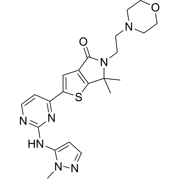
HY-101494
TemuterkibERK1/ERK2 Inhibitor -
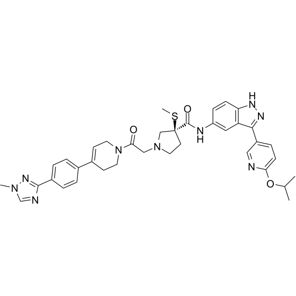
HY-111407
MK-8353ERK1/2 Inhibitor
Publications Citing Use of MedChemExpress (MCE) EF24
MoreAll Caspase Isoforms
More

Biological Activity
Description
Cellular Effect
|
Cell Line

|
Type | Value | Description | References |
|---|---|---|---|---|
| 518A2 | IC50 |
1.8 μM
Compound: 2; EF24
|
Cytotoxicity against human 518A2 cells after 72 hrs by MTT assay
Cytotoxicity against human 518A2 cells after 72 hrs by MTT assay
|
[PMID: 28774574] |
| A2780 | IC50 |
0.5 μM
Compound: EF24
|
Cell viability of human cisplatin-sensitive human A2780 cells after 24 hrs by MTT assay
Cell viability of human cisplatin-sensitive human A2780 cells after 24 hrs by MTT assay
|
[PMID: 17684018] |
| A2780 | IC50 |
0.65 μM
Compound: EF24
|
Cell viability of cisplatin-resistant human A2780 cells after 24 hrs by MTT assay
Cell viability of cisplatin-resistant human A2780 cells after 24 hrs by MTT assay
|
[PMID: 17684018] |
| A549 | IC50 |
1.3 μM
Compound: 1, EF24
|
Inhibition of NF-kappaB nuclear translocation in human A549 cells
Inhibition of NF-kappaB nuclear translocation in human A549 cells
|
[PMID: 19854644] |
| A549 | IC50 |
2.4 μM
Compound: EF24
|
Cytotoxicity against human A549 cells assessed as reduction in cell viability after 72 hrs by MTT assay
Cytotoxicity against human A549 cells assessed as reduction in cell viability after 72 hrs by MTT assay
|
[PMID: 29655083] |
| A549 | IC50 |
2.8 μM
Compound: A1; EF24
|
Antiproliferative activity against human A549 cells after 72 hrs by MTT assay
Antiproliferative activity against human A549 cells after 72 hrs by MTT assay
|
[PMID: 29351887] |
| A549 | IC50 |
7.1 μM
Compound: EF24
|
Cytotoxicity against human A549 cells measured after 72 hrs by MTT assay
Cytotoxicity against human A549 cells measured after 72 hrs by MTT assay
|
[PMID: 27886548] |
| CHO | IC50 |
4.6 μM
Compound: EF24
|
Cell viability of CHO cells after 24 hrs by MTT assay
Cell viability of CHO cells after 24 hrs by MTT assay
|
[PMID: 17684018] |
| DLD-1 | IC50 |
1.3 μM
Compound: 2; EF24
|
Cytotoxicity against human DLD1 cells after 72 hrs by MTT assay
Cytotoxicity against human DLD1 cells after 72 hrs by MTT assay
|
[PMID: 28774574] |
| HCT-116 | IC50 |
1.5 μM
Compound: 2; EF24
|
Cytotoxicity against human HCT116 cells after 72 hrs by MTT assay
Cytotoxicity against human HCT116 cells after 72 hrs by MTT assay
|
[PMID: 28774574] |
| HEK293 | CC50 |
3.58 μM
Compound: 33
|
Cytotoxicity against human HEK293 cells assessed as reduction in cell viability at 20 uM measured after 48 hrs by resazurin reagent based multilabel reader analysis
Cytotoxicity against human HEK293 cells assessed as reduction in cell viability at 20 uM measured after 48 hrs by resazurin reagent based multilabel reader analysis
|
[PMID: 36608774] |
| HeLa 229 | IC50 |
1.9 μM
Compound: A1; EF24
|
Antiproliferative activity against human HeLa 229 cells after 72 hrs by MTT assay
Antiproliferative activity against human HeLa 229 cells after 72 hrs by MTT assay
|
[PMID: 29351887] |
| HepG2 | IC50 |
3.33 μM
Compound: EF24
|
Cytotoxicity against human HepG2 cells assessed as reduction in cell viability incubated for 24 hrs by MTT assay
Cytotoxicity against human HepG2 cells assessed as reduction in cell viability incubated for 24 hrs by MTT assay
|
[PMID: 30771605] |
| HT-29 | IC50 |
1.6 μM
Compound: 2; EF24
|
Cytotoxicity against human HT-29 cells after 72 hrs by MTT assay
Cytotoxicity against human HT-29 cells after 72 hrs by MTT assay
|
[PMID: 28774574] |
| L02 | IC50 |
10.2 μM
Compound: EF24
|
Cytotoxicity against human LO2 cells assessed as reduction in cell viability incubated for 24 hrs by MTT assay
Cytotoxicity against human LO2 cells assessed as reduction in cell viability incubated for 24 hrs by MTT assay
|
[PMID: 30771605] |
| L02 | IC50 |
5.1 μM
Compound: EF24
|
Cytotoxicity against human HL7702 cells assessed as reduction in cell viability after 72 hrs by MTT assay
Cytotoxicity against human HL7702 cells assessed as reduction in cell viability after 72 hrs by MTT assay
|
[PMID: 29655083] |
| L02 | IC50 |
5.8 μM
Compound: EF24
|
Antiproliferative activity against human HL7702 cells by MTT assay
Antiproliferative activity against human HL7702 cells by MTT assay
|
[PMID: 31336310] |
| Lewis lung carcinoma cell line | IC50 |
8.4 μM
Compound: EF24
|
Cytotoxicity against mouse LLC cells measured after 72 hrs by MTT assay
Cytotoxicity against mouse LLC cells measured after 72 hrs by MTT assay
|
[PMID: 27886548] |
| MDA-MB-231 | IC50 |
5.6 μM
Compound: A1
|
Antiproliferative activity against human MDA-MB-231 cells assessed as inhibition of cell proliferation by CCK-8 assay
Antiproliferative activity against human MDA-MB-231 cells assessed as inhibition of cell proliferation by CCK-8 assay
|
[PMID: 38746900] |
| MDA-MB-435 | IC50 |
1.5 μM
Compound: 1, EF24
|
Cytotoxicity against human MDA-MB-435 cells
Cytotoxicity against human MDA-MB-435 cells
|
[PMID: 19854644] |
| MGC-803 | IC50 |
2 μM
Compound: A1; EF24
|
Antiproliferative activity against human MGC803 cells after 72 hrs by MTT assay
Antiproliferative activity against human MGC803 cells after 72 hrs by MTT assay
|
[PMID: 29351887] |
| NB-4 | IC50 |
0.29 μM
Compound: 42
|
Cytotoxicity against human NB4 cells assessed as reduction in cell viability after 72 hrs by MTS assay
Cytotoxicity against human NB4 cells assessed as reduction in cell viability after 72 hrs by MTS assay
|
[PMID: 24960549] |
| NCI-H1650 | IC50 |
1.7 μM
Compound: EF24
|
Cytotoxicity against human NCI-H1650 cells assessed as reduction in cell viability after 72 hrs by MTT assay
Cytotoxicity against human NCI-H1650 cells assessed as reduction in cell viability after 72 hrs by MTT assay
|
[PMID: 29655083] |
| NCI-H1650 | IC50 |
14.6 μM
Compound: EF24
|
Cytotoxicity against human NCI-H1650 cells measured after 72 hrs by MTT assay
Cytotoxicity against human NCI-H1650 cells measured after 72 hrs by MTT assay
|
[PMID: 27886548] |
| NCI-H1975 | IC50 |
1.3 μM
Compound: EF24
|
Cytotoxicity against human NCI-H1975 cells assessed as reduction in cell viability after 72 hrs by MTT assay
Cytotoxicity against human NCI-H1975 cells assessed as reduction in cell viability after 72 hrs by MTT assay
|
[PMID: 29655083] |
| NCI-H441 | IC50 |
44 μM
Compound: 1
|
Antiproliferative activity against human H441 cells assessed as decrease of hexosaminidase activity after 24 hrs
Antiproliferative activity against human H441 cells assessed as decrease of hexosaminidase activity after 24 hrs
|
[PMID: 20638855] |
| NCI-H460 | IC50 |
1.2 μM
Compound: EF24
|
Cytotoxicity against human H460 cells assessed as reduction in cell viability after 72 hrs by MTT assay
Cytotoxicity against human H460 cells assessed as reduction in cell viability after 72 hrs by MTT assay
|
[PMID: 29655083] |
| NCI-H460 | IC50 |
6 μM
Compound: EF24
|
Antiproliferative activity against human H460 cells by MTT assay
Antiproliferative activity against human H460 cells by MTT assay
|
[PMID: 31336310] |
| PANC-1 | IC50 |
1.5 μM
Compound: 2; EF24
|
Cytotoxicity against human PANC1 cells after 72 hrs by MTT assay
Cytotoxicity against human PANC1 cells after 72 hrs by MTT assay
|
[PMID: 28774574] |
| QGY-7703 | IC50 |
4.97 μM
Compound: EF24
|
Cytotoxicity against human QGY7703 cells assessed as reduction in cell viability incubated for 24 hrs by MTT assay
Cytotoxicity against human QGY7703 cells assessed as reduction in cell viability incubated for 24 hrs by MTT assay
|
[PMID: 30771605] |
| SGC-7901 | IC50 |
1 μM
Compound: A1; EF24
|
Antiproliferative activity against human SGC7901 cells after 72 hrs by MTT assay
Antiproliferative activity against human SGC7901 cells after 72 hrs by MTT assay
|
[PMID: 29351887] |
| SGC-7901 | IC50 |
6.5 μM
Compound: EF24
|
Antiproliferative activity against human SGC7901 cells by MTT assay
Antiproliferative activity against human SGC7901 cells by MTT assay
|
[PMID: 31336310] |
| SMMC-7721 | IC50 |
8.8 μM
Compound: EF24
|
Cytotoxicity against human SMMC7721 cells assessed as reduction in cell viability incubated for 24 hrs by MTT assay
Cytotoxicity against human SMMC7721 cells assessed as reduction in cell viability incubated for 24 hrs by MTT assay
|
[PMID: 30771605] |
| U-251 | IC50 |
4.5 μM
Compound: EF24
|
Antiproliferative activity against human U251 cells by MTT assay
Antiproliferative activity against human U251 cells by MTT assay
|
[PMID: 31336310] |
| U-87MG ATCC | IC50 |
1.4 μM
Compound: 2; EF24
|
Cytotoxicity against human U87 cells after 72 hrs by MTT assay
Cytotoxicity against human U87 cells after 72 hrs by MTT assay
|
[PMID: 28774574] |
In Vitro
EF24 (0-10 μM) exhibits cytotoxicity against human breast cancer cells in a dose-dependent increase[2].
EF24 (0.1-100 μM, 6-72 h) effectively inhibits the active synthesis of DNA in both DU-145 prostate cancer cells and MDA-MB-231 breast cancer cells[3].
EF24 (20 μM, 24-72 h) induces cell cycle arrest and apoptosis by means of a redox-dependent mechanism and produces a time-dependent increase in intracellular ROS in MDA-MB-231 human breast cancer cells and DU-145 human prostate cancer cells[3].
MedChemExpress (MCE) has not independently confirmed the accuracy of these methods. They are for reference only. Further protocols information, click here.
-
Cell Line:DU-145 prostate cancer cells and MDA-MB-231 breast cancer cells
-
Concentration:0.1-100 μM
-
Incubation Time:6-72 h
-
Result:Demonstrates approximately 40% inhibition at 1 μM (10-6 M) after a 72-h treatment in the breast cancer cells.
-
Cell Line:DU-145 prostate cancer cells and MDA-MB-231 breast cancer cells
-
Concentration:20 μM
-
Incubation Time:24-72 h
-
Result:Caused a G2/M arrest in both cell lines.
In Vivo
MedChemExpress (MCE) has not independently confirmed the accuracy of these methods. They are for reference only.
-
Animal Model:Rat model of hemorrhage[4]
-
Dosage:0.4 mg/kg
-
Administration:i.p., a single dose for 6 h
-
Result:Prevented the hemorrhage-induced increase in myeloperoxidase activity and phospho-NF-kB p65 in hemorrhaged lung tissue.


Chemical Information
-
CAS No. 342808-40-6
-
Appearance Solid
-
Molecular Weight 311.33
-
Formula C19H15F2NO
-
Color White to yellow
-
SMILES
O=C1/C(CNC/C1=C\C2=C(C=CC=C2)F)=C/C3=C(C=CC=C3)F
-
Shipping
Room temperature in continental US; may vary elsewhere.
-
Storage
Powder -20°C 3 years 4°C 2 years In solvent -80°C 6 months -20°C 1 month


Publications (2)
-
Journal Impact Factor
-
Most Recent
-
Phytomedicine
Curcumin analogue EF24 prevents alveolar epithelial cell senescence to ameliorate idiopathic pulmonary fibrosis via activation of PTEN. [Abstract]2024 Oct:133:155882. PMID: 39096545 -


Solvent & Solubility
In Vitro:
DMSO : 50 mg/mL (160.60 mM; Need ultrasonic; Hygroscopic DMSO has a significant impact on the solubility of product, please use newly opened DMSO)
Please refer to the solubility information to select the appropriate solvent. Once prepared, please aliquot and store the solution to prevent product inactivation from repeated freeze-thaw cycles.
Storage method and period of stock solution: -80°C, 6 months; -20°C, 1 month. When stored at -80°C, please use it within 6 months. When stored at -20°C, please use it within 1 month.
Please refer to the solubility information to select the appropriate solvent. Once prepared, please aliquot and store the solution to prevent product inactivation from repeated freeze-thaw cycles.
Storage method and period of stock solution: -80°C, 6 months; -20°C, 1 month. When stored at -80°C, please use it within 6 months. When stored at -20°C, please use it within 1 month.
Concentration (start) × Volume (start) = Concentration (final) × Volume (final)
In Vivo:
Select the appropriate dissolution method based on your experimental animal and administration route.
- For the following dissolution methods, please ensure to first prepare a clear stock solution using an In Vitro approach and then sequentially add co-solvents:
- To ensure reliable experimental results, the clarified stock solution can be appropriately stored based on storage conditions. As for the working solution for In Vivo experiments, it is recommended to prepare freshly and use it on the same day.
- The percentages shown for the solvents indicate their volumetric ratio in the final prepared solution. If precipitation or phase separation occurs during preparation, heat and/or sonication can be used to aid dissolution.
Add each solvent one by one: 10% DMSO 40% PEG300 5% Tween-80 45% Saline
Solubility: 2.5 mg/mL (8.03 mM); Suspended solution; Need ultrasonic
This protocol yields a suspended solution of 2.5 mg/mL. Suspended solution can be used for oral and intraperitoneal injection.
Taking 1 mL working solution as an example, add 100 μL DMSO stock solution (25.0 mg/mL) to 400 μL PEG300, and mix evenly; then add 50 μL Tween-80 and mix evenly; then add 450 μL Saline to adjust the volume to 1 mL.
Preparation of Saline: Dissolve 0.9 g sodium chloride in ddH₂O and dilute to 100 mL to obtain a clear Saline solution.
Add each solvent one by one: 10% DMSO 90% (20% SBE-β-CD in Saline)
Solubility: 2.5 mg/mL (8.03 mM); Suspended solution; Need ultrasonic
This protocol yields a suspended solution of 2.5 mg/mL. Suspended solution can be used for oral and intraperitoneal injection.
Taking 1 mL working solution as an example, add 100 μL DMSO stock solution (25.0 mg/mL) to 900 μL 20% SBE-β-CD in Saline, and mix evenly.
Preparation of 20% SBE-β-CD in Saline (4°C, storage for one week): 2 g SBE-β-CD powder is dissolved in 10 mL Saline, completely dissolve until clear.
In Vivo Dissolution Calculator
Please enter the basic information of animal experiments:
-
-
-
-
Recommended: Prepare an additional quantity of animals to account for potential losses during experiments.
Please enter your animal formula composition:
-
%DMSO +
Recommended: Keep the proportion of DMSO in working solution below 2% if your animal is weak.
-
%+
-
+%Tween-80 + +
-
%Saline +
The co-solvents required include: DMSO, . All of co-solvents are available by MedChemExpress (MCE). , Tween 80. All of co-solvents are available by MedChemExpress (MCE).
Working solution concentration: 0.22 mg/mL
Method for preparing stock solution: mg drug dissolved in μL DMSO. Stock solution concentration: mg/mL.
1. Take μL DMSO stock solution;
2. Add μL .
μL , mix evenly;
3. Then add μL Tween 80, mix evenly;
4. Then add μL
Please ensure that the stock solution in the first step is dissolved to a clear state, and add co-solvents in sequence. You can use ultrasonic heating (ultrasonic cleaner, recommended frequency 20-40 kHz), vortexing, etc. to assist dissolution.

Protocols
-
Apoptosis
Apoptosis, also called programmed cell death, is generally characterized by distinct morphological characteristics.
-
TUNEL staining for apoptotic DNA fragmentation
TUNEL staining detects DNA strand breaks by using terminal deoxynucleotidyl transferase to add labeled nucleotides to exposed 3′-OH DNA termini, generating either microscopic staining in fixed cells or tissue sections, or fluorescence/cytometric signal in cell suspensions. TUNEL positivity reflects DNA fragmentation but should not be interpreted alone as definitive apoptosis, because TUNEL can also label necrotic, autolytic, mechanically damaged, or DNA-repair-associated DNA breaks.
-
Flow cytometric DNA-content cell-cycle staining
Flow cytometric DNA-content cell-cycle staining measures the fluorescence intensity of DNA-bound fluorochromes in single cells or nuclei to estimate DNA content distributions, allowing assignment of populations to G0/G1, S, and G2/M phases by DNA histogram deconvolution. Propidium iodide (PI) intercalates into DNA, and PI fluorescence is proportional to cellular DNA content when staining is performed under conditions that make DNA accessible and minimize non-DNA signal. Cells with G2/M DNA content are expected to show approximately twice the fluorescence intensity of G0/G1 cells, while S-phase cells occupy intermediate fluorescence values. PI-based DNA-content analysis can also detect cells with fractional DNA content, often reported as sub-G1, when DNA fragmentation and extraction during staining reduce retained DNA signal in apoptotic cells. DAPI is an alternative DNA fluorochrome for univariate DNA-content analysis, while bivariate approaches combining DNA content with proliferation
-
Annexin V plus membrane-impermeant dye apoptosis staining
Annexin V-based apoptosis assays rely on the detection of phosphatidylserine (PS) externalization from the inner leaflet of the plasma membrane to the outer leaflet, an early biochemical hallmark of apoptosis. Fluorescently labeled Annexin V binds PS in a calcium-dependent manner, enabling identification of early apoptotic cells by flow cytometry or fluorescence microscopy. When combined with a membrane-impermeant DNA-binding dye (e. g. , propidium iodide), this approach allows discrimination between viable (Annexin V−/dye−), early apoptotic (Annexin V+/dye−), and late apoptotic or necrotic (Annexin V+/dye+) cell populations by assessing membrane integrity and PS exposure.
-
BrdU Incorporation Assay
Bromodeoxyuridine (BrdU) incorporation assay is based on the principle that BrdU, a thymidine analog, is incorporated into newly synthesized DNA during the S phase of the cell cycle, thereby serving as a marker of DNA replication and cellular proliferation. Incorporated BrdU can be detected using anti-BrdU antibodies following DNA denaturation, enabling visualization or quantification of proliferating cells through immunochemical detection methods such as immunofluorescence or immunohistochemistry.
-
Protocol for Pharmacokinetic Study
Pharmacokinetic studies quantify how an organism handles a drug over time through absorption, distribution, metabolism, and excretion, and the core experimental readout is the concentration-time profile of parent drug and, when relevant, metabolites in biological matrices such as plasma, whole blood, urine, bile, or tissue. Pharmacokinetic analysis links dose, route, exposure, clearance, half-life, distribution, bioavailability, and systemic exposure to drug efficacy and toxicity hypotheses rather than measuring a signaling pathway directly. The literature links pharmacokinetics to drug-development phenotypes by showing that drug metabolism and pharmacokinetics influence compound progression, exposure-response interpretation, safety margins, dosing strategy, and failure risk during discovery and development. DMPK science contributes to compound optimization by integrating physicochemical properties, in vitro metabolism, transporter behavior, in vivo exposure, and pharmacodynamic contex
-
Apoptosis Solutions
Apoptosis is a regulated, generally non-lytic cell-death pathway that removes unwanted, damaged, infected, or abnormal cells through coordinated morphological changes, caspase activation, DNA fragmentation, and membrane remodeling. The intrinsic apoptosis pathway is controlled mainly by mitochondrial outer membrane permeabilization, BCL-2 family proteins, cytochrome c release, apoptosome formation, caspase-9 activation, and downstream executioner caspase-3/7 activation. The extrinsic apoptosis pathway is initiated by death receptors such as Fas, TNFR, and TRAIL receptors, which recruit adaptor proteins and activate caspase-8 before engaging executioner caspases or mitochondrial amplification through BID cleavage. Apoptosis is linked to many phenotypes, including cancer cell killing, tissue homeostasis, immune regulation, neurodegeneration, infection response, and treatment-induced cytotoxicity; unresolved questions include how apoptosis interacts with necroptosis, pyroptosis, ferroptos
-
Protocol for Cell Cycle
Cell-cycle analysis by flow cytometry measures DNA content in single cells to estimate the fraction of cells in G0/G1, S, and G2/M phases. Propidium iodide intercalates into DNA, and after RNA removal with RNase, fluorescence intensity reflects cellular DNA content: 2N cells are assigned to G0/G1, cells between 2N and 4N to S phase, and 4N cells to G2/M. DNA-content analysis alone cannot reliably separate G0 from G1 or G2 from M. Ki-67 can distinguish quiescent G0 cells from cycling cells, EdU or BrdU incorporation marks active DNA synthesis in S phase, and phospho-histone H3 staining identifies mitotic cells within the 4N population.
-
Breast Cancer Modeling
Breast cancer is a heterogeneous cancer, and it has been distinguished into four subtypes: luminal A, luminal B, HER2-positive and basal-like. Molecular mutations, epigenetic alterations, hormone exposure and immune microenvironment are related to the progression of breast cancer.


Purity & Documentation
-
Data Sheet (278 KB)
-
SDS (393 KB)
- English - EN (393 KB)
- Français - FR (393 KB)
- Deutsch - DE (393 KB)
- Norwegian - NO (393 KB)
- Español - ES (393 KB)
- Swedish - SV (393 KB)
- Italian - IT (393 KB)
- Korean - KR (393 KB)
- Portuguese - PT (393 KB)
-
Handling Instructions (2659 KB)
References
[1]. Lelli D, et al. Curcumin and treatment of melanoma: The potential role of microRNAs. Biomed Pharmacother. 2017 Apr;88:832-834. [Content Brief]
[2]. Mosley CA, et al. Highly active anticancer curcumin analogues. Adv Exp Med Biol. 2007;595:77-103. [Content Brief]
[3]. Adams BK, et al. EF24, a novel synthetic curcumin analog, induces apoptosis in cancer cells via a redox-dependent mechanism. Anticancer Drugs. 2005 Mar;16(3):263-75. [Content Brief]
[4]. Yadav VR, et al. Pharmacologic suppression of inflammation by a diphenyldifluoroketone, EF24, in a rat model of fixed-volume hemorrhage improves survival. J Pharmacol Exp Ther. 2013 Nov;347(2):346-56. [Content Brief]


Complete Stock Solution Preparation Table
Please refer to the solubility information to select the appropriate solvent. Once prepared, please aliquot and store the solution to prevent product inactivation from repeated freeze-thaw cycles.
Storage method and period of stock solution: -80°C, 6 months; -20°C, 1 month. When stored at -80°C, please use it within 6 months. When stored at -20°C, please use it within 1 month.
| Optional Solvent | Concentration Solvent Mass | 1 mg | 5 mg | 10 mg | 25 mg |
|---|---|---|---|---|---|
| DMSO | 1 mM | 3.2120 mL | 16.0601 mL | 32.1203 mL | 80.3006 mL |
| 5 mM | 0.6424 mL | 3.2120 mL | 6.4241 mL | 16.0601 mL | |
| 10 mM | 0.3212 mL | 1.6060 mL | 3.2120 mL | 8.0301 mL | |
| 15 mM | 0.2141 mL | 1.0707 mL | 2.1414 mL | 5.3534 mL | |
| 20 mM | 0.1606 mL | 0.8030 mL | 1.6060 mL | 4.0150 mL | |
| 25 mM | 0.1285 mL | 0.6424 mL | 1.2848 mL | 3.2120 mL | |
| 30 mM | 0.1071 mL | 0.5353 mL | 1.0707 mL | 2.6767 mL | |
| 40 mM | 0.0803 mL | 0.4015 mL | 0.8030 mL | 2.0075 mL | |
| 50 mM | 0.0642 mL | 0.3212 mL | 0.6424 mL | 1.6060 mL | |
| 60 mM | 0.0535 mL | 0.2677 mL | 0.5353 mL | 1.3383 mL | |
| 80 mM | 0.0402 mL | 0.2008 mL | 0.4015 mL | 1.0038 mL | |
| 100 mM | 0.0321 mL | 0.1606 mL | 0.3212 mL | 0.8030 mL |


EF24 Related Classifications
HY-119272 Related Classifications
 Powered by Bioz