



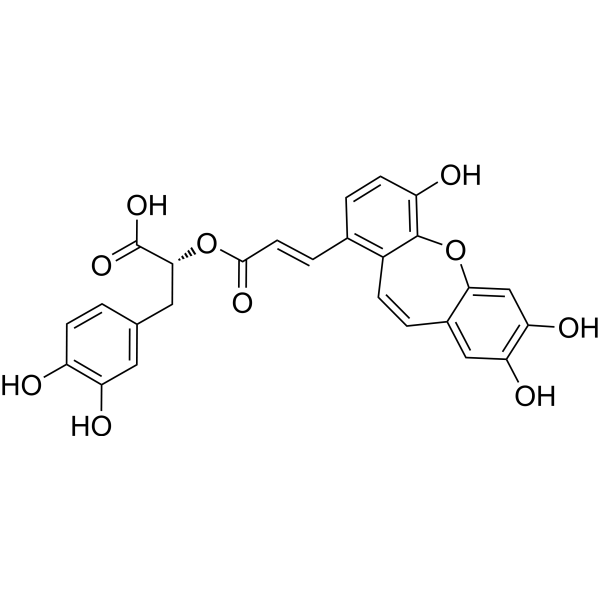
Isosalvianolic acid C
Isosalvianolic acid C is a naturally derived phenolic compound that acts as a signal modulator, antioxidant, and enzyme inhibitor. Isosalvianolic acid C induces the phosphorylation of PLC-γ, IP3R, PKC, and P38, triggering Ca2+ mobilization, degranulation, and chemokine release. Isosalvianolic acid C activates MRGPRX2 and its murine homolog MrgprB2, leading to pseudo-allergic reactions in vivo. Isosalvianolic acid C inhibits Cu2+-induced LDL peroxidation. Isosalvianolic acid C can be used in studies related to pseudo-allergy, diabetes, and atherosclerosis.
For research use only. We do not sell to patients.
- CAS No.: 142115-17-1
- Formula: C26H20O10
- Molecular Weight:492.43
-
Storage:
Please store the product under the recommended conditions in the Certificate of Analysis.

To place orders, for customer services and technical support, please contact: MedChemExpress USA
Tel: 609-228-6898 E-mail: [email protected] [email protected]


-
 Biological Activity
-
Chemical Information
- Protocols
- Purity & Documentation
- References
-
 Help & FAQs


All Phospholipase Isoforms
More

Biological Activity
Description
IC50 & Target
[1]|
p38 |
PKC |
In Vitro
Isosalvianolic acid C (50-200 μM; 30 min) induces degranulation of the human mast cell line LAD2 in a dose-dependent manner, and stimulates histamine release from the human mast cell line LAD2 in a dose-dependent manner[1].
Isosalvianolic acid C (50-200 μM; 12 h) dose-dependently induces MCP-1 release from human mast cell line LAD2[1].
Isosalvianolic acid C (50-200 μM; acute bath application) induces dose-dependent intracellular Ca2+ mobilization in HEK293-MRGPRX2 and HEK293-MrgprB2 cells[1].
Isosalvianolic acid C binds to the MRGPRX2 receptor with a KD value of 12.77×10⁻⁵ M[1].
Isosalvianolic acid C (50-200 μM; 16 h) dose-dependently activates the PLCγ, IP3R, PKC and P38 signaling pathways in LAD2 human mast cells[1].
Isosalvianolic acid C (50-200 μM; 30 min) induces degranulation, MCP-1 release and histamine release in human LAD2 mast cells in an MRGPRX2-dependent manner[1].
Isosalvianolic acid C methyl ester, namely (8'R)-Isosalvianolic acid C methyl ester, exerts potent inhibitory activity against purified α-glucosidase with an IC50 of 0.11 μM, and also blocks AGE formation in the bovine serum albumin-glucose reaction system with an IC50 of 0.07 μM[2].
Isosalvianolic acid C (compound 6) (1 h preincubation; 2 h Cu2+ incubation) inhibits Cu2+-induced peroxidation of human low-density lipoprotein (LDL), with an IC50 value of 2.72 μM[3].
MedChemExpress (MCE) has not independently confirmed the accuracy of these methods. They are for reference only. Further protocols information, click here.
-
Cell Line:LAD2 human mast cells
-
Concentration:50, 100, 200 μM
-
Incubation Time:16 h
-
Result:Significantly increased the levels of phosphorylated PLCγ1, phosphorylated IP3R, phosphorylated PKC, and phosphorylated P38 in a dose-dependent manner, with all phosphorylated protein levels significantly higher than control levels.
In Vivo
MedChemExpress (MCE) has not independently confirmed the accuracy of these methods. They are for reference only.
-
Animal Model:C57BL/6 (WT, adult, 25-30 g)
C57BL/6-MrgprB2 knockout (MUT, adult, 25-30 g)
C57BL/6-KitW-sh/W-sh (adult, 25-30 g)[1] -
Dosage:0.21, 0.52, 1.04 mg/kg (systemic anaphylaxis)
0.5, 1, 2.0 mg/mL (hindpaw inflammation) -
Administration:i.v. via tail vein; single dose (systemic anaphylaxis)
intraplantar; single dose (hindpaw inflammation) -
Result:Increased Evans blue leakage into ears in a dose-dependent manner in systemic anaphylaxis model treated with 0.52 mg/kg reagent.
Triggered dose-dependent hindpaw swelling with paw thickness increase rates of 15%, 16%, and 18% respectively at 0.5 mg/mL, 1.0 mg/mL, and 2.0 mg/mL doses.
Induced dose-dependent Evans blue extravasation with OD620 nm per g paw weight values of 2.5, 2.5, and 3.5 respectively at 0.5 mg/mL, 1.0 mg/mL, and 2.0 mg/mL doses.
Almost completely eliminated hindpaw inflammation in mast cell-deficient C57BL/6-KitW-sh/W-sh mice.
Resulted in almost no hindpaw inflammation in MrgprB2 knockout mice treated with 0.5 mg/mL reagent.


Chemical Information
-
CAS No. 142115-17-1
-
Molecular Weight 492.43
-
Formula C26H20O10
-
SMILES
OC(C=C1C[C@H](C(O)=O)OC(/C=C/C2=C3C(OC4=CC(O)=C(O)C=C4C=C3)=C(C=C2)O)=O)=C(C=C1)O
-
Structure Classification
-
Shipping
Room temperature in continental US; may vary elsewhere.
-
Storage
Please store the product under the recommended conditions in the Certificate of Analysis.

Protocols
-
Kinase activity and phosphorylation assays
Kinase activity assays measure the ability of kinases to transfer phosphate groups from ATP to specific substrates, while phosphorylation assays detect the presence and levels of phosphorylated proteins. Common methods include radiolabeled ATP incorporation (e. g. ,), ADP release detection via bioluminescence (e. g. ,[3]), enzyme-linked immunosorbent assays (ELISA) for phospho-specific epitopes (e. g. ,[6]), and microtiter-based formats for high-throughput screening (e. g. ,[8]). The ADP-Glo assay quantifies kinase activity by measuring ADP produced during phosphorylation using a luciferase-based system. Radiometric assays involve autoradiography or scintillation counting after incorporation of 32P-labeled ATP into substrate proteins. ELISA-based approaches rely on phospho-specific antibodies to detect activated kinases in cell lysates or purified samples.
-
Western Blot
Western blotting (WB) is a commonly used experimental method in molecular biology, biochemistry, and immunogenetics for identifying and quantifying target proteins. It combines gel electrophoresis with immunoassay, enabling researchers to analyze protein expression, post-translational modifications, and molecular weight.
-
Research Protocol for Cardiovascular Diseases
Cardiovascular disease can be modeled as maladaptive cardiac remodeling, where ischemic injury or pressure overload activates inflammatory signaling, fibroblast activation, extracellular-matrix deposition, cardiomyocyte hypertrophy, vascular remodeling, and progressive ventricular dysfunction. The TGF-β/SMAD axis is a central profibrotic pathway after myocardial injury and pressure overload, while innate immune and cytokine pathways regulate leukocyte recruitment, scar formation, and adverse remodeling. Key unresolved questions include which inflammatory signals are reparative versus harmful, when fibrosis is protective versus maladaptive, and whether pathway inhibition improves function without weakening necessary infarct healing or compensatory remodeling.
-
Protocol for Kinase activity and phosphorylation assays
Kinase activity assays measure transfer of phosphate from ATP to a protein or peptide substrate, generating phosphorylated substrate, ADP, or incorporated radiolabeled phosphate as the readout; phosphorylation assays measure site-specific phosphorylation in cells or tissues as a proxy for kinase-pathway activation, inhibition, or substrate regulation. Phosphorylation can be detected by phospho-specific Western blot, immunoprecipitation kinase assay, phospho-immunofluorescence, phospho-flow cytometry, luminescent ADP detection, radiolabeled ATP incorporation, or reporter-based pathway assays, and these readouts can be applied to cancer cells, primary neurons, mouse tumors, organoids, inflammatory macrophages, ferroptosis studies, and mitophagy studies when the kinase target is biologically relevant.
-
Ca2+ Staining Technique
Ca2+ staining is an experimental technique that utilizes specific fluorescent probes (such as Fluo-4 AM, Fura-2, etc.) to qualitatively or quantitatively detect dynamic changes in intracellular Ca2+ concentrations; this is achieved by monitoring the changes in fluorescent signals generated when these probes bind to free intracellular calcium ions. The underlying principle relies primarily on the presence of chelating groups within the probe's molecular structure that possess high affinity for calcium ions.
-
Research Protocol for Metabolic Diseases
AMP-activated protein kinase, AMPK, is a conserved cellular energy sensor that responds to reduced cellular energy status and coordinates metabolism by increasing ATP-generating catabolic pathways while suppressing ATP-consuming anabolic processes. In metabolic disease research, the AMPK pathway is experimentally relevant because it regulates hepatic lipid synthesis, fatty acid oxidation, glucose production, skeletal-muscle glucose disposal, mTORC1-linked biosynthesis, autophagy, mitochondrial homeostasis, and whole-body energy balance. The central pathway logic is that energy stress, metformin, exercise-like stimulation, or direct AMPK activators increase AMPKα Thr172 phosphorylation and downstream substrate phosphorylation, including ACC and RAPTOR. Phosphorylation of ACC suppresses lipogenesis and supports fatty acid oxidation, whereas phosphorylation of RAPTOR suppresses mTORC1 signaling and links cellular energy status to growth and protein synthesis control. The pathway is linked


Purity & Documentation
References
[1]. Lin Y, et al. Isosalvianolic acid C-induced pseudo-allergic reactions via the mast cell specific receptor MRGPRX2. International immunopharmacology. 2019 Jun;71:22-31. [Content Brief]
[2]. Ma HY, et al. Constituents with α-glucosidase and advanced glycation end-product formation inhibitory activities from Salvia miltiorrhiza Bge. Journal of natural medicines. 2011 Jan;65(1):37-42. [Content Brief]
[3]. Lin YL, et al. Anti-lipid-peroxidative principles from Tournefortia sarmentosa. Journal of natural products. 2002 May;65(5):745-7. [Content Brief]

Calculators
Concentration (start) × Volume (start) = Concentration (final) × Volume (final)


Isosalvianolic acid C Related Classifications
HY-122967 Related Classifications
 Powered by Bioz
Keywords
- Isosalvianolic acid C
- 142115-17-1
- PKC
- Mas-related G-protein-coupled Receptor (MRGPR)
- p38 MAPK
- Phospholipase
- LDL peroxidation
- HEK293-MRGPRX2 cells
- LAD2 human mast cells
- pseudo-allergic reactions
- α-glucosidase
- MRGPRX2
- HEK293-MrgprB2 cells
- MrgprB2
- advanced glycation end-product
- diabetes mellitus
- Inhibitor
- inhibitor
- inhibit