



BPKDi

Based on 1 Customer Validation
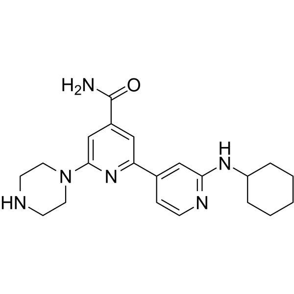
BPKDi is an inhibitor of PKD. BPKDi inhibits three members of the PKD family, PKD1, PKD2, and PKD3, with IC50 values of 1 nM, 9 nM, and 1 nM, respectively. BPKDi blocks signal-dependent phosphorylation and nuclear export of class IIa HDACs in cardiomyocytes and concomitantly suppresses hypertrophy of these cells.
연구목적의 판매만을 진행합니다. 환자를 대상으로 한 판매는 하지 않습니다.
- Purity : 99.74%
- CAS No.: 1201673-28-0
- 화학식: C21H28N6O
- 분자량:380.49
-
보관:
4°C, sealed storage, away from moisture and light
* In solvent : -80°C, 6 months; -20°C, 1 month (sealed storage, away from moisture and light)

To place orders, for customer services and technical support, please contact: MedChemExpress USA
Tel: 609-228-6898 E-mail: [email protected] [email protected]


-
 Biological Activity
-
Chemical Information
-
용액&용해도
- Protocol
- 순도&문서
- References
-
 Help & FAQs


-
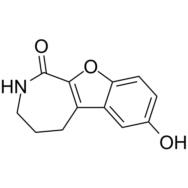
HY-12239
CID755673PKD Inhibitor -
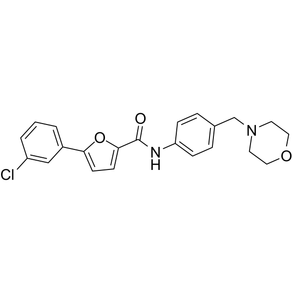
HY-13454
CID 2011756PKD Inhibitor -

HY-101495
18-HEPEEster Product -
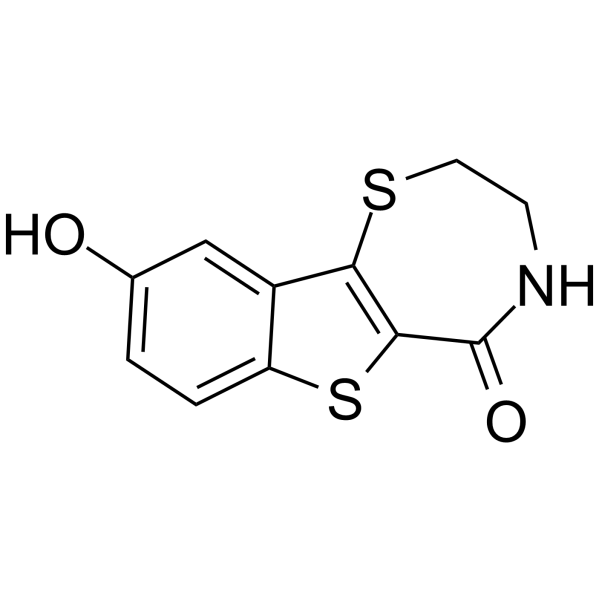
HY-15528
kb NB 142-70PKD Inhibitor -
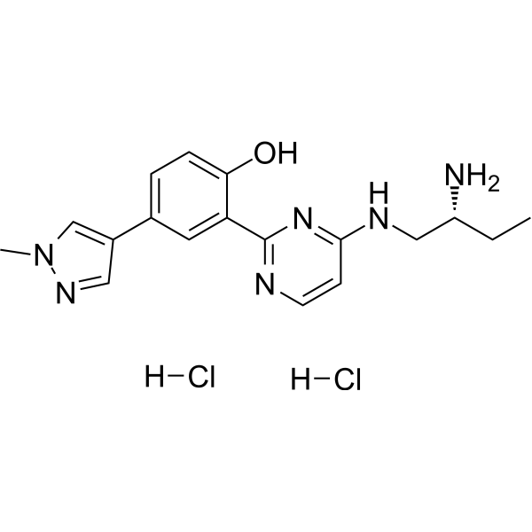
HY-15698A
CRT0066101 dihydrochloridePKD/PIM2 Inhibitor -
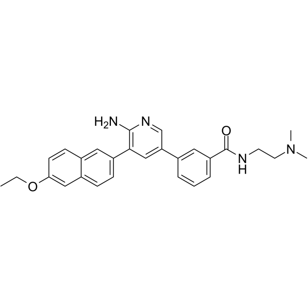
HY-112547
CRT5PKD Inhibitor -
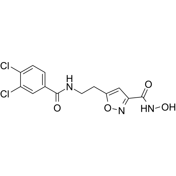
HY-126330
SS-208HDAC Inhibitor -
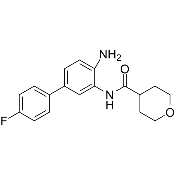
HY-102083
BRD4884HDAC Inhibitor -

HY-134043
Chlorthalidone Impurity G -

HY-15528R
kb NB 142-70 (Standard)PKD Inhibitor -
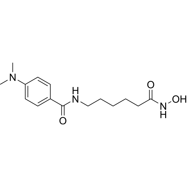
HY-117394
MD 85HDAC Inhibitor -
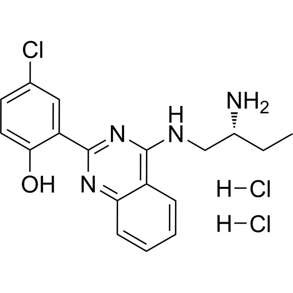
HY-131962A
PKD-IN-1 dihydrochloridePDK-1 Inhibitor


Biological Activity
제품 설명
IC50 & Target
[1]|
PKD1/PKCμ 1 nM (IC50) |
PKD2 9 nM (IC50) |
PKD3 1 nM (IC50) |
In Vitro
BPKDi (1 μM, 30 min) significantly attenuates PMA (HY-18739) (25 nM)-induced auto-phosphorylation of PKD1 in neonatal rat ventricular myocytes (NRVMs)[1].
BPKDi (1 μM, 72 h) completely blocks agonist-dependent induction of myomaxin mRNA expression, supporting the notion that PKD derepresses MEF2 target genes via class IIa HDACs in myocytes[1].
MedChemExpress (MCE) has not independently confirmed the accuracy of these methods. They are for reference only.
-
Cell Line:Neonatal rat ventricular myocytes
-
Concentration:1 μM
-
Incubation Time:30 min
-
Result:Efficiently blocked PDK1 auto-phosphorylation in response to independent G protein-coupled receptor (GPCR) agonists.
Significantly attenuated the agonist-dependent phosphorylation of HDAC5 and HDAC4.
-
Cell Line:Neonatal rat ventricular myocytes
-
Concentration:1 μM
-
Incubation Time:30 min
-
Result:Completely blocked agonist-dependent nuclear export of HDAC5.


Chemical Information
-
CAS No. 1201673-28-0
-
Appearance Solid
-
분자량 380.49
-
화학식 C21H28N6O
-
Color Off-white to light yellow
-
SMILES
O=C(N)C1=CC(C2=CC(NC3CCCCC3)=NC=C2)=NC(N4CCNCC4)=C1
-
선적
Room temperature in continental US; may vary elsewhere.
-
보관
4°C, sealed storage, away from moisture and light
* In solvent : -80°C, 6 months; -20°C, 1 month (sealed storage, away from moisture and light)


용액&용해도
In Vitro:
DMSO : 50 mg/mL (131.41 mM; ultrasonic and warming and heat to 60°C; Hygroscopic DMSO has a significant impact on the solubility of product, please use newly opened DMSO)
Please refer to the solubility information to select the appropriate solvent. Once prepared, please aliquot and store the solution to prevent product inactivation from repeated freeze-thaw cycles.
Storage method and period of stock solution: -80°C, 6 months; -20°C, 1 month (sealed storage, away from moisture and light). When stored at -80°C, please use it within 6 months. When stored at -20°C, please use it within 1 month.
Please refer to the solubility information to select the appropriate solvent. Once prepared, please aliquot and store the solution to prevent product inactivation from repeated freeze-thaw cycles.
Storage method and period of stock solution: -80°C, 6 months; -20°C, 1 month (sealed storage, away from moisture and light). When stored at -80°C, please use it within 6 months. When stored at -20°C, please use it within 1 month.
Concentration (start) × Volume (start) = Concentration (final) × Volume (final)


순도&문서
-
Data Sheet (272 KB)
-
SDS (252 KB)
- English - EN (252 KB)
- Français - FR (252 KB)
- Deutsch - DE (252 KB)
- Norwegian - NO (252 KB)
- Español - ES (252 KB)
- Swedish - SV (252 KB)
- Italian - IT (252 KB)
- Korean - KR (252 KB)
- Portuguese - PT (252 KB)
-
Handling Instructions (2659 KB)
References


Complete Stock Solution Preparation Table
Please refer to the solubility information to select the appropriate solvent. Once prepared, please aliquot and store the solution to prevent product inactivation from repeated freeze-thaw cycles.
Storage method and period of stock solution: -80°C, 6 months; -20°C, 1 month (sealed storage, away from moisture and light). When stored at -80°C, please use it within 6 months. When stored at -20°C, please use it within 1 month.
| Optional Solvent | Concentration Solvent Mass | 1 mg | 5 mg | 10 mg | 25 mg |
|---|---|---|---|---|---|
| DMSO | 1 mM | 2.6282 mL | 13.1410 mL | 26.2819 mL | 65.7048 mL |
| 5 mM | 0.5256 mL | 2.6282 mL | 5.2564 mL | 13.1410 mL | |
| 10 mM | 0.2628 mL | 1.3141 mL | 2.6282 mL | 6.5705 mL | |
| 15 mM | 0.1752 mL | 0.8761 mL | 1.7521 mL | 4.3803 mL | |
| 20 mM | 0.1314 mL | 0.6570 mL | 1.3141 mL | 3.2852 mL | |
| 25 mM | 0.1051 mL | 0.5256 mL | 1.0513 mL | 2.6282 mL | |
| 30 mM | 0.0876 mL | 0.4380 mL | 0.8761 mL | 2.1902 mL | |
| 40 mM | 0.0657 mL | 0.3285 mL | 0.6570 mL | 1.6426 mL | |
| 50 mM | 0.0526 mL | 0.2628 mL | 0.5256 mL | 1.3141 mL | |
| 60 mM | 0.0438 mL | 0.2190 mL | 0.4380 mL | 1.0951 mL | |
| 80 mM | 0.0329 mL | 0.1643 mL | 0.3285 mL | 0.8213 mL | |
| 100 mM | 0.0263 mL | 0.1314 mL | 0.2628 mL | 0.6570 mL |


BPKDi Related Classifications
HY-118052 Related Classifications
 Powered by Bioz