



Lobaplatin
Based on 5 publication(s) in Google Scholar
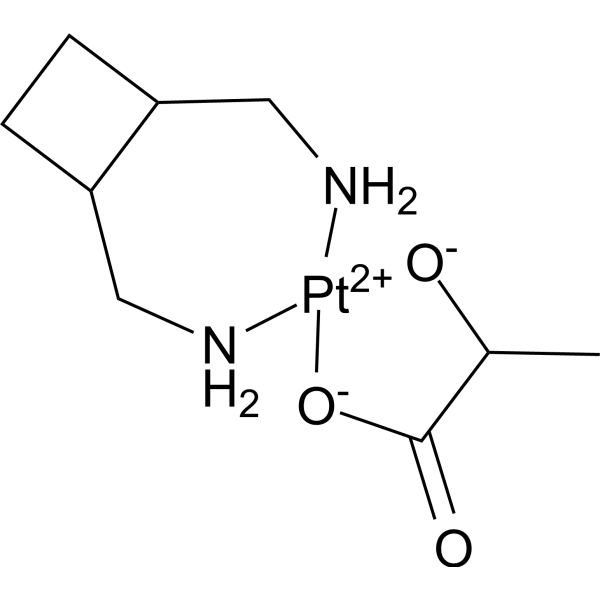
Lobaplatin (D-19466) is a diastereometric mixture of platinum(II) complexe. Lobaplatin arrests cell cycle at G1 and G2/M phase. Lobaplatin induces apoptosis by increasing expressions of caspase and Bax, decreasing expression of Bcl-2. Lobaplatin can be used for research of cancer.
For research use only. We do not sell to patients.
- Purity : 98.0%
- CAS No.: 135558-11-1
- Formula: C9H18N2O3Pt
- Molecular Weight:397.33
-
Storage:Powder -20°C, 3 years , 4°C, 2 years ; In solvent -80°C, 6 months , -20°C, 1 month

To place orders, for customer services and technical support, please contact: MedChemExpress USA
Tel: 609-228-6898 E-mail: [email protected] [email protected]


-
 Biological Activity
-
Chemical Information
-
Solvent & Solubility
- Protocols
- Purity & Documentation
- References
-
 Help & FAQs


-
HY-17371
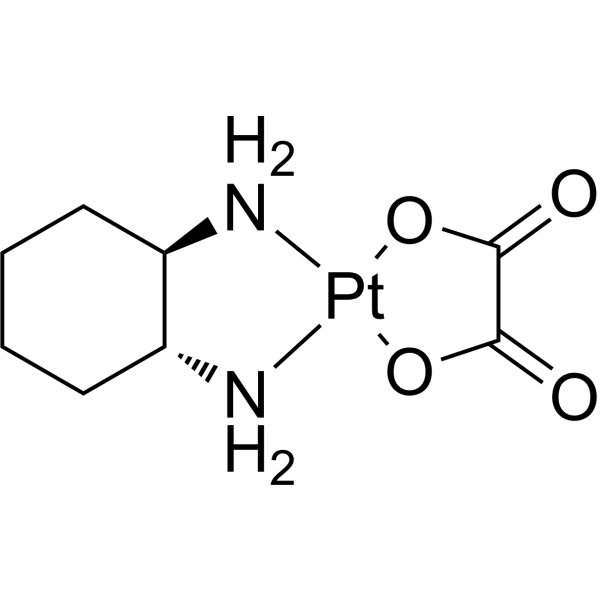
OxaliplatinDNA Synthesis Inhibitor -
HY-10087
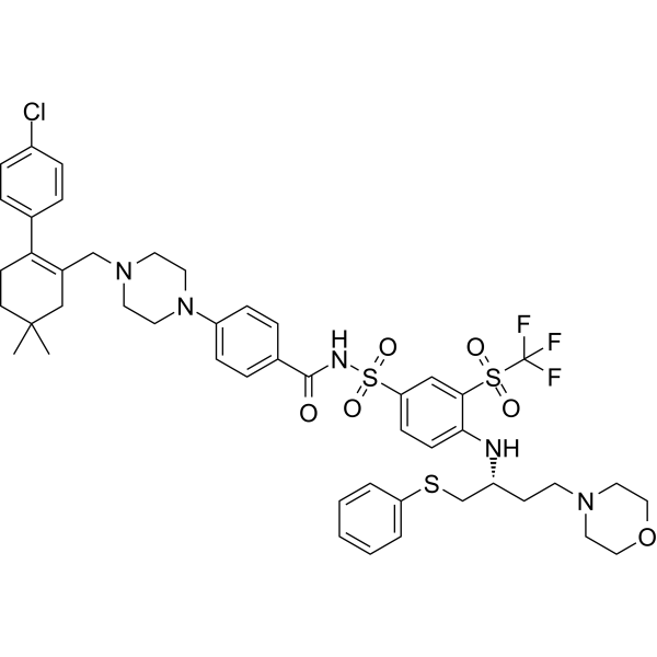
NavitoclaxBcl-2 Inhibitor -
HY-103243
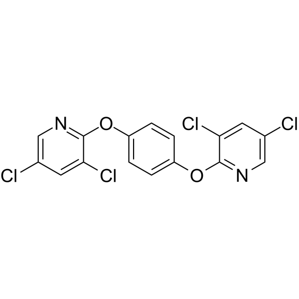
TCPOBOPCAR Agonist -
HY-N0087
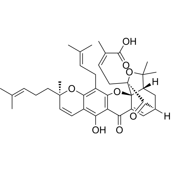
Gambogic AcidBcl-2 Family Inhibitor -
HY-100741
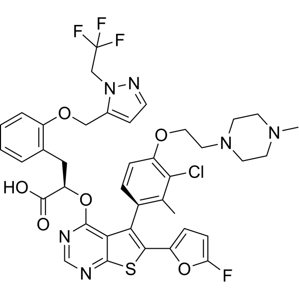
S63845MCL1 Inhibitor -
HY-19741
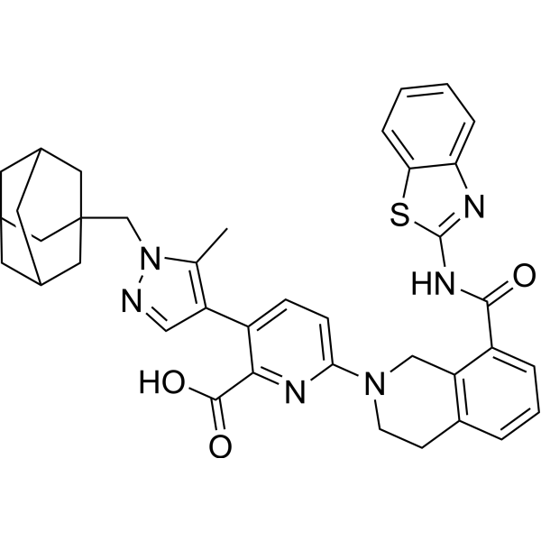
A-1331852Bcl-xL Inhibitor -
HY-N0831
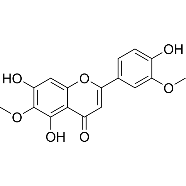
JaceosidinBax Activator -
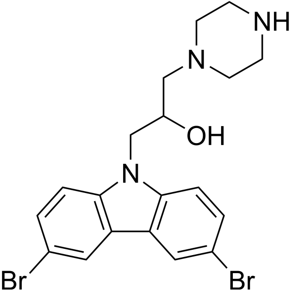
HY-103269
BAI1BAX Inhibitor -
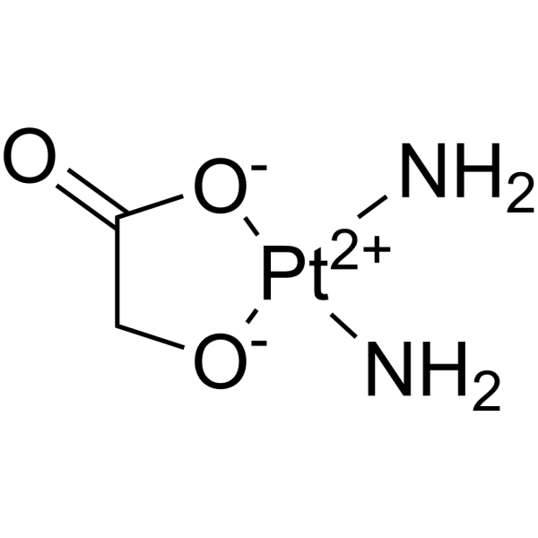
HY-13700
NedaplatinDNA/RNA Synthesis Inhibitor -
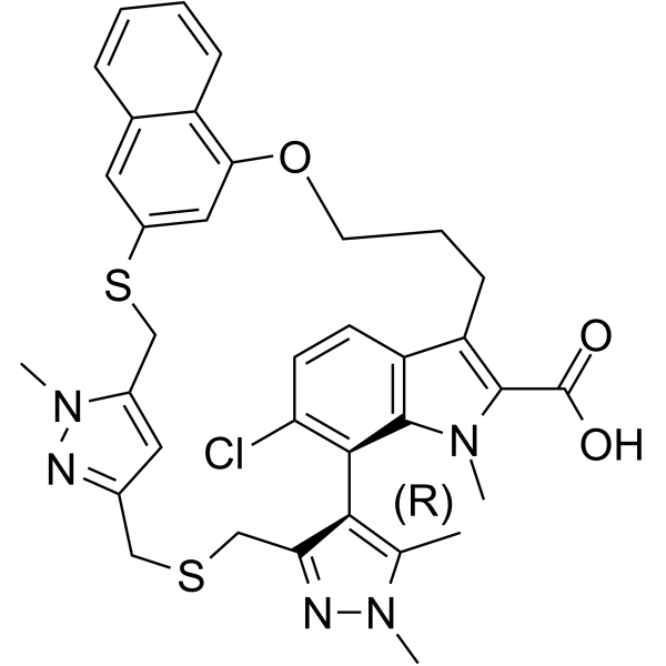
HY-101533
AZD-5991Mcl-1 Inhibitor -
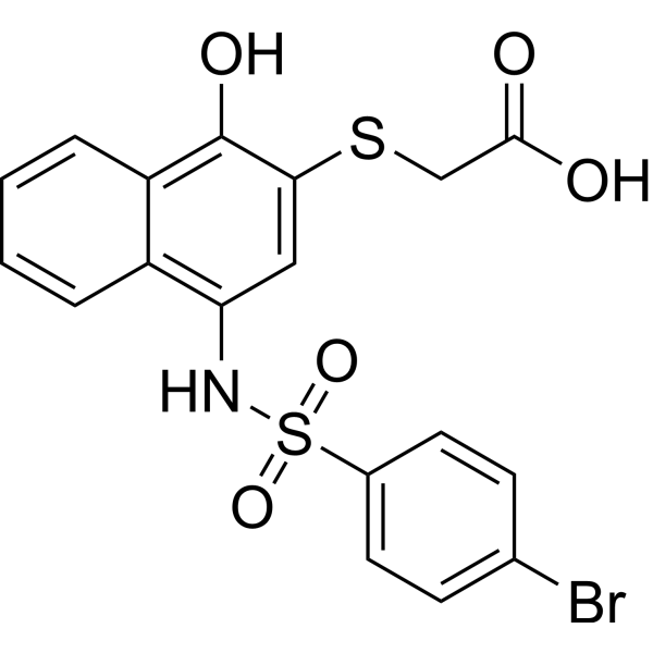
HY-18628
UMI-77Bcl-2 Family Inhibitor -

HY-10087R
Navitoclax (Standard)Reference Standard
Publications Citing Use of MedChemExpress (MCE) Lobaplatin
MoreAll Caspase Isoforms
More

Biological Activity
Description
In Vitro
Lobaplatin (D-19466, 0.25-32 μg/mL; 24-72 h) exhibits anti-proliferative activities against esophageal squamous cell carcinoma (ESCC) cell lines[1].
Lobaplatin (0-16 μg/mL; 48 h) induces esophageal squamous cell carcinoma (ESCC) apoptosis and modulates expression of apoptosis-related proteins[1].
Lobaplatin (1.45 μg/mL; 0-48 h; SMMC-7721 cells) arrests cell cycle progression at G1 and G2/M phases in a time-dependent manner[2].
Lobaplatin (1.45 μg/mL; 0-48 h; SMMC-7721 cells) inhibits the mRNA levels of cyclin B, CDK1, and CDC25C phosphatase, down-regulates Rb/E2F complexes and up-regulates of CDK inhibitors[2].
MedChemExpress (MCE) has not independently confirmed the accuracy of these methods. They are for reference only.
-
Cell Line:KYSE-410 cells and EC-109 cells
-
Concentration:0.25, 0.5, 1, 2, 4, 8, 16 and 32 μg/mL
-
Incubation Time:24, 48 and 72 hours
-
Result:Inhibited the growth of KYSE-410 and EC-109 cells in a dose- and time-dependent manner.
Inhibited the clone formation activity of KYSE-410 and EC-109 cells in a dosedependent manner.
-
Cell Line:KYSE-410 cells and EC-109 cells
-
Concentration:0.25, 1, 4 and 16 μg/mL
-
Incubation Time:48 hours
-
Result:Increased the percentage of apoptotic cells in a dose-dependent manner.
-
Cell Line:KYSE-410 cells and EC-109 cells
-
Concentration:0, 1, 4 and 16 μg/mL
-
Incubation Time:48 hours
-
Result:Increased expressions of cleaved-caspase-3, cleaved-caspase-8, cleaved-caspase-9 and Bax, while decreased expression of Bcl-2.
-
Cell Line:SMMC-7721 cells
-
Concentration:1.45 μg/mL
-
Incubation Time:0, 24, 36 and 48 hours
-
Result:Arrested the proportions of G1, S, and G2/M phases in cells were 45.31, 22.88, and 31.81% at 0 h, 59.91, 11.92, and 28.17% at 24 h, 56.89, 2.83, and 40.28% at 36 h, and 53.80, 2.07, and 44.13% at 48 h, respectively.
-
Cell Line:SMMC-7721 cells
-
Concentration:1.45 μg/mL
-
Incubation Time:0, 24, 36 and 48 hours
-
Result:Down-regulated cyclin B, CDK1, CDC25C, phosphorylated CDK1 (pCDK1), pCDK4, Rb, E2F, and pRb, and up-regulated p53, p21, and p27.
In Vivo
MedChemExpress (MCE) has not independently confirmed the accuracy of these methods. They are for reference only.
-
Animal Model:Male BALB/c nude mice (4-6 weeks) with ESCC xenograft[1]
-
Dosage:Intraperitoneal injection; once a week, for 3 weeks
-
Administration:5 and 10 mg/kg
-
Result:Suppressed tumor volumes in a dose-dependent manner.
Increased expressions of Bax and decreased expressions of Bcl-2.
Clinical Trial
| NCT Number | Sponsor | Condition | Start Date |
Phase

|
|---|---|---|---|---|
| NCT01329991 | 2011-05 | PHASE1 |


Chemical Information
-
CAS No. 135558-11-1
-
Appearance Solid
-
Molecular Weight 397.33
-
Formula C9H18N2O3Pt
-
Color White to off-white
-
SMILES
CC1[O-][Pt+2]2([O-]C1=O)[NH2]CC(CC3)C3C[NH2]2
-
Synonyms
D-19466
-
Shipping
Room temperature in continental US; may vary elsewhere.
-
Storage
Powder -20°C 3 years 4°C 2 years In solvent -80°C 6 months -20°C 1 month


Publications (5)
-
Journal Impact Factor
-
Most Recent
-
Mol Cancer
Organoids derived from patients provide a new opportunity for research and individualized treatment of malignant peritoneal mesothelioma. [Abstract]2024 Jan 10;23(1):12. PMID: 38200517 -
Sci Adv
2025 Oct 31;11(44):eadx6809. PMID: 41160705 -
Cancer Cell Int
Quantitative phosphoproteomic analysis reveals chemoresistance-related proteins and signaling pathways induced by rhIL-6 in human osteosarcoma cells. [Abstract]2021 Oct 30;21(1):581. PMID: 34717622 -
Sci Rep
Nrf2 inhibition increases sensitivity to chemotherapy of colorectal cancer by promoting ferroptosis and pyroptosis. [Abstract]2023 Sep 1;13(1):14359. PMID: 37658132 -
Hum Cell
EP300 promotes tumor stemness via epigenetic activation of CRISP3 leading to lobaplatin resistance in triple-negative breast cancer. [Abstract]2024 Sep;37(5):1475-1488. PMID: 38879857


Solvent & Solubility
In Vitro:
H2O : 50 mg/mL (125.84 mM; ultrasonic and warming and heat to 60°C; DMSO can inactivate Lobaplatin's activity)
Please refer to the solubility information to select the appropriate solvent. Once prepared, please aliquot and store the solution to prevent product inactivation from repeated freeze-thaw cycles.
Storage method and period of stock solution: -80°C, 6 months; -20°C, 1 month. When stored at -80°C, please use it within 6 months. When stored at -20°C, please use it within 1 month.
* Note: If you choose water as the stock solution, please dilute it to the working solution, then filter and sterilize it with a 0.22 μm filter before use.
Please refer to the solubility information to select the appropriate solvent. Once prepared, please aliquot and store the solution to prevent product inactivation from repeated freeze-thaw cycles.
Storage method and period of stock solution: -80°C, 6 months; -20°C, 1 month. When stored at -80°C, please use it within 6 months. When stored at -20°C, please use it within 1 month.
* Note: If you choose water as the stock solution, please dilute it to the working solution, then filter and sterilize it with a 0.22 μm filter before use.
Concentration (start) × Volume (start) = Concentration (final) × Volume (final)
In Vivo:
For the following dissolution methods, please prepare the working solution directly:
It is recommended to prepare fresh solutions and use them promptly within a short period of time.
The percentages shown for the solvents indicate their volumetric ratio in the final prepared solution. If precipitation or phase separation occurs during preparation, heat and/or sonication can be used to aid dissolution.
Add each solvent one by one: PBS
Solubility: 100 mg/mL (251.68 mM); Clear solution; Need ultrasonic


Purity & Documentation
-
Data Sheet (283 KB)
-
SDS (393 KB)
- English - EN (393 KB)
- Français - FR (393 KB)
- Deutsch - DE (393 KB)
- Norwegian - NO (393 KB)
- Español - ES (393 KB)
- Swedish - SV (393 KB)
- Italian - IT (393 KB)
- Korean - KR (393 KB)
- Portuguese - PT (393 KB)
-
Handling Instructions (2659 KB)
References
[1]. Du L, et, al. Antitumor activity of Lobaplatin against esophageal squamous cell carcinoma through caspase-dependent apoptosis and increasing the Bax/Bcl-2 ratio. Biomed Pharmacother. 2017 Nov;95:447-452. [Content Brief]
[2]. Wu Q, et, al. Lobaplatin arrests cell cycle progression in human hepatocellular carcinoma cells. J Hematol Oncol. 2010 Oct 31;3:43. [Content Brief]
[3]. McKeage MJ. Lobaplatin: a new antitumour platinum drug. Expert Opin Investig Drugs. 2001 Jan;10(1):119-28. [Content Brief]


Complete Stock Solution Preparation Table
Please refer to the solubility information to select the appropriate solvent. Once prepared, please aliquot and store the solution to prevent product inactivation from repeated freeze-thaw cycles.
Storage method and period of stock solution: -80°C, 6 months; -20°C, 1 month. When stored at -80°C, please use it within 6 months. When stored at -20°C, please use it within 1 month.
| Optional Solvent | Concentration Solvent Mass | 1 mg | 5 mg | 10 mg | 25 mg |
|---|---|---|---|---|---|
| H2O | 1 mM | 2.5168 mL | 12.5840 mL | 25.1680 mL | 62.9200 mL |
| 5 mM | 0.5034 mL | 2.5168 mL | 5.0336 mL | 12.5840 mL | |
| 10 mM | 0.2517 mL | 1.2584 mL | 2.5168 mL | 6.2920 mL | |
| 15 mM | 0.1678 mL | 0.8389 mL | 1.6779 mL | 4.1947 mL | |
| 20 mM | 0.1258 mL | 0.6292 mL | 1.2584 mL | 3.1460 mL | |
| 25 mM | 0.1007 mL | 0.5034 mL | 1.0067 mL | 2.5168 mL | |
| 30 mM | 0.0839 mL | 0.4195 mL | 0.8389 mL | 2.0973 mL | |
| 40 mM | 0.0629 mL | 0.3146 mL | 0.6292 mL | 1.5730 mL | |
| 50 mM | 0.0503 mL | 0.2517 mL | 0.5034 mL | 1.2584 mL | |
| 60 mM | 0.0419 mL | 0.2097 mL | 0.4195 mL | 1.0487 mL | |
| 80 mM | 0.0315 mL | 0.1573 mL | 0.3146 mL | 0.7865 mL | |
| 100 mM | 0.0252 mL | 0.1258 mL | 0.2517 mL | 0.6292 mL |
* Note: If you choose water as the stock solution, please dilute it to the working solution, then filter and sterilize it with a 0.22 μm filter before use.


Lobaplatin Related Classifications
HY-105930 Related Classifications
 Powered by Bioz