



Imofinostat

Based on 1 Customer Validation
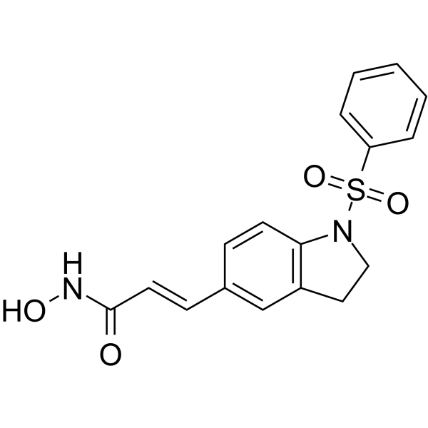
Imofinostat (ABT-301; MPT0E028) is an orally active and selective HDAC inhibitor with IC50s of 53.0 nM, 106.2 nM, 29.5 nM for HDAC1, HDAC2 and HDAC6, respectively. Imofinostat has a weak inhibitory effect on HDAC8 (IC50 of 2.5 μM), but no inhibitory effect on HDAC4 (IC50>10 μM). Imofinostat reduces the viability of B-cell lymphomas by inducing apoptosis and possesses potent direct Akt targeting ability and reduces Akt phosphorylation in B-cell lymphoma. Imofinostat has a broad-spectrum antitumor activity, including colorectal cancer, B-cell lymphoma, non-small cell lung carcinoma (NSCLC), and pancreatic cancer, while also showing therapeutic potential in non-tumor diseases like emphysema and pulmonary fibrosis.
For research use only. We do not sell to patients.
- Purity : 99.40%
- CAS No.: 1338320-94-7
- Formula: C17H16N2O4S
- Molecular Weight:344.38
-
Storage:
4°C, sealed storage, away from moisture and light
* In solvent : -80°C, 6 months; -20°C, 1 month (sealed storage, away from moisture and light)

To place orders, for customer services and technical support, please contact: MedChemExpress USA
Tel: 609-228-6898 E-mail: [email protected] [email protected]


-
 Biological Activity
-
Chemical Information
-
Solvent & Solubility
- Protocols
- Purity & Documentation
- References
-
 Help & FAQs




Biological Activity
Description
IC50 & Target
[1,2]|
HDAC1 53.0 nM (IC50) |
HDAC2 106.2 nM (IC50) |
HDAC6 29.5 nM (IC50) |
HDAC8 2532.6 nM (IC50) |
In Vitro
Imofinostat (MPT0E028) (0-10 μM; 48 h) significantly inhibits cell proliferation and induces apoptosis in HCT116 cells[1].
Imofinostat (0-10 μM; 24 h) inhibits cell growth in a concentration-dependent manner. Imofinostat increases the number of cells in the sub-G1 phase of the cell cycle. Imofinostat induces caspase 3 and PARP activation in a concentrationdependent manner, and this apoptosis[1].
Imofinostat (0.3-100 μM; 24 h) induces significant concentration-dependent growth inhibition in Ramos and BJAB cells. Imofinostat increases the subG1 phase population in a time- and concentration-dependent manner. Imofinostat induces caspase-3, -6, -7, -8, -9 activation and PARP cleavage[2].
Imofinostat (0.01-1 μM) pretreated human lung fibroblasts (WI-38) for 30 minutes and then treated with stimulants, which inhibits the expression of CTGF induced by TGF-β, thrombin, and ET-1[5].
MedChemExpress (MCE) has not independently confirmed the accuracy of these methods. They are for reference only.
-
Cell Line:HCT116 cells
-
Concentration:0-10 μM
-
Incubation Time:48 h
-
Result:Inhibited cell growth in a concentration-dependent manner.
-
Cell Line:HCT116 cells
-
Concentration:0.3 μM, 1 μM, 3 μM, 10 μM
-
Incubation Time:24 h
-
Result:Increased the number of cells in the sub-G1 phase of the cell cycle.
-
Cell Line:HCT116 cells
-
Concentration:1 μM, 3 μM, 10 μM
-
Incubation Time:24 h
-
Result:Induced caspase 3 and PARP activation in a concentration-dependent manner.
Induced hyperacetylation of α-tubulin and histone H3.
In Vivo
Imofinostat (100 mg/kg; oral gavage; once daily;) significantly prolongs survival in NOD/SCID mice bearing human B-cell lymphoma Ramos cells[2].
Imofinostat (50-200 mg/kg; oral gavage; once daily; 31 days) inhibits tumor growth in a dose-dependent manner, activates caspase 3 and PARP, and increases acetylation levels of histone H3 and α-tubulin in nude mice xenografted with BJAB cells, without significant body weight changes[2].
Imofinostat (25-100 mg/kg; oral gavage; once daily; 20 days) reduces pulmonary fibrosis in a dose-dependent manner, decreases expression of CTGF, fibronectin, α-SMA, and collagen, and inhibits phosphorylation of ERK, JNK, and p38 in Bleomycin (HY-17565A)-induced C57BL/6 mouse model[5].
Imofinostat (25 mg/kg; oral gavage; once daily; continuous administration) significantly reduces tumor volume, increases cleaved caspase-3 levels, downregulates EGFR expression, and causes no weight loss or adverse effects in AsPC-1 pancreatic cancer xenograft nude mice[6].
MedChemExpress (MCE) has not independently confirmed the accuracy of these methods. They are for reference only.
-
Animal Model:Nude mice (female, 17.2-22.0 g, 8 weeks old) subcutaneously injected with HCT116 cells in the right flank[1].
-
Dosage:50, 100, or 200 mg/kg
-
Administration:Oral gavage; once a day; for 15 days
-
Result:Tumor growth was significantly delayed and inhibited in a dose-dependent manner.
No significant body weight changes or adverse effects were observed in treatment groups.
-
Animal Model:NOD/SCID mice (male, 5 weeks old) engrafted with Ramos cells via tail vein injection[2].
-
Dosage:100 mg/kg
-
Administration:Oral gavage, daily until study end.
-
Result:Prolonged survival compared to the control group.
-
Animal Model:Nude mice (male, 20-24 g, 5 weeks old) subcutaneously injected with BJAB cells mixed with Matrigel[2].
-
Dosage:50, 100, or 200 mg/kg
-
Administration:Oral gavage, daily for 31 days.
-
Result:Tumor growth was inhibited in a dose-dependent manner. Caspase 3 and PARP were activated, and acetylation of histone H3 and α-tubulin was increased in treated groups. Akt phosphorylation was inhibited, and no significant body weight changes were observed.
-
Animal Model:C57BL/6 mice (female, 8 weeks old) intratracheally administered bleomycin (0.3 U/kg) to induce pulmonary fibrosis[5].
-
Dosage:25, 50, or 100 mg/kg
-
Administration:Oral gavage, daily from day 1 to day 20.
-
Result:Reduced fibrosis score, decreased expression of CTGF, fibronectin, α-SMA, and collagen, and inhibited phosphorylation of ERK, JNK, and p38 in a dose-dependent manner.
-
Animal Model:Balb/c-nude mice (male, 4 weeks old) subcutaneously injected with AsPC-1 cells[6].
-
Dosage:25 mg/kg
-
Administration:Oral gavage, daily, for 25 days
-
Result:Tumor volume was significantly reduced. Cleaved caspase-3 levels were increased, and EGFR expression was downregulated in tumor tissues. No weight loss or adverse effects were observed.
- Kinase activity and phosphorylation assays
- Fibrosis/Collagen Morphometry
- Western Blot
- Collagen: Sirius Red Staining
- Detection of Apoptosis
- TUNEL staining for apoptotic DNA fragmentation
- Annexin V plus membrane-impermeant dye apoptosis staining
- Connective Tissue: Masson's Trichrome/Collagen Trichrome Staining
- Apoptosis Solutions
- Protocol for Kinase activity and phosphorylation assays
- Subcutaneous Cell-Line-Derived Xenograft
- Cell Viability Determination by MTT Colorimetric Assay
Clinical Trial
| NCT Number | Sponsor | Condition | Start Date |
Phase

|
|---|---|---|---|---|
| NCT01329991 | 2011-05 | PHASE1 |


Chemical Information
-
CAS No. 1338320-94-7
-
Appearance Solid
-
Molecular Weight 344.38
-
Formula C17H16N2O4S
-
Color White to off-white
-
SMILES
O=C(/C=C/C1=CC2=C(C=C1)N(CC2)S(=O)(C3=CC=CC=C3)=O)NO
-
Synonyms
ABT-301; MPT0E028; TMU-C-0012
-
Shipping
Room temperature in continental US; may vary elsewhere.
-
Storage
4°C, sealed storage, away from moisture and light
* In solvent : -80°C, 6 months; -20°C, 1 month (sealed storage, away from moisture and light)


Solvent & Solubility
In Vitro:
DMSO : 100 mg/mL (290.38 mM; Need ultrasonic; Hygroscopic DMSO has a significant impact on the solubility of product, please use newly opened DMSO)
Please refer to the solubility information to select the appropriate solvent. Once prepared, please aliquot and store the solution to prevent product inactivation from repeated freeze-thaw cycles.
Storage method and period of stock solution: -80°C, 6 months; -20°C, 1 month (sealed storage, away from moisture and light). When stored at -80°C, please use it within 6 months. When stored at -20°C, please use it within 1 month.
Please refer to the solubility information to select the appropriate solvent. Once prepared, please aliquot and store the solution to prevent product inactivation from repeated freeze-thaw cycles.
Storage method and period of stock solution: -80°C, 6 months; -20°C, 1 month (sealed storage, away from moisture and light). When stored at -80°C, please use it within 6 months. When stored at -20°C, please use it within 1 month.
Concentration (start) × Volume (start) = Concentration (final) × Volume (final)
In Vivo:
Select the appropriate dissolution method based on your experimental animal and administration route.
- For the following dissolution methods, please ensure to first prepare a clear stock solution using an In Vitro approach and then sequentially add co-solvents:
- To ensure reliable experimental results, the clarified stock solution can be appropriately stored based on storage conditions. As for the working solution for In Vivo experiments, it is recommended to prepare freshly and use it on the same day.
- The percentages shown for the solvents indicate their volumetric ratio in the final prepared solution. If precipitation or phase separation occurs during preparation, heat and/or sonication can be used to aid dissolution.
Add each solvent one by one: 10% DMSO 40% PEG300 5% Tween-80 45% Saline
Solubility: ≥ 2.5 mg/mL (7.26 mM); Clear solution
This protocol yields a clear solution of ≥ 2.5 mg/mL (saturation unknown).
Taking 1 mL working solution as an example, add 100 μL DMSO stock solution (25.0 mg/mL) to 400 μL PEG300, and mix evenly; then add 50 μL Tween-80 and mix evenly; then add 450 μL Saline to adjust the volume to 1 mL.
Preparation of Saline: Dissolve 0.9 g sodium chloride in ddH₂O and dilute to 100 mL to obtain a clear Saline solution.
Add each solvent one by one: 10% DMSO 90% (20% SBE-β-CD in Saline)
Solubility: ≥ 2.5 mg/mL (7.26 mM); Clear solution
This protocol yields a clear solution of ≥ 2.5 mg/mL (saturation unknown).
Taking 1 mL working solution as an example, add 100 μL DMSO stock solution (25.0 mg/mL) to 900 μL 20% SBE-β-CD in Saline, and mix evenly.
Preparation of 20% SBE-β-CD in Saline (4°C, storage for one week): 2 g SBE-β-CD powder is dissolved in 10 mL Saline, completely dissolve until clear.
In Vivo Dissolution Calculator
Please enter the basic information of animal experiments:
-
-
-
-
Recommended: Prepare an additional quantity of animals to account for potential losses during experiments.
Please enter your animal formula composition:
-
%DMSO +
Recommended: Keep the proportion of DMSO in working solution below 2% if your animal is weak.
-
%+
-
+%Tween-80 + +
-
%Saline +
The co-solvents required include: DMSO, . All of co-solvents are available by MedChemExpress (MCE). , Tween 80. All of co-solvents are available by MedChemExpress (MCE).
Working solution concentration: 0.22 mg/mL
Method for preparing stock solution: mg drug dissolved in μL DMSO. Stock solution concentration: mg/mL. * In solvent : -80°C, 6 months; -20°C, 1 month (sealed storage, away from moisture and light)
1. Take μL DMSO stock solution;
2. Add μL .
μL , mix evenly;
3. Then add μL Tween 80, mix evenly;
4. Then add μL
Please ensure that the stock solution in the first step is dissolved to a clear state, and add co-solvents in sequence. You can use ultrasonic heating (ultrasonic cleaner, recommended frequency 20-40 kHz), vortexing, etc. to assist dissolution.


Purity & Documentation
-
Data Sheet (287 KB)
-
SDS (252 KB)
- English - EN (252 KB)
- Français - FR (252 KB)
- Deutsch - DE (252 KB)
- Norwegian - NO (252 KB)
- Español - ES (252 KB)
- Swedish - SV (252 KB)
- Italian - IT (252 KB)
- Korean - KR (252 KB)
- Portuguese - PT (252 KB)
-
Handling Instructions (2659 KB)
References
[1]. Huang HL, et al. Anticancer activity of MPT0E028, a novel potent histone deacetylase inhibitor, in human colorectal cancer HCT116 cells in vitro and in vivo. PLoS One. 2012;7(8):e43645. [Content Brief]
[2]. Huang HL, et al. Novel oral histone deacetylase inhibitor, MPT0E028, displays potent growth-inhibitory activity against human B-cell lymphoma in vitro and in vivo. Oncotarget. 2015 Mar 10;6(7):4976-91. [Content Brief]
[3]. Chen MC, et al. The HDAC inhibitor, MPT0E028, enhances erlotinib-induced cell death in EGFR-TKI-resistant NSCLC cells. Cell Death Dis. 2013 Sep 19;4(9):e810. doi: 10.1038/cddis.2013.330. Erratum in: Cell Death Dis. 2024 Jul 9;15(7):490. [Content Brief]
[4]. Yeh LY, et al. A Potent Histone Deacetylase Inhibitor MPT0E028 Mitigates Emphysema Severity via Components of the Hippo Signaling Pathway in an Emphysematous Mouse Model. Front Med (Lausanne). 2022 May 18;9:794025. [Content Brief]
[5]. Liu CH, et al. MPT0E028, a novel pan-HDAC inhibitor, prevents pulmonary fibrosis through inhibition of TGF-β-induced CTGF expression in human lung fibroblasts: Involvement of MKP-1 activation. Eur J Pharmacol. 2024 Aug 15;977:176711. [Content Brief]
[6]. Chao MW, et al. Combination treatment strategy for pancreatic cancer involving the novel HDAC inhibitor MPT0E028 with a MEK inhibitor beyond K-Ras status. Clin Epigenetics. 2019 May 29;11(1):85. [Content Brief]


Complete Stock Solution Preparation Table
Please refer to the solubility information to select the appropriate solvent. Once prepared, please aliquot and store the solution to prevent product inactivation from repeated freeze-thaw cycles.
Storage method and period of stock solution: -80°C, 6 months; -20°C, 1 month (sealed storage, away from moisture and light). When stored at -80°C, please use it within 6 months. When stored at -20°C, please use it within 1 month.
| Optional Solvent | Concentration Solvent Mass | 1 mg | 5 mg | 10 mg | 25 mg |
|---|---|---|---|---|---|
| DMSO | 1 mM | 2.9038 mL | 14.5188 mL | 29.0377 mL | 72.5942 mL |
| 5 mM | 0.5808 mL | 2.9038 mL | 5.8075 mL | 14.5188 mL | |
| 10 mM | 0.2904 mL | 1.4519 mL | 2.9038 mL | 7.2594 mL | |
| 15 mM | 0.1936 mL | 0.9679 mL | 1.9358 mL | 4.8396 mL | |
| 20 mM | 0.1452 mL | 0.7259 mL | 1.4519 mL | 3.6297 mL | |
| 25 mM | 0.1162 mL | 0.5808 mL | 1.1615 mL | 2.9038 mL | |
| 30 mM | 0.0968 mL | 0.4840 mL | 0.9679 mL | 2.4198 mL | |
| 40 mM | 0.0726 mL | 0.3630 mL | 0.7259 mL | 1.8149 mL | |
| 50 mM | 0.0581 mL | 0.2904 mL | 0.5808 mL | 1.4519 mL | |
| 60 mM | 0.0484 mL | 0.2420 mL | 0.4840 mL | 1.2099 mL | |
| 80 mM | 0.0363 mL | 0.1815 mL | 0.3630 mL | 0.9074 mL | |
| 100 mM | 0.0290 mL | 0.1452 mL | 0.2904 mL | 0.7259 mL |


Imofinostat Related Classifications
HY-124295 Related Classifications
 Powered by Bioz