



Rigosertib
Based on 6 publication(s) in Google Scholar
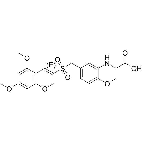
Rigosertib (ON-01910) is a multi-kinase inhibitor and a selective anti-cancer agent, which induces apoptosis by inhibition the PI3 kinase/Akt pathway, promots the phosphorylation of histone H2AX and induces G2/M arrest in cell cycle. Rigosertib is a selective and non-ATP-competitive inhibitor of PLK1 with an IC50 of 9 nM.
For research use only. We do not sell to patients.
- Purity : 97.14%
- CAS No.: 592542-59-1
- Formula: C21H25NO8S
- Molecular Weight:451.49
-
Storage:
-20°C, protect from light, stored under nitrogen
* The compound is unstable in solutions, freshly prepared is recommended.

To place orders, for customer services and technical support, please contact: MedChemExpress USA
Tel: 609-228-6898 E-mail: [email protected] [email protected]


-
 Biological Activity
-
Chemical Information
-
Solvent & Solubility
- Protocols
- Purity & Documentation
- References
-
 Help & FAQs


-
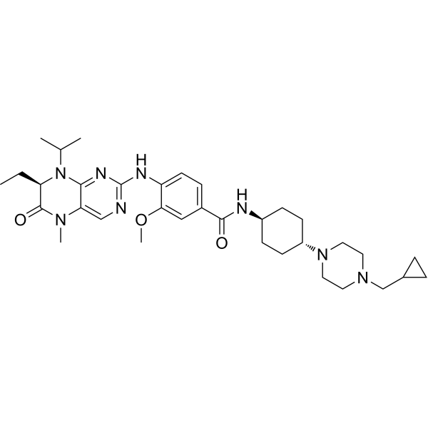
HY-12137
VolasertibPLK1 Inhibitor -
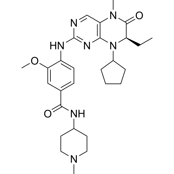
HY-50698
BI 2536PLK1 Inhibitor -
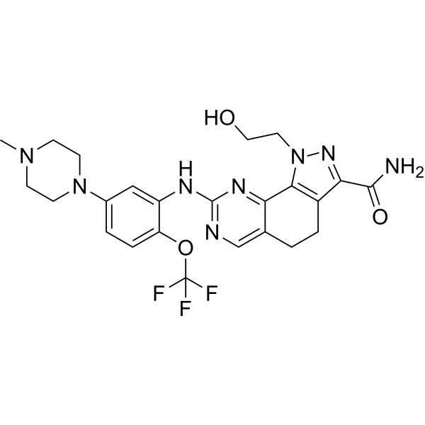
HY-15828
OnvansertibPLK1 Inhibitor -
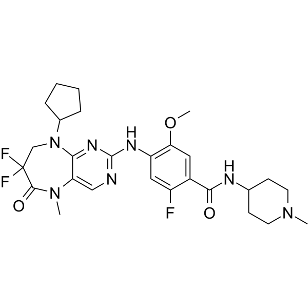
HY-15160
TAK-960PLK1 Inhibitor -
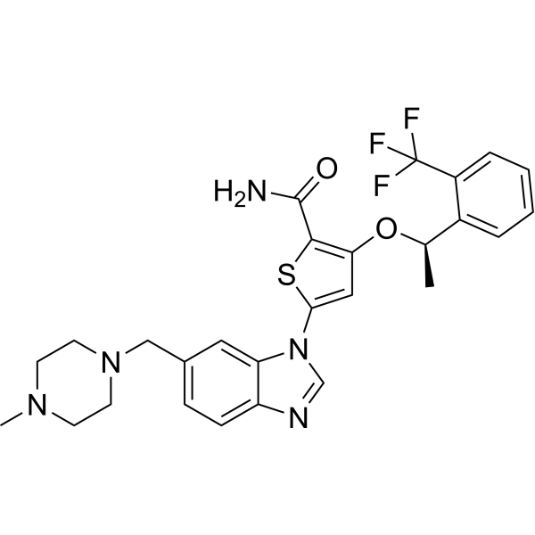
HY-50877
GSK461364PLK1 Inhibitor -
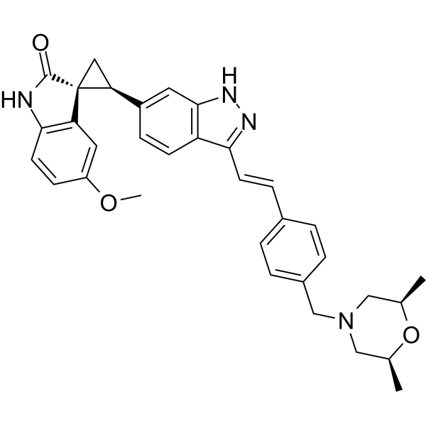
HY-12300
OcifisertibPLK4 Inhibitor -
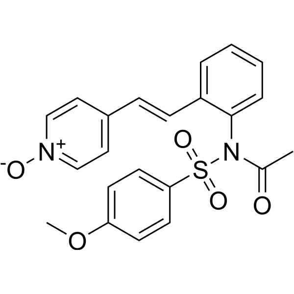
HY-12045
HMN-214PLK1 Inhibitor -
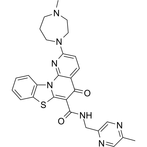
HY-13323
CX-5461RNA Polymerase I Inhibitor -
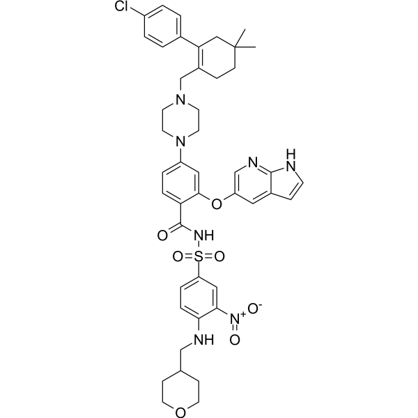
HY-15531
VenetoclaxBCL-2 Inhibitor -
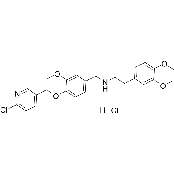
HY-15158
SBE13 HydrochloridePlk1 Inhibitor -
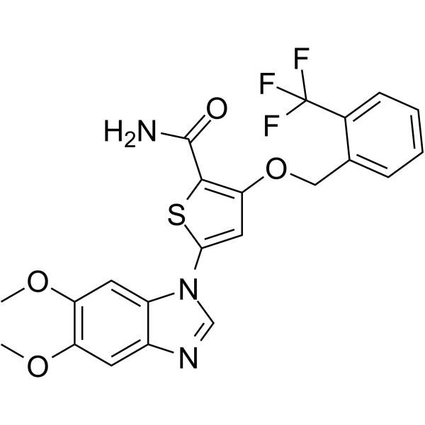
HY-11003
GW843682XPLK Inhibitor -
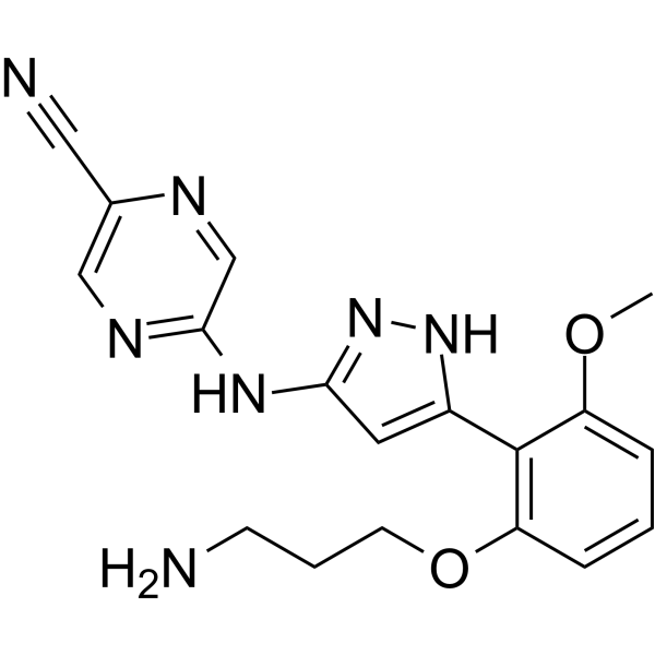
HY-18174
PrexasertibCHK1 Inhibitor
Publications Citing Use of MedChemExpress (MCE) Rigosertib
More

Biological Activity
Description
IC50 & Target
[1]|
PLK1 9 nM (IC50) |
PLK2 260 nM (IC50) |
PDGFR 18 nM (IC50) |
Src 155 nM (IC50) |
BCR-ABL 32 nM (IC50) |
Cdk1 260 nM (IC50) |
Flt1 42 nM (IC50) |
Fyn 182 nM (IC50) |
Cellular Effect
|
Cell Line

|
Type | Value | Description | References |
|---|---|---|---|---|
| A2780 | GI50 |
0.062 μM
Compound: ON01910
|
Antiproliferative activity against human A2780 cells assessed as cell growth inhibition after 72 hrs by MTT assay
Antiproliferative activity against human A2780 cells assessed as cell growth inhibition after 72 hrs by MTT assay
|
[PMID: 24471873] |
| A-431 | IC50 |
0.36 μM
Compound: Rigosertib
|
Cytotoxicity against human A-431 cells assessed as inhibition of cell growth incubated for 72 hrs by MTT assay
Cytotoxicity against human A-431 cells assessed as inhibition of cell growth incubated for 72 hrs by MTT assay
|
[PMID: 38261411] |
| A549 | IC50 |
0.37 μM
Compound: Rigosertib
|
Cytotoxicity against human A549 cells assessed as inhibition of cell growth incubated for 72 hrs by MTT assay
Cytotoxicity against human A549 cells assessed as inhibition of cell growth incubated for 72 hrs by MTT assay
|
[PMID: 38261411] |
| DU-145 | GI50 |
0.075 μM
Compound: ON01910
|
Antiproliferative activity against AR negative human DU145 cells assessed as cell growth inhibition after 72 hrs by MTT assay
Antiproliferative activity against AR negative human DU145 cells assessed as cell growth inhibition after 72 hrs by MTT assay
|
[PMID: 24471873] |
| DU-145 | IC50 |
0.075 μM
Compound: 27a
|
Cytotoxicity against human DU145 cells after 96 hrs by trypan blue exclusion assay
Cytotoxicity against human DU145 cells after 96 hrs by trypan blue exclusion assay
|
[PMID: 21812421] |
| HCT-116 | GI50 |
0.05 μM
Compound: 3j
|
Cytotoxicity against human HCT116 cells after 72 hrs by MTT assay
Cytotoxicity against human HCT116 cells after 72 hrs by MTT assay
|
[PMID: 21463944] |
| HCT-116 | GI50 |
0.07 μM
Compound: ON01910
|
Antiproliferative activity against human HCT116 cells assessed as cell growth inhibition after 72 hrs by MTT assay
Antiproliferative activity against human HCT116 cells assessed as cell growth inhibition after 72 hrs by MTT assay
|
[PMID: 24471873] |
| HeLa | GI50 |
0.012 μM
Compound: ON01910
|
Antiproliferative activity against human HeLa cells assessed as cell growth inhibition after 72 hrs by MTT assay
Antiproliferative activity against human HeLa cells assessed as cell growth inhibition after 72 hrs by MTT assay
|
[PMID: 24471873] |
| HeLa S3 | EC50 |
45 nM
Compound: Chemical probe : ON01910
|
Antiproliferative activity against human HeLa S3 cells assessed as inhibition in cell growth incubated for 72 hrs by Alamar Blue dye based fluorescence spectrophotometric analysis
Antiproliferative activity against human HeLa S3 cells assessed as inhibition in cell growth incubated for 72 hrs by Alamar Blue dye based fluorescence spectrophotometric analysis
|
[PMID: 17291758] |
| HepG2 | IC50 |
1.34 μM
Compound: Rigosertib
|
Cytotoxicity against human HepG2 cells assessed as inhibition of cell growth incubated for 72 hrs by MTT assay
Cytotoxicity against human HepG2 cells assessed as inhibition of cell growth incubated for 72 hrs by MTT assay
|
[PMID: 38261411] |
| HT-29 | IC50 |
0.16 μM
Compound: Rigosertib
|
Cytotoxicity against human HT-29 cells assessed as inhibition of cell growth incubated for 72 hrs by MTT assay
Cytotoxicity against human HT-29 cells assessed as inhibition of cell growth incubated for 72 hrs by MTT assay
|
[PMID: 38261411] |
| K562 | IC50 |
0.0075 μM
Compound: 27a
|
Cytotoxicity against human K562 cells after 96 hrs by trypan blue exclusion assay
Cytotoxicity against human K562 cells after 96 hrs by trypan blue exclusion assay
|
[PMID: 21812421] |
| K562 | IC50 |
0.15 μM
Compound: Rigosertib
|
Cytotoxicity against human K562 cells assessed as inhibition of cell growth incubated for 72 hrs by MTT assay
Cytotoxicity against human K562 cells assessed as inhibition of cell growth incubated for 72 hrs by MTT assay
|
[PMID: 38261411] |
| L02 | IC50 |
>40 μM
Compound: Rigosertib
|
Cytotoxicity against human L02 cells assessed as inhibition of cell growth incubated for 72 hrs by MTT assay
Cytotoxicity against human L02 cells assessed as inhibition of cell growth incubated for 72 hrs by MTT assay
|
[PMID: 38261411] |
| LNCaP | GI50 |
0.025 μM
Compound: ON01910
|
Antiproliferative activity against AR positive human LNCAP cells assessed as cell growth inhibition after 72 hrs by MTT assay
Antiproliferative activity against AR positive human LNCAP cells assessed as cell growth inhibition after 72 hrs by MTT assay
|
[PMID: 24471873] |
| MCF7 | GI50 |
0.05 μM
Compound: 3j
|
Cytotoxicity against human MCF7 cells after 72 hrs by MTT assay
Cytotoxicity against human MCF7 cells after 72 hrs by MTT assay
|
[PMID: 21463944] |
| MCF7 | GI50 |
0.05 μM
Compound: ON01910
|
Antiproliferative activity against ER positive human MCF7 cells assessed as cell growth inhibition after 72 hrs by MTT assay
Antiproliferative activity against ER positive human MCF7 cells assessed as cell growth inhibition after 72 hrs by MTT assay
|
[PMID: 24471873] |
| MCF7 | GI50 |
0.05 μM
Compound: ON01910
|
Antiproliferative activity against human MCF7 cells assessed as cell growth inhibition after 72 hrs by MTT assay
Antiproliferative activity against human MCF7 cells assessed as cell growth inhibition after 72 hrs by MTT assay
|
[PMID: 24471873] |
| MCF7 | IC50 |
0.26 μM
Compound: Rigosertib
|
Cytotoxicity against human MCF7 cells assessed as inhibition of cell growth incubated for 72 hrs by MTT assay
Cytotoxicity against human MCF7 cells assessed as inhibition of cell growth incubated for 72 hrs by MTT assay
|
[PMID: 38261411] |
| MDA-MB-231 | GI50 |
0.057 μM
Compound: ON01910
|
Antiproliferative activity against ER negative human MDA-MB-231 cells assessed as cell growth inhibition after 72 hrs by MTT assay
Antiproliferative activity against ER negative human MDA-MB-231 cells assessed as cell growth inhibition after 72 hrs by MTT assay
|
[PMID: 24471873] |
| MDA-MB-468 | GI50 |
0.02 μM
Compound: 3j
|
Cytotoxicity against human MDA468 cells after 72 hrs by MTT assay
Cytotoxicity against human MDA468 cells after 72 hrs by MTT assay
|
[PMID: 21463944] |
| MDA-MB-468 | GI50 |
0.302 μM
Compound: 3j
|
Cytotoxicity against human MDA468 cells after 48 hrs by MTT assay
Cytotoxicity against human MDA468 cells after 48 hrs by MTT assay
|
[PMID: 21463944] |
| MDA-MB-468 | GI50 |
0.601 μM
Compound: 3j
|
Cytotoxicity against human MDA468 cells after 24 hrs by MTT assay
Cytotoxicity against human MDA468 cells after 24 hrs by MTT assay
|
[PMID: 21463944] |
| MRC5 | GI50 |
0.71 μM
Compound: 3j
|
Cytotoxicity against human MRC5 cells after 72 hrs by MTT assay
Cytotoxicity against human MRC5 cells after 72 hrs by MTT assay
|
[PMID: 21463944] |
| MV4-11 | IC50 |
1.15 μM
Compound: Rigosertib
|
Cytotoxicity against human MV4-11 cells assessed as inhibition of cell growth incubated for 72 hrs by MTT assay
Cytotoxicity against human MV4-11 cells assessed as inhibition of cell growth incubated for 72 hrs by MTT assay
|
[PMID: 38261411] |
| NCI-H1975 | IC50 |
0.23 μM
Compound: Rigosertib
|
Cytotoxicity against human NCI-H1975 cells assessed as inhibition of cell growth incubated for 72 hrs by MTT assay
Cytotoxicity against human NCI-H1975 cells assessed as inhibition of cell growth incubated for 72 hrs by MTT assay
|
[PMID: 38261411] |
| PANC-1 | GI50 |
0.039 μM
Compound: ON01910
|
Antiproliferative activity against human PANC1 cells assessed as cell growth inhibition after 72 hrs by MTT assay
Antiproliferative activity against human PANC1 cells assessed as cell growth inhibition after 72 hrs by MTT assay
|
[PMID: 24471873] |
| PANC-1 | IC50 |
0.26 μM
Compound: Rigosertib
|
Cytotoxicity against human PANC-1 cells assessed as inhibition of cell growth incubated for 72 hrs by MTT assay
Cytotoxicity against human PANC-1 cells assessed as inhibition of cell growth incubated for 72 hrs by MTT assay
|
[PMID: 38261411] |
| SGC-7901 | IC50 |
0.25 μM
Compound: Rigosertib
|
Cytotoxicity against human SGC-7901 cells assessed as inhibition of cell growth incubated for 72 hrs by MTT assay
Cytotoxicity against human SGC-7901 cells assessed as inhibition of cell growth incubated for 72 hrs by MTT assay
|
[PMID: 38261411] |
| T47D | GI50 |
<0.01 μM
Compound: 3j
|
Cytotoxicity against human T47D cells after 72 hrs by MTT assay
Cytotoxicity against human T47D cells after 72 hrs by MTT assay
|
[PMID: 21463944] |
| WI-38 | GI50 |
>10 μM
Compound: ON01910
|
Antiproliferative activity against human WI38 cells assessed as cell growth inhibition after 72 hrs by MTT assay
Antiproliferative activity against human WI38 cells assessed as cell growth inhibition after 72 hrs by MTT assay
|
[PMID: 24471873] |
In Vitro
Rigosertib is non-ATP-competitive inhibitor of PLK1 with IC50 of 9 nM. Rigosertib also exhibits inhibition of PLK2, PDGFR, Flt1, BCR-ABL, Fyn, Src, and CDK1, with IC50 of 18-260 nM. Rigosertib shows cell killing activity against 94 different tumor cell lines with IC50 of 50-250 nM, including BT27, MCF-7, DU145, PC3, U87, A549, H187, RF1, HCT15, SW480, and KB cells. While in normal cells, such as HFL, PrEC, HMEC, and HUVEC, Rigosertib has little or no effect unless its concentration is greater than 5-10 μM. In HeLa cells, Rigosertib (100-250 nM) induces spindle abnormalities and apoptosis[3]. Rigosertib also inhibits several multidrug resistant tumor cell lines, including MES-SA, MES-SA/DX5a, CEM, and CEM/C2a, with IC50 of 50-100 nM. In DU145 cells, Rigosertib (0.25-5 μM) blocks cell cycle progression in G2/M phase, results in an accumulation of cells containing subG1 content of DNA, and activates apoptotic pathways. In A549 cells, Rigosertib (50 nM-0.5 μM) induces loss of viability and caspase 3/7 activation[4]. Rigosertib sodium (2 μM) induces apoptosis in chronic lymphocytic leukemia (CLL) cells without toxicity against T-cells or normal B-cells. Rigosertib sodium (2 μM) also abrogates the pro-survival effect of follicular dendritic cells on CLL cells and reduces SDF-1-induced migration of leukemic cells[5].
MedChemExpress (MCE) has not independently confirmed the accuracy of these methods. They are for reference only.
In Vivo
MedChemExpress (MCE) has not independently confirmed the accuracy of these methods. They are for reference only.
- Kinase activity and phosphorylation assays
- Western Blot
- Detection of Apoptosis
- TUNEL staining for apoptotic DNA fragmentation
- Flow cytometric DNA-content cell-cycle staining
- Annexin V plus membrane-impermeant dye apoptosis staining
- BrdU Incorporation Assay
- Apoptosis Solutions
- Protocol for Cell Cycle
- Protocol for Kinase activity and phosphorylation assays
- Directly Induced Neuron Culture


Chemical Information
-
CAS No. 592542-59-1
-
Appearance Solid
-
Molecular Weight 451.49
-
Formula C21H25NO8S
-
Color White to light yellow
-
SMILES
COC1=CC=C(C=C1NCC(O)=O)CS(/C=C/C2=C(C=C(C=C2OC)OC)OC)(=O)=O
-
Synonyms
ON-01910
-
Shipping
Room temperature in continental US; may vary elsewhere.
-
Storage
-20°C, protect from light, stored under nitrogen
* The compound is unstable in solutions, freshly prepared is recommended.


Publications (6)
-
Journal Impact Factor
-
Most Recent
-
Sci Transl Med
PP2A inhibition is a druggable MEK inhibitor resistance mechanism in KRAS-mutant lung cancer cells. [Abstract]2018 Jul 18;10(450):eaaq1093. PMID: 30021885 -
Cancer Lett
GPI inactivation mediates pentose phosphate pathway flux switch-on inducing temozolomide resistance in glioma stem cell. [Abstract]2026 May 1:645:218384. PMID: 41763452 -
Int J Biol Sci
Glucocorticoids improve severe or critical COVID-19 by activating ACE2 and reducing IL-6 levels. [Abstract]2020 Jun 27;16(13):2382-2391. PMID: 32760206 -
Oncogenesis
STAG2 expression imparts distinct therapeutic vulnerabilities in muscle-invasive bladder cancer cells. [Abstract]2025 Mar 1;14(1):4. PMID: 40025053 -
Sci Rep
Identification of Polo-like kinases as potential novel drug targets for influenza A virus. [Abstract]2017 Aug 17;7(1):8629. PMID: 28819179 -
Oncol Res
Therapeutic Targeting PLK1 by ON-01910.Na Is Effective in Local Treatment of Retinoblastoma. [Abstract]2021 Sep 7;28(7):745-761. PMID: 33573708


Solvent & Solubility
In Vitro:
DMSO : 75 mg/mL (166.12 mM; Need ultrasonic; Hygroscopic DMSO has a significant impact on the solubility of product, please use newly opened DMSO)
Please refer to the solubility information to select the appropriate solvent. The compound is unstable in solutions, freshly prepared is recommended.
Please refer to the solubility information to select the appropriate solvent. The compound is unstable in solutions, freshly prepared is recommended.
Concentration (start) × Volume (start) = Concentration (final) × Volume (final)
In Vivo:
Select the appropriate dissolution method based on your experimental animal and administration route.
- For the following dissolution methods, please ensure to first prepare a clear stock solution using an In Vitro approach and then sequentially add co-solvents:
- To ensure reliable experimental results, the clarified stock solution can be appropriately stored based on storage conditions. As for the working solution for In Vivo experiments, it is recommended to prepare freshly and use it on the same day.
- The percentages shown for the solvents indicate their volumetric ratio in the final prepared solution. If precipitation or phase separation occurs during preparation, heat and/or sonication can be used to aid dissolution.
Add each solvent one by one: 10% DMSO 40% PEG300 5% Tween-80 45% Saline
Solubility: ≥ 2.5 mg/mL (5.54 mM); Clear solution
This protocol yields a clear solution of ≥ 2.5 mg/mL (saturation unknown).
Taking 1 mL working solution as an example, add 100 μL DMSO stock solution (25.0 mg/mL) to 400 μL PEG300, and mix evenly; then add 50 μL Tween-80 and mix evenly; then add 450 μL Saline to adjust the volume to 1 mL.
Preparation of Saline: Dissolve 0.9 g sodium chloride in ddH₂O and dilute to 100 mL to obtain a clear Saline solution.
Add each solvent one by one: 10% DMSO 90% (20% SBE-β-CD in Saline)
Solubility: 2.5 mg/mL (5.54 mM); Suspended solution; Need ultrasonic
This protocol yields a suspended solution of 2.5 mg/mL. Suspended solution can be used for oral and intraperitoneal injection.
Taking 1 mL working solution as an example, add 100 μL DMSO stock solution (25.0 mg/mL) to 900 μL 20% SBE-β-CD in Saline, and mix evenly.
Preparation of 20% SBE-β-CD in Saline (4°C, storage for one week): 2 g SBE-β-CD powder is dissolved in 10 mL Saline, completely dissolve until clear.
In Vivo Dissolution Calculator
Please enter the basic information of animal experiments:
-
-
-
-
Recommended: Prepare an additional quantity of animals to account for potential losses during experiments.
Please enter your animal formula composition:
-
%DMSO +
Recommended: Keep the proportion of DMSO in working solution below 2% if your animal is weak.
-
%+
-
+%Tween-80 + +
-
%Saline +
The co-solvents required include: DMSO, . All of co-solvents are available by MedChemExpress (MCE). , Tween 80. All of co-solvents are available by MedChemExpress (MCE).
Working solution concentration: 0.22 mg/mL
Method for preparing stock solution: mg drug dissolved in μL DMSO. Stock solution concentration: mg/mL. * The compound is unstable in solutions, freshly prepared is recommended.
1. Take μL DMSO stock solution;
2. Add μL .
μL , mix evenly;
3. Then add μL Tween 80, mix evenly;
4. Then add μL
Please ensure that the stock solution in the first step is dissolved to a clear state, and add co-solvents in sequence. You can use ultrasonic heating (ultrasonic cleaner, recommended frequency 20-40 kHz), vortexing, etc. to assist dissolution.


Purity & Documentation
-
Data Sheet (280 KB)
-
SDS (394 KB)
- English - EN (394 KB)
- Français - FR (394 KB)
- Deutsch - DE (394 KB)
- Norwegian - NO (394 KB)
- Español - ES (394 KB)
- Swedish - SV (394 KB)
- Italian - IT (394 KB)
- Korean - KR (394 KB)
- Portuguese - PT (394 KB)
-
Handling Instructions (2659 KB)
References
[1]. Xu F, et al. Rigosertib as a selective anti-tumor agent can ameliorate multiple dysregulated signalingtransduction pathways in high-grade myelodysplastic syndrome. Sci Rep. 2014 Dec 4;4:7310. [Content Brief]
[2]. Hyoda T, et al. Rigosertib induces cell death of a myelodysplastic syndrome-derived cell line by DNA damage-induced G2/M arrest. Cancer Sci. 2015 Mar;106(3):287-93. [Content Brief]
[3]. Gumireddy K, et al. ON01910, a non-ATP-competitive small molecule inhibitor of Plk1, is a potent anticancer agent. Cancer Cell. 2005 Mar;7(3):275-86. [Content Brief]
[4]. Reddy MV, et al. Discovery of a clinical stage multi-kinase inhibitor sodium (E)-2-{2-methoxy-5-[(2',4',6'-trimethoxystyrylsulfonyl)methyl]phenylamino}acetate (ON 01910.Na): synthesis, structure-activity relationship, and biological activity. J Med Chem. 2011 Sep 22;54(18):6254-76. [Content Brief]
[5]. Chapman CM, et al. ON 01910.Na is selectively cytotoxic for chronic lymphocytic leukemia cells through a dual mechanism of action involving PI3K/AKT inhibition and induction of oxidative stress. Clin Cancer Res. 2012 Apr 1;18(7):1979-91 [Content Brief]


Complete Stock Solution Preparation Table
Please refer to the solubility information to select the appropriate solvent. The compound is unstable in solutions, freshly prepared is recommended.
| Optional Solvent | Concentration Solvent Mass | 1 mg | 5 mg | 10 mg | 25 mg |
|---|---|---|---|---|---|
| DMSO | 1 mM | 2.2149 mL | 11.0744 mL | 22.1489 mL | 55.3722 mL |
| 5 mM | 0.4430 mL | 2.2149 mL | 4.4298 mL | 11.0744 mL | |
| 10 mM | 0.2215 mL | 1.1074 mL | 2.2149 mL | 5.5372 mL | |
| 15 mM | 0.1477 mL | 0.7383 mL | 1.4766 mL | 3.6915 mL | |
| 20 mM | 0.1107 mL | 0.5537 mL | 1.1074 mL | 2.7686 mL | |
| 25 mM | 0.0886 mL | 0.4430 mL | 0.8860 mL | 2.2149 mL | |
| 30 mM | 0.0738 mL | 0.3691 mL | 0.7383 mL | 1.8457 mL | |
| 40 mM | 0.0554 mL | 0.2769 mL | 0.5537 mL | 1.3843 mL | |
| 50 mM | 0.0443 mL | 0.2215 mL | 0.4430 mL | 1.1074 mL | |
| 60 mM | 0.0369 mL | 0.1846 mL | 0.3691 mL | 0.9229 mL | |
| 80 mM | 0.0277 mL | 0.1384 mL | 0.2769 mL | 0.6922 mL | |
| 100 mM | 0.0221 mL | 0.1107 mL | 0.2215 mL | 0.5537 mL |


Rigosertib Related Classifications
HY-12037A Related Classifications
 Powered by Bioz