



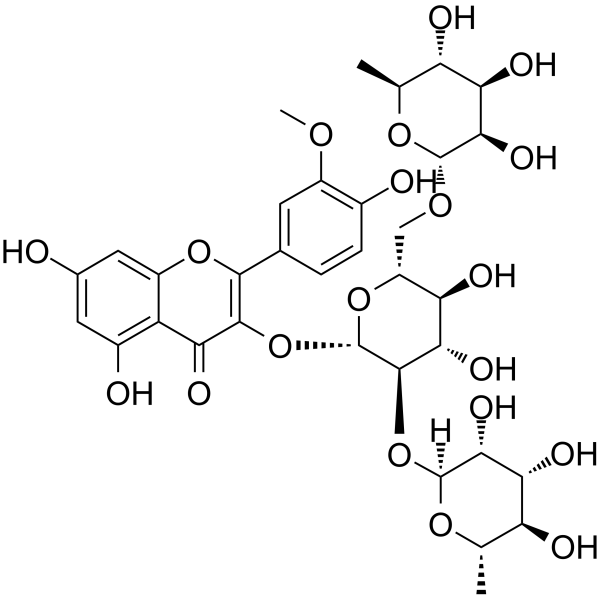
Typhaneoside
Based on 4 publication(s) in Google Scholar
Typhaneoside is an orally active activator of PI3K/Akt/mTOR and farnesoid X receptor. Typhaneoside promotes the activation of AMPK and Caspase-3, induces apoptosis, ferroptosis, autophagy, ROS accumulation, cell cycle arrest at the G2/M phase, and reduces cancer cell viability. Typhaneoside improves glucose and lipid metabolism, alleviates inflammatory responses, oxidative stress and hepatic lipid accumulation, and exerts hepatoprotective effects. Typhaneoside can be used in research related to heart failure after myocardial infarction, acute myeloid leukemia, non-alcoholic fatty liver disease and neurological disorders.
For research use only. We do not sell to patients.
- Purity : 99.74%
- CAS No.: 104472-68-6
- Formula: C34H42O20
- Molecular Weight:770.69
-
Storage:
4°C, protect from light
* In solvent : -80°C, 6 months; -20°C, 1 month (protect from light)

To place orders, for customer services and technical support, please contact: MedChemExpress USA
Tel: 609-228-6898 E-mail: [email protected] [email protected]


-
 Biological Activity
-
Chemical Information
-
Solvent & Solubility
- Protocols
- Purity & Documentation
- References
-
 Help & FAQs


-
HY-N0527
PentagalloylglucoseAntioxidant -
HY-13653
(-)-Epigallocatechin GallateAnticancer Agent -
HY-N1362
Salvianolic acid BAntioxidant -
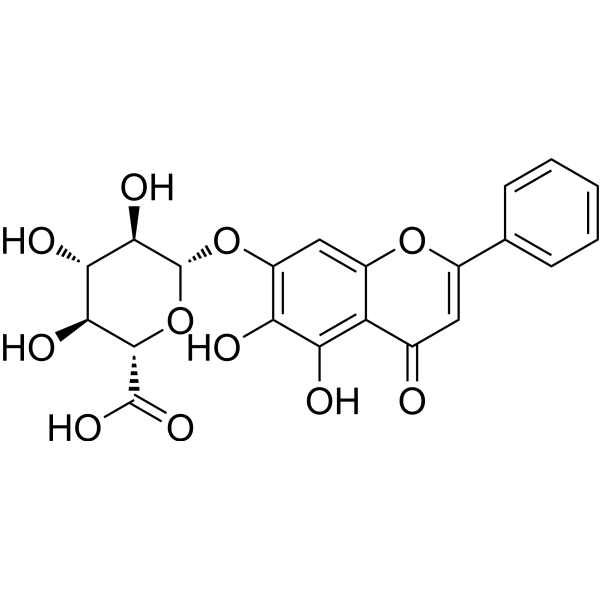
HY-N0197
BaicalinAutophagy Inducer -
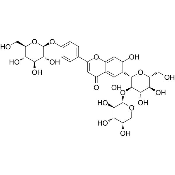
HY-N1419
VaccarinAKT/ERK/AMPK Agonists -
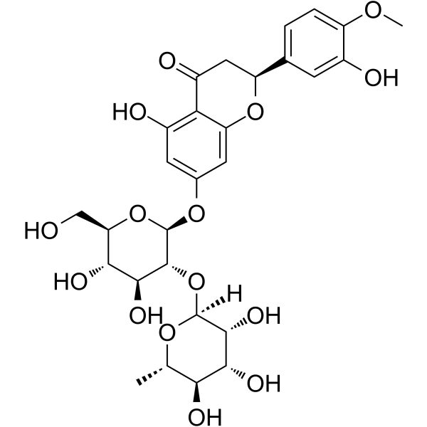
HY-N0101
NeohesperidinWnt/β-Catenin Activator -
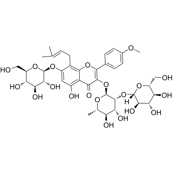
HY-N0257
Epimedin ANatural Product -
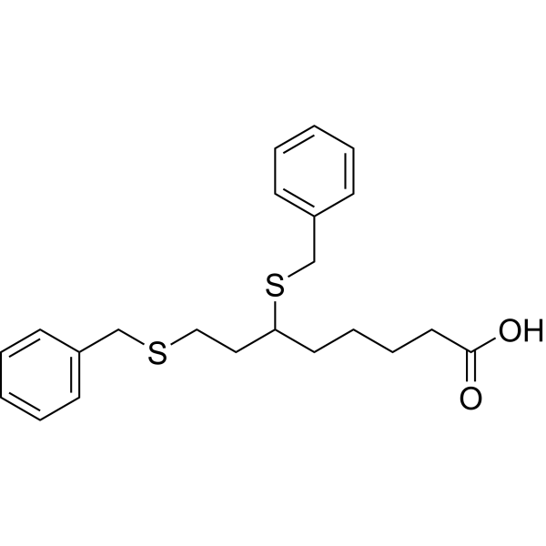
HY-15453
DevimistatMitochondrial Metabolism Inhibitor -

HY-N0712R
Typhaneoside (Standard)Autophagy Inhibitor -
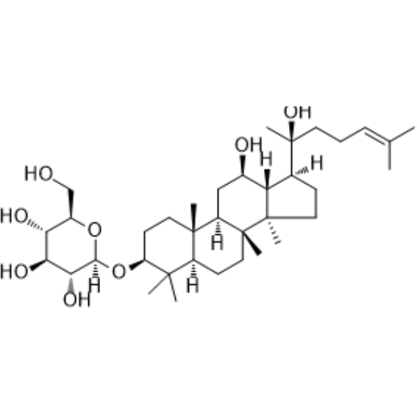
HY-N0605
Ginsenoside Rh2Caspase Activator -
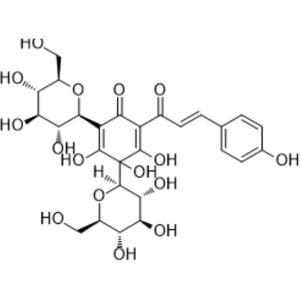
HY-N0567
Hydroxysafflor yellow AAnticancer Agent -
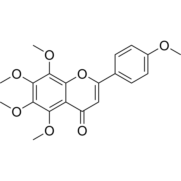
HY-N0133
TangeretinApoptosis Inducer
Publications Citing Use of MedChemExpress (MCE) Typhaneoside
MoreAll Calcium Channel Isoforms
More

Biological Activity
Description
Cellular Effect
|
Cell Line

|
Type | Value | Description | References |
|---|---|---|---|---|
| N9 | IC50 |
>100 μM
Compound: 10
|
Antineuroinflammatory activity in mouse N9 cells assessed as inhibition of LPS-induced nitric oxide production after 24 hrs by Griess assay
Antineuroinflammatory activity in mouse N9 cells assessed as inhibition of LPS-induced nitric oxide production after 24 hrs by Griess assay
|
[PMID: 28073678] |
| Raji | IC50 |
592 molar ratio
Compound: 12
|
Inhibition of TPA-induced EBV-early antigen activation in human Raji cells relative to TPA
Inhibition of TPA-induced EBV-early antigen activation in human Raji cells relative to TPA
|
[PMID: 17190444] |
In Vitro
Typhaneoside (0-50 μM; 12-48 h) significantly reduces the viability of Kas-1, HL60 and NB4 acute myeloid leukemia cells in a time- and dose-dependent manner, while exerts no significant effect on K562 acute myeloid leukemia cells or normal 293T cells[2].
Typhaneoside (20-40 μM; 24 h) induces apoptosis in Kas-1, HL60 and NB4 acute myeloid leukemia cells in a dose-dependent manner, with the highest apoptosis rate induced by treatment at 40 μM[2].
Typhaneoside (20-40 μM; 24 h) dose-dependently reduces the mRNA expression levels of genes associated with mitochondrial dysfunction (NDUFS3, SDHB, UQCRFS1, TFAM, ClpP) in Kas-1, HL60 and NB4 acute myeloid leukemia cells[2].
The effects of typhaneoside (40 μM; 24 h) on inducing reactive oxygen species production and apoptosis in Kas-1, HL60 and NB4 acute myeloid leukemia cells are significantly reversed by pre-treatment with NAC or DFO[2].
Typhaneoside (40 μM; 24 h) upregulates autophagy-related proteins (ATG5, ATG7, Beclin 1, LC3) in Kas-1, HL60, and NB4 acute myeloid leukemia cells, and this effect is reversed by pre-treatment with 5 mM NAC for 2 h[2].
Typhaneoside (12.5-50 μM; 24 h) activates FXR-dependent BSEP promoter activity in HEK293T cells, with significant activation observed at the concentration of 50 μM after 24 h[3].
Typhaneoside (12.5-50 μM; 24 h) alleviates OAPA-induced lipid accumulation, oxidative stress, inflammatory response and glucose metabolism disorder in HepG2 cells by activating the AKT/GSK3β signaling pathway[3].
Typhaneoside (5-50 μM; 10 min) concentration-dependently inhibits 4-aminopyridine (HY-B0604)-induced glutamate release from rat cerebral cortical synaptosomes, with an IC50 of 20 μM[4].
Typhaneoside (20 μM; 10 min)-mediated inhibition of 4-aminopyridine-induced glutamate release from rat cerebral cortex synaptosomes targets Ca2+-dependent vesicular exocytosis, rather than reverse transport via Ca2+-independent glutamate transporters[4].
Typhaneoside (20 μM) inhibits KCl-induced glutamate release from rat cerebral cortex synaptosomes[4].
Typhaneoside (20 μM; 10 min)-mediated inhibition of 4-aminopyridine-induced glutamate release from rat cerebral cortex synaptosomes depends on N-type (Cav2.2) voltage-dependent Ca2+ channels, and does not involve intracellular Ca2+ release or mitochondrial Na+/Ca2+ exchange[4].
The inhibitory effect of Typhaneoside (20 μM; 10 min) on 4-aminopyridine-induced glutamate release from rat cerebral cortex synaptosomes depends on the MAPK/ERK signaling pathway, and does not involve PKA or PKC[4].
Typhaneoside (20 μM; 10 min) reduces the phosphorylation levels of 4-aminopyridine-induced ERK1/2 and its presynaptic target synapsin I in rat cerebral cortex synaptosomes, but exerts no such effect on JNK or p38[4].
MedChemExpress (MCE) has not independently confirmed the accuracy of these methods. They are for reference only. Further protocols information, click here.
-
Cell Line:Kas-1, HL60, NB4, K562, 293T
-
Concentration:0, 10, 20, 30, 40 and 50 μM
-
Incubation Time:12 h, 24 h, 48 h
-
Result:Reduced cell viability of Kas-1, HL60, and NB4 cells in a time- and dose-dependent manner.
Showed no significant effect on cell viability of K562 or 293T cells.
-
Cell Line:Kas-1, HL60, NB4
-
Concentration:20, 30 and 40 μM
-
Incubation Time:24 h
-
Result:Dose-dependently arrested Kas-1, HL60, and NB4 cells at the G2/M phase.
Decreased G1/G0 phase cell percentage in a dose-dependent manner for all three cell lines.
Decreased S phase cell percentage in a dose-dependent manner for all three cell lines.
-
Cell Line:Kas-1, HL60, NB4
-
Concentration:20, 30 and 40 μM
-
Incubation Time:24 h
-
Result:Dose-dependently reduced protein expression of Cyclin B1 and p-Cdc2 in Kas-1, HL60, and NB4 cells.
Dose-dependently enhanced protein expression of p53 and p27 in Kas-1, HL60, and NB4 cells.\nDose-dependently reduced protein expression of anti-apoptotic Bcl-2 in Kas-1, HL60, and NB4 cells.
Dose-dependently increased protein expression of pro-apoptotic Bax and cleaved Caspase-3 in Kas-1, HL60, and NB4 cells.\nDose-dependently increased protein expression of p-AMPK in Kas-1, HL60, and NB4 cells.
Dose-dependently decreased protein expression of p-mTOR in Kas-1, HL60, and NB4 cells.\nDose-dependently increased protein expression of ATG5, ATG7, Beclin 1, and LC3 in Kas-1, HL60, and NB4 cells.
-
Cell Line:Kas-1, HL60, NB4
-
Concentration:40 μM
-
Incubation Time:24 h (with 2 h pre-treatment of 5 mM NAC)
-
Result:Induced upregulation of ATG5, ATG7, Beclin 1, and LC3 protein expression that was reversed by pretreatment with 5 mM NAC in Kas-1, HL60, and NB4 cells.
In Vivo
Typhaneoside (10-30 mg/kg; i.p.; daily; 30 days) dose-dependently suppresses AML tumor growth in BALB/c nude mice, with the 30 mg/kg dose achieving the greatest tumor volume reduction and the highest 30-day survival rate of ~50%, while showing no overt tissue toxicity[2].
Typhaneoside (30 mg/kg; i.p.; daily; 30 days) causes no overt tissue toxicity in healthy BALB/c nude mice[2].
Typhaneoside (15-60 mg/kg/day; p.o.; daily; 8 weeks) dose-dependently alleviates HFD-induced NAFLD in male C57BL/6 mice by activating FXR signaling, reducing body and adipose tissue weight, improving lipid and glucose homeostasis, mitigating liver injury, oxidative stress and inflammation, and enhancing BAT thermogenesis and energy expenditure[3].
MedChemExpress (MCE) has not independently confirmed the accuracy of these methods. They are for reference only.
-
Animal Model:BALB/c nude (4-week-old male; AML model via subcutaneous injection of 1×10⁷ HL60 cells, tumor grown to 50 mm3 before treatment)[2]
-
Dosage:10 mg/kg; 20 mg/kg; 30 mg/kg
-
Administration:i.p.; daily; 30 days
-
Result:Significantly reduced mean tumor volume to ~1000 mm3 (10 mg/kg), ~800 mm3 (20 mg/kg), and ~500 mm3 (30 mg/kg) at day 30 compared to controls.
Improved 30-day survival rate to ~20% (10 mg/kg), ~30% (20 mg/kg), and ~50% (30 mg/kg) in a dose-dependent manner.
Significantly reduced white blood cell counts in a dose-dependent manner compared to controls.
-
Animal Model:C57BL/6 (male, 8-week-old, 20-22 g, SPF, HFD-induced NAFLD)[3]
-
Dosage:15 mg/kg/day; 30 mg/kg/day; 60 mg/kg/day
-
Administration:p.o.; daily; 8 weeks
-
Result:Reduced HFD-induced body weight gain in a dose-dependent manner, with a significant decrease in area under the curve for body weight across all doses.
Dose-dependently reduced liver weight,
Dose-dependently reversed HFD-induced serum and hepatic lipid abnormalities: decreased serum triglyceride (TG), low-density lipoprotein cholesterol (LDL-C), and free fatty acid (FFA) levels; increased serum high-density lipoprotein cholesterol (HDL-C) levels; decreased hepatic TG levels.
Dose-dependently reversed HFD-induced liver injury, oxidative stress, and inflammation: decreased serum and hepatic AST, ALT, and MDA levels; increased serum and hepatic SOD levels; suppressed HFD-induced upregulation of proinflammatory mRNA (IL-6, IL-1β, NF-κB, TNF-α) in liver and WAT.
Dose-dependently upregulated mRNA expression of FXR, SHP, BSEP, and TGR5 in liver, and increased hepatic FXR protein expression detected by immunofluorescence.


Chemical Information
-
CAS No. 104472-68-6
-
Appearance Solid
-
Molecular Weight 770.69
-
Formula C34H42O20
-
Color Light yellow to yellow
-
SMILES
O=C1C(O[C@H](O[C@H](CO[C@H](O[C@@H](C)[C@H](O)[C@H]2O)[C@@H]2O)[C@@H](O)[C@@H]3O)[C@@H]3O[C@@](O[C@@H](C)[C@H](O)[C@H]4O)([H])[C@@H]4O)=C(C5=CC(OC)=C(O)C=C5)OC6=CC(O)=CC(O)=C16
-
Structure Classification
-
Initial Source
-
Shipping
Room temperature in continental US; may vary elsewhere.
-
Storage
4°C, protect from light
* In solvent : -80°C, 6 months; -20°C, 1 month (protect from light)


Publications (4)
-
Journal Impact Factor
-
Most Recent
-
Microbiome
Desulfovibrio vulgaris interacts with novel gut epithelial immune receptor LRRC19 and exacerbates colitis. [Abstract]2024 Jan 3;12(1):4. PMID: 38172943 -
Food Chem
Effects of sun drying combined with baking processes on the flavor quality of Chongqing Tuocha raw tea. [Abstract]2025 Dec 30:497:146992. PMID: 41285060 -
Food Chem
Flavonoid-mediated metabolic underpinning quality variation in red bud-sport pear mutants. [Abstract]2025 Oct 15:489:144992. PMID: 40466530 -


Solvent & Solubility
In Vitro:
DMSO : 250 mg/mL (324.38 mM; Need ultrasonic; Hygroscopic DMSO has a significant impact on the solubility of product, please use newly opened DMSO)
Please refer to the solubility information to select the appropriate solvent. Once prepared, please aliquot and store the solution to prevent product inactivation from repeated freeze-thaw cycles.
Storage method and period of stock solution: -80°C, 6 months; -20°C, 1 month (protect from light). When stored at -80°C, please use it within 6 months. When stored at -20°C, please use it within 1 month.
Please refer to the solubility information to select the appropriate solvent. Once prepared, please aliquot and store the solution to prevent product inactivation from repeated freeze-thaw cycles.
Storage method and period of stock solution: -80°C, 6 months; -20°C, 1 month (protect from light). When stored at -80°C, please use it within 6 months. When stored at -20°C, please use it within 1 month.
Concentration (start) × Volume (start) = Concentration (final) × Volume (final)
In Vivo:
Select the appropriate dissolution method based on your experimental animal and administration route.
- For the following dissolution methods, please ensure to first prepare a clear stock solution using an In Vitro approach and then sequentially add co-solvents:
- To ensure reliable experimental results, the clarified stock solution can be appropriately stored based on storage conditions. As for the working solution for In Vivo experiments, it is recommended to prepare freshly and use it on the same day.
- The percentages shown for the solvents indicate their volumetric ratio in the final prepared solution. If precipitation or phase separation occurs during preparation, heat and/or sonication can be used to aid dissolution.
Add each solvent one by one: 10% DMSO 40% PEG300 5% Tween-80 45% Saline
Solubility: ≥ 2.08 mg/mL (2.70 mM); Clear solution
This protocol yields a clear solution of ≥ 2.08 mg/mL (saturation unknown).
Taking 1 mL working solution as an example, add 100 μL DMSO stock solution (20.8 mg/mL) to 400 μL PEG300, and mix evenly; then add 50 μL Tween-80 and mix evenly; then add 450 μL Saline to adjust the volume to 1 mL.
Preparation of Saline: Dissolve 0.9 g sodium chloride in ddH₂O and dilute to 100 mL to obtain a clear Saline solution.
Add each solvent one by one: 10% DMSO 90% (20% SBE-β-CD in Saline)
Solubility: ≥ 2.08 mg/mL (2.70 mM); Clear solution
This protocol yields a clear solution of ≥ 2.08 mg/mL (saturation unknown).
Taking 1 mL working solution as an example, add 100 μL DMSO stock solution (20.8 mg/mL) to 900 μL 20% SBE-β-CD in Saline, and mix evenly.
Preparation of 20% SBE-β-CD in Saline (4°C, storage for one week): 2 g SBE-β-CD powder is dissolved in 10 mL Saline, completely dissolve until clear.
In Vivo Dissolution Calculator
Please enter the basic information of animal experiments:
-
-
-
-
Recommended: Prepare an additional quantity of animals to account for potential losses during experiments.
Please enter your animal formula composition:
-
%DMSO +
Recommended: Keep the proportion of DMSO in working solution below 2% if your animal is weak.
-
%+
-
+%Tween-80 + +
-
%Saline +
The co-solvents required include: DMSO, . All of co-solvents are available by MedChemExpress (MCE). , Tween 80. All of co-solvents are available by MedChemExpress (MCE).
Working solution concentration: 0.22 mg/mL
Method for preparing stock solution: mg drug dissolved in μL DMSO. Stock solution concentration: mg/mL. * In solvent : -80°C, 6 months; -20°C, 1 month (protect from light)
1. Take μL DMSO stock solution;
2. Add μL .
μL , mix evenly;
3. Then add μL Tween 80, mix evenly;
4. Then add μL
Please ensure that the stock solution in the first step is dissolved to a clear state, and add co-solvents in sequence. You can use ultrasonic heating (ultrasonic cleaner, recommended frequency 20-40 kHz), vortexing, etc. to assist dissolution.

Protocols
-
Mammalian live/dead viability and cytotoxicity staining
Live/dead viability and cytotoxicity staining assays are based on the simultaneous detection of intracellular esterase activity in metabolically active (viable) cells and membrane integrity loss in non-viable cells. In commonly used dual-staining approaches, membrane-permeant fluorogenic substrates are converted by intracellular esterases into fluorescent products in live cells, while impermeant DNA-binding dyes selectively enter cells with compromised plasma membranes and label nucleic acids in dead or dying cells, enabling discrimination between viable and non-viable populations by fluorescence microscopy or flow cytometry.
-
Annexin V plus membrane-impermeant dye apoptosis staining
Annexin V-based apoptosis assays rely on the detection of phosphatidylserine (PS) externalization from the inner leaflet of the plasma membrane to the outer leaflet, an early biochemical hallmark of apoptosis. Fluorescently labeled Annexin V binds PS in a calcium-dependent manner, enabling identification of early apoptotic cells by flow cytometry or fluorescence microscopy. When combined with a membrane-impermeant DNA-binding dye (e. g. , propidium iodide), this approach allows discrimination between viable (Annexin V−/dye−), early apoptotic (Annexin V+/dye−), and late apoptotic or necrotic (Annexin V+/dye+) cell populations by assessing membrane integrity and PS exposure.
-
ROS/oxidative-stress fluorescent staining
ROS/oxidative-stress fluorescent staining uses cell-permeant fluorogenic probes that become fluorescent after oxidation inside cells or tissues; commonly used examples include DCFH-DA/DCFDA for broad cellular oxidant detection, DHE for superoxide-related signal detection, MitoSOX for mitochondrial superoxide-related signal detection, and CellROX probes for oxidative-stress-associated fluorescence readouts. The assay detects probe oxidation rather than a single ROS species unless the probe and analysis method have been chemically validated for that species. DCFH-DA enters cells, is deacetylated by intracellular esterases to DCFH, and produces fluorescent DCF after oxidation, so the readout is used as an operational measure of total cellular oxidative stress rather than a species-specific ROS measurement. DHE and MitoSOX can report superoxide-related oxidation, but red fluorescence alone can include non-specific ethidium-like oxidation products; HPLC or optimized spectral approaches are
-
BrdU Incorporation Assay
Bromodeoxyuridine (BrdU) incorporation assay is based on the principle that BrdU, a thymidine analog, is incorporated into newly synthesized DNA during the S phase of the cell cycle, thereby serving as a marker of DNA replication and cellular proliferation. Incorporated BrdU can be detected using anti-BrdU antibodies following DNA denaturation, enabling visualization or quantification of proliferating cells through immunochemical detection methods such as immunofluorescence or immunohistochemistry.
-
Protocol for Cell Cycle
Cell-cycle analysis by flow cytometry measures DNA content in single cells to estimate the fraction of cells in G0/G1, S, and G2/M phases. Propidium iodide intercalates into DNA, and after RNA removal with RNase, fluorescence intensity reflects cellular DNA content: 2N cells are assigned to G0/G1, cells between 2N and 4N to S phase, and 4N cells to G2/M. DNA-content analysis alone cannot reliably separate G0 from G1 or G2 from M. Ki-67 can distinguish quiescent G0 cells from cycling cells, EdU or BrdU incorporation marks active DNA synthesis in S phase, and phospho-histone H3 staining identifies mitotic cells within the 4N population.
-
CCK-8/WST-8 Cell Proliferation Assay
The CCK-8/WST-8 assay is based on the reduction of the water-soluble tetrazolium salt WST-8 to a water-soluble formazan product by cellular dehydrogenases in metabolically active cells, where the generated formazan amount is proportional to the number of living cells and is quantified by measuring absorbance in the visible range, providing a colorimetric readout for cell viability and proliferation assessment. This class of tetrazolium-based assays improves upon earlier MTT-based systems by producing a water-soluble formazan, eliminating the need for organic solubilization steps and enabling direct spectrophotometric measurement in culture medium.
-
Research Protocol for Cardiovascular Diseases
Cardiovascular disease can be modeled as maladaptive cardiac remodeling, where ischemic injury or pressure overload activates inflammatory signaling, fibroblast activation, extracellular-matrix deposition, cardiomyocyte hypertrophy, vascular remodeling, and progressive ventricular dysfunction. The TGF-β/SMAD axis is a central profibrotic pathway after myocardial injury and pressure overload, while innate immune and cytokine pathways regulate leukocyte recruitment, scar formation, and adverse remodeling. Key unresolved questions include which inflammatory signals are reparative versus harmful, when fibrosis is protective versus maladaptive, and whether pathway inhibition improves function without weakening necessary infarct healing or compensatory remodeling.
-
Research Protocol for Metabolic Diseases
AMP-activated protein kinase, AMPK, is a conserved cellular energy sensor that responds to reduced cellular energy status and coordinates metabolism by increasing ATP-generating catabolic pathways while suppressing ATP-consuming anabolic processes. In metabolic disease research, the AMPK pathway is experimentally relevant because it regulates hepatic lipid synthesis, fatty acid oxidation, glucose production, skeletal-muscle glucose disposal, mTORC1-linked biosynthesis, autophagy, mitochondrial homeostasis, and whole-body energy balance. The central pathway logic is that energy stress, metformin, exercise-like stimulation, or direct AMPK activators increase AMPKα Thr172 phosphorylation and downstream substrate phosphorylation, including ACC and RAPTOR. Phosphorylation of ACC suppresses lipogenesis and supports fatty acid oxidation, whereas phosphorylation of RAPTOR suppresses mTORC1 signaling and links cellular energy status to growth and protein synthesis control. The pathway is linked
-
Cell Viability Determination by MTT Colorimetric Assay
The following protocol uses the MTT colorimetric assay as a classic literature-established method for assessing cell viability/metabolic activity in cultured mammalian cells. MTT[3-(4,5-dimethylthiazol-2-yl)-2,5-diphenyltetrazolium bromide] is reduced by metabolically active cells to a colored formazan product; the amount of formazan is quantified spectrophotometrically and provides an indirect measure of metabolically active viable cells. Importantly, MTT reduction reflects cellular oxidoreductase/metabolic activity rather than an absolute direct count of living cells, so changes in cellular metabolism can alter the signal independently of cell number.
-
3T3-L1 preadipocyte-to-adipocyte differentiation
3T3-L1 preadipocytes are induced to differentiate after growth arrest using adipogenic media containing insulin, dexamethasone, and IBMX; differentiation is assessed by lipid-droplet accumulation, triglyceride increase, Oil Red O staining, and adipocyte-marker induction such as PPARγ and C/EBPα.
-
Apoptosis
Apoptosis, also called programmed cell death, is generally characterized by distinct morphological characteristics.
-
TUNEL staining for apoptotic DNA fragmentation
TUNEL staining detects DNA strand breaks by using terminal deoxynucleotidyl transferase to add labeled nucleotides to exposed 3′-OH DNA termini, generating either microscopic staining in fixed cells or tissue sections, or fluorescence/cytometric signal in cell suspensions. TUNEL positivity reflects DNA fragmentation but should not be interpreted alone as definitive apoptosis, because TUNEL can also label necrotic, autolytic, mechanically damaged, or DNA-repair-associated DNA breaks.
-
Flow cytometric DNA-content cell-cycle staining
Flow cytometric DNA-content cell-cycle staining measures the fluorescence intensity of DNA-bound fluorochromes in single cells or nuclei to estimate DNA content distributions, allowing assignment of populations to G0/G1, S, and G2/M phases by DNA histogram deconvolution. Propidium iodide (PI) intercalates into DNA, and PI fluorescence is proportional to cellular DNA content when staining is performed under conditions that make DNA accessible and minimize non-DNA signal. Cells with G2/M DNA content are expected to show approximately twice the fluorescence intensity of G0/G1 cells, while S-phase cells occupy intermediate fluorescence values. PI-based DNA-content analysis can also detect cells with fractional DNA content, often reported as sub-G1, when DNA fragmentation and extraction during staining reduce retained DNA signal in apoptotic cells. DAPI is an alternative DNA fluorochrome for univariate DNA-content analysis, while bivariate approaches combining DNA content with proliferation
-
Cell Cytotoxicity Assay
Cytotoxicity assays are usually based on the assessment of cell membrane damage, which can also be indirectly detected by measuring cell viability. Detection methods include MTT assay, CKK-8 assay, LDH assay and ATP assay, etc.
-
Autophagy
Autophagy is a process in which eukaryotic cells use lysosomes to degrade their own cytoplasmic proteins and damaged organelles under the regulation of autophagy related gene (Atg). Microtubule-associated proteins light chain 3 (LC3) is recognized as autophagy marker, which transfers from cytoplasmic LC3 (LC3-I) to membrane type (LC3-II). LC3-II/I ratio could be detected by Western Blot and fluorescence microscopy.
-
Apoptosis Solutions
Apoptosis is a regulated, generally non-lytic cell-death pathway that removes unwanted, damaged, infected, or abnormal cells through coordinated morphological changes, caspase activation, DNA fragmentation, and membrane remodeling. The intrinsic apoptosis pathway is controlled mainly by mitochondrial outer membrane permeabilization, BCL-2 family proteins, cytochrome c release, apoptosome formation, caspase-9 activation, and downstream executioner caspase-3/7 activation. The extrinsic apoptosis pathway is initiated by death receptors such as Fas, TNFR, and TRAIL receptors, which recruit adaptor proteins and activate caspase-8 before engaging executioner caspases or mitochondrial amplification through BID cleavage. Apoptosis is linked to many phenotypes, including cancer cell killing, tissue homeostasis, immune regulation, neurodegeneration, infection response, and treatment-induced cytotoxicity; unresolved questions include how apoptosis interacts with necroptosis, pyroptosis, ferroptos
-
Ferroptosis Solutions
Ferroptosis is an iron-dependent, non-apoptotic form of regulated cell death characterized by lethal lipid peroxidation and sensitivity to suppression by iron chelators or lipophilic radical-trapping antioxidants. The core pathway links cystine uptake through system Xc−, glutathione availability, GPX4-dependent detoxification of phospholipid hydroperoxides, iron-dependent oxidative reactions, and polyunsaturated-phospholipid metabolism into a cell-death program that is biochemically and morphologically distinct from apoptosis, necrosis, and autophagy. The ferroptosis pathway is experimentally linked to phenotype through chemical and genetic perturbation. Erastin induces ferroptosis by inhibiting cystine uptake through system Xc− and weakening antioxidant defenses, while GPX4 inhibition or depletion causes lipid peroxide accumulation and ferroptotic cancer-cell death. ACSL4 and oxidizable arachidonoyl- or adrenoyl-containing phosphatidylethanolamines shape ferroptosis sensitivity by con
-
Lysosome and acidic-vesicle live-cell staining
Lysosome and acidic-vesicle live-cell staining detects acidic intracellular compartments by using membrane-permeant acidotropic probes that accumulate in low-pH vesicles, including lysosomes, late endosomes, autolysosomes, and acidic phagosomes. LysoTracker staining is commonly used as an intensity-based readout of acidic lysosomal compartment abundance or enlargement, while acridine orange produces green fluorescence in less concentrated compartments and red fluorescence after concentration-dependent accumulation in acidic vesicular organelles. Loss or reduction of acridine-orange red signal can be used as a readout of lysosomal membrane permeabilization or reduced acidic-vesicle integrity. This protocol is designed for live cultured cells and can be adapted for fluorescence microscopy, high-content imaging, plate-reader readout, or flow cytometry when the selected literature supports the readout. Because these dyes report acidotropic accumulation rather than lysosome identity alone,
-
MTT Cell Proliferation Assay
The MTT assay is a colorimetric endpoint assay for estimating viable cell number, cell growth, cytotoxicity, or cell activation in cultured mammalian cells. Living cells reduce the yellow tetrazolium salt MTT into purple/blue formazan, while dead cells do not generate the same signal; the resulting color can be quantified with a multiwell spectrophotometer. MTT reduction is commonly interpreted as a readout of metabolic activity that often correlates with viable cell number, but it should not be treated as a direct cell-counting method unless the assay is optimized for the cell type and experimental condition. Studies show that MTT reduction can involve mitochondrial and non-mitochondrial reducing systems, and formazan may accumulate in intracellular lipid droplets rather than simply marking mitochondria.
-
Macroautophagy Solutions
Macroautophagy is a conserved lysosome-dependent degradation pathway in which cytoplasmic material is sequestered into double-membrane autophagosomes and delivered to lysosomes for degradation and recycling. The pathway supports cellular homeostasis during nutrient limitation, organelle stress, protein-aggregate accumulation, infection, differentiation, and tissue remodeling by coupling cargo sequestration, autophagosome maturation, lysosomal fusion, and degradation of cargo-derived macromolecules. The core molecular sequence includes initiation by nutrient- and stress-regulated autophagy machinery, autophagosome nucleation, LC3/ATG8-family conjugation to autophagosomal membranes, cargo selection through receptors such as SQSTM1/p62, autophagosome-lysosome fusion, and lysosomal degradation. LC3 was identified as a mammalian homolog of yeast Atg8 that localizes to autophagosomal membranes after processing, and p62/SQSTM1 was shown to connect ubiquitinated cargo with autophagic degradati
-
Lipid Droplets: Oil Red O/Sudan Dye Lipid Staining
Lipid droplets are intracellular organelles with a neutral-lipid core that stores triacylglycerols and sterol esters, and Oil Red O or Sudan dyes detect these hydrophobic lipid deposits by partitioning into retained lipids in fresh or frozen specimens. Oil Red O stains neutral triglycerides and lipids in frozen tissue sections or air-dried cytologic preparations, while Sudan Black B has also been used as a histochemical fat stain for lipid-rich tissue structures.


Purity & Documentation
-
Data Sheet (296 KB)
-
SDS (393 KB)
- English - EN (393 KB)
- Français - FR (393 KB)
- Deutsch - DE (393 KB)
- Norwegian - NO (393 KB)
- Español - ES (393 KB)
- Swedish - SV (393 KB)
- Italian - IT (393 KB)
- Korean - KR (393 KB)
- Portuguese - PT (393 KB)
-
Handling Instructions (2659 KB)
References
[1]. Zhang X, et al. Effect of typhaneoside on ventricular remodeling and regulation of PI3K/Akt/mTOR pathway. Wirkung von Typhaneosid auf ventrikuläres Remodeling und Regulierung des PI3K/Akt/mTOR-Signalwegs. Herz. 2020;45(Suppl 1):113-122. [Content Brief]
[2]. Zhu HY, et al. Typhaneoside prevents acute myeloid leukemia (AML) through suppressing proliferation and inducing ferroptosis associated with autophagy. Biochem Biophys Res Commun. 2019;516(4):1265-1271. [Content Brief]
[3]. Zheng Y, et al. Hepatoprotective effect of Typhaneoside on non-alcoholic fatty liver disease via farnesoid X receptor in vivo and in vitro. Biomed Pharmacother. 2023;164:114957. [Content Brief]
[4]. Chiu KM, et al. Typhaneoside Suppresses Glutamate Release Through Inhibition of Voltage-Dependent Calcium Entry in Rat Cerebrocortical Nerve Terminals. Chem Res Toxicol. 2021;34(5):1286-1295. [Content Brief]


Complete Stock Solution Preparation Table
Please refer to the solubility information to select the appropriate solvent. Once prepared, please aliquot and store the solution to prevent product inactivation from repeated freeze-thaw cycles.
Storage method and period of stock solution: -80°C, 6 months; -20°C, 1 month (protect from light). When stored at -80°C, please use it within 6 months. When stored at -20°C, please use it within 1 month.
| Optional Solvent | Concentration Solvent Mass | 1 mg | 5 mg | 10 mg | 25 mg |
|---|---|---|---|---|---|
| DMSO | 1 mM | 1.2975 mL | 6.4877 mL | 12.9754 mL | 32.4385 mL |
| 5 mM | 0.2595 mL | 1.2975 mL | 2.5951 mL | 6.4877 mL | |
| 10 mM | 0.1298 mL | 0.6488 mL | 1.2975 mL | 3.2438 mL | |
| 15 mM | 0.0865 mL | 0.4325 mL | 0.8650 mL | 2.1626 mL | |
| 20 mM | 0.0649 mL | 0.3244 mL | 0.6488 mL | 1.6219 mL | |
| 25 mM | 0.0519 mL | 0.2595 mL | 0.5190 mL | 1.2975 mL | |
| 30 mM | 0.0433 mL | 0.2163 mL | 0.4325 mL | 1.0813 mL | |
| 40 mM | 0.0324 mL | 0.1622 mL | 0.3244 mL | 0.8110 mL | |
| 50 mM | 0.0260 mL | 0.1298 mL | 0.2595 mL | 0.6488 mL | |
| 60 mM | 0.0216 mL | 0.1081 mL | 0.2163 mL | 0.5406 mL | |
| 80 mM | 0.0162 mL | 0.0811 mL | 0.1622 mL | 0.4055 mL | |
| 100 mM | 0.0130 mL | 0.0649 mL | 0.1298 mL | 0.3244 mL |


Typhaneoside Related Classifications
HY-N0712 Related Classifications
 Powered by Bioz
Keywords
- Typhaneoside
- 104472-68-6
- mTOR
- Akt
- FXR
- PI3K
- Autophagy
- Ferroptosis
- Apoptosis
- Reactive Oxygen Species (ROS)
- Calcium Channel
- PI3K/Akt/mTOR autophagy transduction pathway
- acute myeloid leukemia
- farnesoid X receptor
- tumor necrosis factor alpha
- matrix metalloproteinase 2
- interleukin-6
- matrix metalloproteinase 9
- cardiomyocyte
- Inhibitor
- inhibitor
- inhibit