



Onvoloxastat
Based on 8 publication(s) in Google Scholar
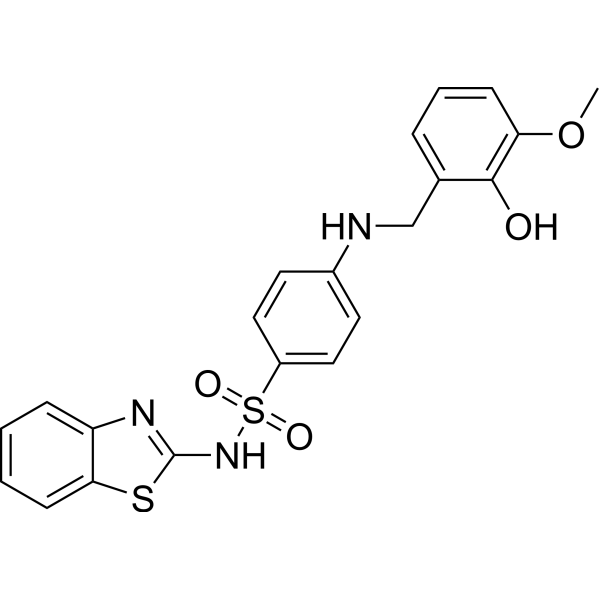
Onvoloxastat (ML355) is an orally active selective 12-LOX inhibitor with an IC50 of 0.34 μM. Onvoloxastat inhibits platelet activation by blocking thrombin- or thromboxane A2-induced phosphorylation of Akt, PI3K and Erk1/2. At low doses (1-20 μM), it mainly acts by reducing ROS levels, while at high doses (50 μM), it functions via activating the cAMP pathway. Onvoloxastat can be used in research related to thrombosis and diabetes.
For research use only. We do not sell to patients.
- Purity : 98.11%
- CAS No.: 1532593-30-8
- Formula: C21H19N3O4S2
- Molecular Weight:441.52
-
Storage:Powder -20°C, 3 years , 4°C, 2 years ; In solvent -80°C, 1 year , -20°C, 6 months

To place orders, for customer services and technical support, please contact: MedChemExpress USA
Tel: 609-228-6898 E-mail: [email protected] [email protected]


-
 Biological Activity
-
Chemical Information
-
Solvent & Solubility
- Protocols
- Purity & Documentation
- References
-
 Help & FAQs


-
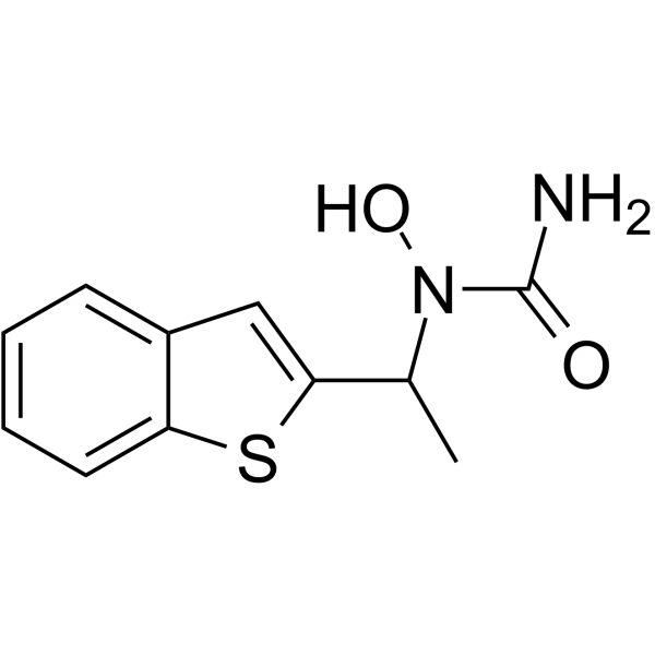
HY-14164
Zileuton5-Lipoxygenase Inhibitor -
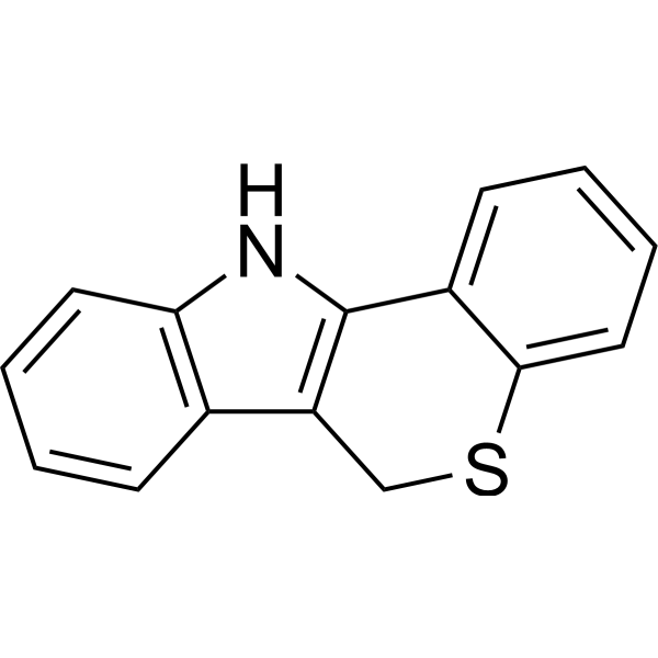
HY-103157
PD14617615-LO Inhibitor -
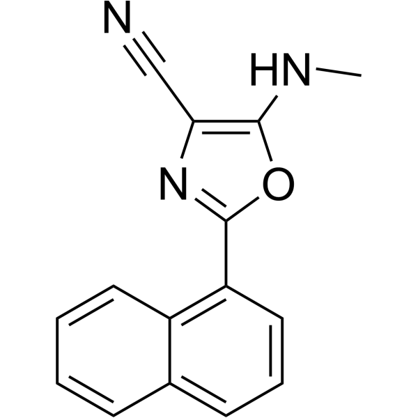
HY-111310
ML35115-LOX-1 Inhibitor -
HY-N0198
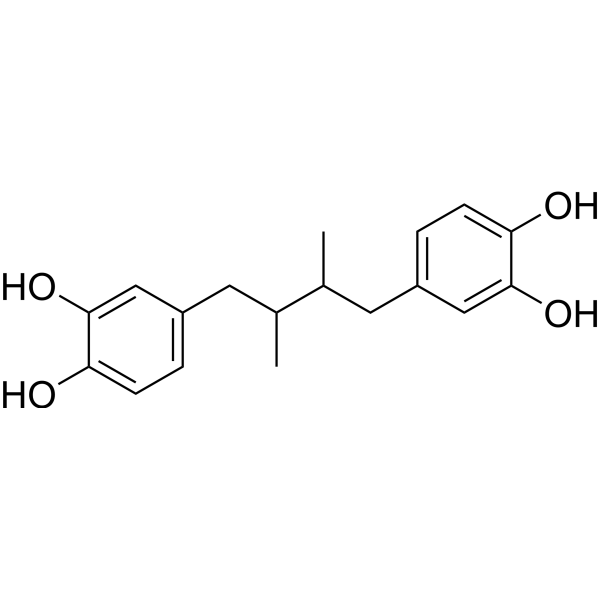
Nordihydroguaiaretic acidAutophagy Inducer -
HY-N0892
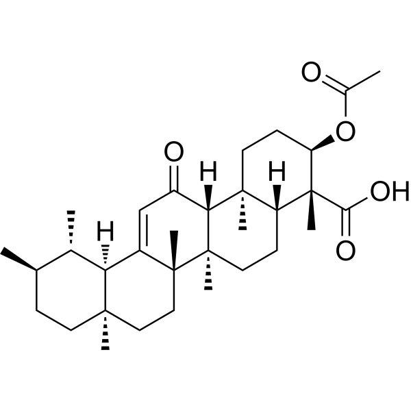
AKBANrf2 Activator -
HY-14166
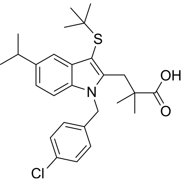
MK-886FLAP Inhibitor -
HY-N0196
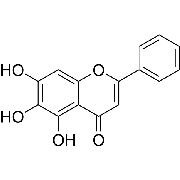
BaicaleinXanthine Oxidase Inhibitor -
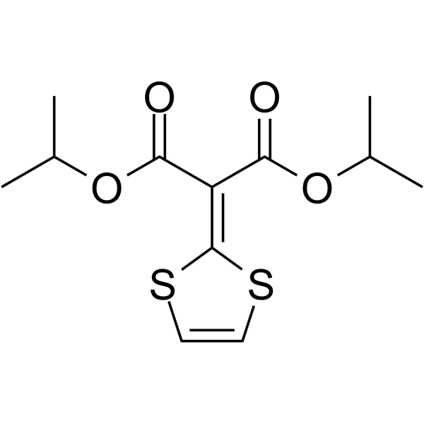
HY-A0060
Malotilate5-LOX Inhibitor -

HY-10071
Y-27632ROCK Inhibitor -
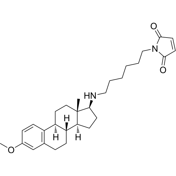
HY-13419
U-73122Phospholipase C Inhibitor -
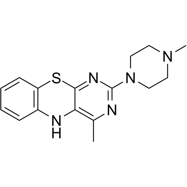
HY-118480
4-MMPB15-lipoxygenase Inhibitor -
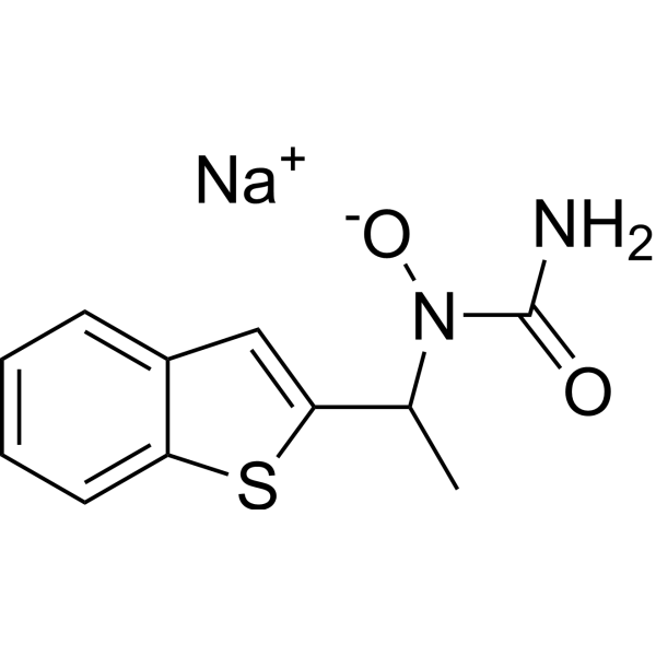
HY-14164A
Zileuton sodium5-lipoxygenase Inhibitor
Publications Citing Use of MedChemExpress (MCE) Onvoloxastat
More- Nat Med. 2018 Jan;24(1):73-83. [Abstract]
- Cell Metab. 2021 Oct 5;33(10):2059-2075.e10. [Abstract]
- Nat Commun. 2024 Sep 10;15(1):7914. [Abstract]
- Cell Commun Signal. 2020 May 4;18(1):70. [Abstract]
- Phytomedicine. 2024 Jul 25:130:155757. [Abstract]
- Free Radic Biol Med. 2024 Oct:223:325-340. [Abstract]
- Int Immunopharmacol. 2024 Oct 7;143(Pt 1):113320. [Abstract]
- Inflammation. 2026 Mar 3;49(1):92. [Abstract]
 Customer Validation & Images

-
Others
-
Flow Cytometry
-
WB


Biological Activity
Description
IC50 & Target
[1]|
12-LOX 0.34 μM (IC50) |
In Vitro
Onvoloxastat (ML355) (1-50 μM; 40 min) dose-dependently inhibits αIIbβ3 integrin activation in washed human platelets induced by Thrombin (HY-114164) and U46619 (HY-108566)[1].
Onvoloxastat (50 μM; 1-60 min) inhibits fibrinogen binding in a time-dependent manner in washed human platelets[1].
Onvoloxastat (1-50 μM; 30 min) induces cAMP-dependent phosphorylation of VASP at Ser157 and Ser239 in washed human platelets without affecting cell viability[1].
Onvoloxastat (0.1-100 μM; 10 min) does not directly affect cGMP-producing activity in recombinant human GC-1 enzyme[1].
Onvoloxastat (25-100 μM; 5 min) dose-dependently inhibits platelet aggregation induced by Thrombin, PAR1-AP, PAR4-AP (HY-P1309) and Collagen (HY-NP003), and reduces 12-HETE production in washed human platelets[2].
Onvoloxastat (25-100 μM; pre-incubated for 5 min) attenuates platelet adhesion, aggregation and thrombosis in human whole blood[2].
Onvoloxastat (1 pM-100 μM; 4 h) inhibits AA/IONO-stimulated 12-HETE production in mouse pancreatic islet β cells (BTC3)[3].
Onvoloxastat (10 μM; 30 min) inhibits AA/IONO-induced 12-HETE expression in human primary islet cells[3].
Onvoloxastat (1-50 μM; 30 min) dose-dependently blocks the phosphorylation of Akt, PI3K and Erk1/2 and reduces reactive oxygen species (ROS) levels in thrombin-activated washed human platelets, but does not affect p38 phosphorylation. It does not inhibit platelet activation in Crp-XL-activated washed human platelets[1].
Onvoloxastat (1-50 μM; 60 min) dose-dependently inhibits ABT-737 (HY-50907)-induced phosphatidylserine (PS) externalization and Caspase-3 cleavage in washed human platelets[1].
MedChemExpress (MCE) has not independently confirmed the accuracy of these methods. They are for reference only. Further protocols information, click here.
-
Cell Line:Washed human platelets
-
Concentration:1, 10, 20, 50 μM
-
Incubation Time:30 min
-
Result:Blocked thrombin-induced PI3K, Akt, and Erk1/2 phosphorylation, but did not affect p38 kinase phosphorylation.
Did not inhibit Crp-XL-induced Syk and PLCγ2 phosphorylation, and enhanced the phosphorylation signals at the 50 μM dose.
Induced cAMP-dependent VASP phosphorylation at Ser157 and Ser239.
-
Cell Line:Washed human platelets
-
Concentration:1, 10, 20, 50 μM
-
Incubation Time:30 min
-
Result:Did not affect the viability signal of the platelets.
-
Cell Line:Washed human platelets
-
Concentration:1, 10, 20, 50 μM
-
Incubation Time:60 min
-
Result:Dose-dependently blocked ABT-737-induced phosphatidylserine (PS) exposure on the platelet surface.
-
Cell Line:Washed human platelets
-
Concentration:1, 10, 20, 50 μM
-
Incubation Time:60 min
-
Result:Prevented ABT-737-induced Caspase-3 cleavage.
-
Cell Line:Mouse beta cells (BTC3)
-
Concentration:1 pM, 0.01 μM, 0.1 μM, 1 μM, 10 μM, 100 μM
-
Incubation Time:4 h
-
Result:Dose-dependently inhibited AA/IONO-stimulated 12-HETE production.
-
Cell Line:Human primary donor islets
-
Concentration:10 μM
-
Incubation Time:30 min
-
Result:Inhibited AA/IONO-induced 12-HETE levels.
Parmacokinetics
In Vivo
Onvoloxastat (1.5 mg/kg; i.v.; single dose; 24 h pretreatment) inhibits platelet recruitment and thrombus growth in a laser-induced cremaster muscle microartery thrombosis model[1].
Onvoloxastat (1.5 mg/kg; i.v.; single administration; 24 h pre-treatment) does not impair normal hemostatic function, prolong bleeding time, or increase blood loss in the tail-clip bleeding model and thromboelastography assay[1].
Onvoloxastat (15-30 mg/kg; i.g.; twice daily; for 2 consecutive days) inhibits thrombus growth and significantly delays the time to complete vascular occlusion in a ferric chloride FeCl3-induced mesenteric arteriolar thrombosis model in wild-type mice[2].
Onvoloxastat (1.88-30 mg/kg; i.g.; twice daily; for 2 consecutive days) potently inhibits thrombosis in a laser-induced cremasteric arteriole thrombosis model in WT mice[2].
Onvoloxastat (15 mg/kg; i.g.; twice daily; for 2 consecutive days; recorded for 5 minutes after laser injury, with repeated injuries at 5- and 10-minute intervals) exerts no significant effect on hemostatic plug formation and platelet plug dynamics in the laser-ablated saphenous vein hemostasis model of WT mice[2].
Onvoloxastat (15 mg/kg; i.g.; twice daily; for 2 consecutive days) does not alter microvascular hemostasis time in laser-induced cremaster muscle microvascular rupture and plasma extravasation models of WT mice[2].
Onvoloxastat (3.5-30 mg/kg; i.g.; twice daily; for 2 consecutive days) does not significantly prolong tail bleeding time or increase total red blood cell loss in the tail amputation bleeding model of WT mice[2].
MedChemExpress (MCE) has not independently confirmed the accuracy of these methods. They are for reference only.
-
Animal Model:C57BL/6 wild-type mice (male,10-12 weeks old) laser-induced cremaster arteriole thrombosis model[1]
-
Dosage:1.5 mg/kg
-
Administration:i.v.; single dose; 24 h pretreatment
-
Result:Reduced dynamic platelet accumulation and recruitment within the thrombus at the injury site.
-
Animal Model:C57BL/6 wild-type mice (male,10-12 weeks old) FeCl3-induced carotid artery thrombosis model[1]
-
Dosage:1.5 mg/kg
-
Administration:i.v.; single dose; 24 h pretreatment
-
Result:Delayed the growth rate of thrombus in the carotid artery.
Significantly prolonged the time to complete vessel occlusion.
-
Animal Model:C57BL/6 wild-type mice (male,10-12 weeks old) tail bleeding model and thromboelastography (TEG) coagulation test[1]
-
Dosage:1.5 mg/kg
-
Administration:i.v.; single dose; 24 h pretreatment
-
Result:Did not cause significant alterations in coagulation parameters.
Did not significantly prolong tail bleeding time or increase total hemoglobin loss.
-
Animal Model:C57BL/6 wild-type mice (male,10-12 weeks old) FeCl3-induced mesenteric arteriole thrombosis (topical application of 30 μL of 250 mM FeCl3 solution)[2]
-
Dosage:15, 30 mg/kg
-
Administration:i.g.; twice daily; for 2 consecutive days
-
Result:Severely impaired platelet adhesion, aggregation, and thrombus formation; the formed thrombi were unstable and frequently embolized
Significantly delayed the time to complete vessel occlusion in mesenteric arterioles, with some mice failing to occlude within 40 minutes.
-
Animal Model:C57BL/6 wild-type mice (male,10-12 weeks old) laser-induced cremaster arteriole thrombosis model[2]
-
Dosage:1.88, 3.75, 7.5, 15, 30 mg/kg
-
Administration:i.g.; twice daily; for 2 consecutive days
-
Result:Strongly inhibited platelet recruitment and fibrin formation at the injury site; thrombi were smaller, unstable, and detached more readily.
At moderate doses (3.75, 7.5 mg/kg), significantly inhibited platelet recruitment, but fibrin content was not significantly.
At the lowest dose (1.88 mg/kg), platelet recruitment and fibrin content were comparable to the control group.
-
Animal Model:C57BL/6 wild-type mice (male,10-12 weeks old) laser ablation saphenous vein hemostasis model (puncturing the vessel wall)[2]
-
Dosage:15 mg/kg
-
Administration:i.g.; twice daily; for 2 consecutive days
-
Result:Did not significantly alter hemostatic plug formation at the site of vascular injury.
Under repeated injury stimulation, the dynamics of the platelet plug and fibrin formation within the hemostatic clot did not significantly differ compared to the control group.
-
Animal Model:C57BL/6 wild-type mice (male,10-12 weeks old) laser-induced cremaster microvasculature (arteriole/venule) rupture plasma extravasation model[2]
-
Dosage:15 mg/kg
-
Administration:i.g.; twice daily; for 2 consecutive days
-
Result:Showed no notable difference in plasma (fluorescent dextran) extravasation or the time required for cessation of extravasation after platelet plug formation compared to controls.
-
Animal Model:C57BL/6 wild-type mice (male,10-12 weeks old) tail-bleeding model (transection of 5mm of the distal tail, submerged in warm saline)[2]
-
Dosage:3.5, 15, 30 mg/kg
-
Administration:i.g.; twice daily; for 2 consecutive days
-
Result:Did not significantly prolong the tail bleeding time of the mice.
Did not increase the total amount of red blood cell loss.


Chemical Information
-
CAS No. 1532593-30-8
-
Appearance Solid
-
Molecular Weight 441.52
-
Formula C21H19N3O4S2
-
Color White to gray
-
SMILES
O=S(NC1=NC2=CC=CC=C2S1)(C3=CC=C(NCC4=CC=CC(OC)=C4O)C=C3)=O
-
Synonyms
ML355
-
Shipping
Room temperature in continental US; may vary elsewhere.
-
Storage
Powder -20°C 3 years 4°C 2 years In solvent -80°C 1 year -20°C 6 months


Publications (8)
-
Journal Impact Factor
-
Most Recent
-
Nat Med
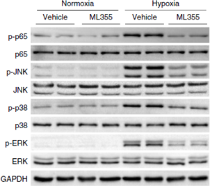
An ALOX12-12-HETE-GPR31 signaling axis is a key mediator of hepatic ischemia-reperfusion injury. [Abstract]2018 Jan;24(1):73-83. PMID: 29227475
Onvoloxastat purchased from MedChemExpress. Usage Cited in: Nat Med. 2018 Jan;24(1):73-83. [Abstract]
The phosphorylated and total levels of p65, JNK, p38 and ERK in primary hepatocytes treated with ML355 (10 μM) under normoxia (Nor) or hypoxia (Hyp) conditions. For western blot analysis, GAPDH served as a loading control.
-
Cell Metab
Pharmacological inhibition of arachidonate 12-lipoxygenase ameliorates myocardial ischemia-reperfusion injury in multiple species. [Abstract]2021 Oct 5;33(10):2059-2075.e10. PMID: 34536344 -
Nat Commun
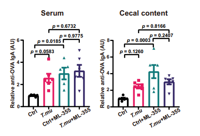
A mouse protozoan boosts antigen-specific mucosal IgA responses in a specific lipid metabolism- and signaling-dependent manner. [Abstract]2024 Sep 10;15(1):7914. PMID: 39256385
Onvoloxastat purchased from MedChemExpress. Usage Cited in: Nat Commun. 2024 Sep 10;15(1):7914. [Abstract]
Starting from day 6, mice were administered the carrier and ML-355 (15 mg/kg) daily via oral gavage. On day 7, mice were challenged by intraperitoneal injection of OVA+CFA. On day 21, the relative levels of anti-OVA IgA in serum and cecal contents were measured.
-
Cell Commun Signal
Cytochrome P450 1A1 enhances inflammatory responses and impedes phagocytosis of bacteria in macrophages during sepsis. [Abstract]2020 May 4;18(1):70. PMID: 32366266 -
Phytomedicine
Baicalein alleviates cisplatin-induced acute kidney injury by inhibiting ALOX12-dependent ferroptosis. [Abstract]2024 Jul 25:130:155757. PMID: 38805781 -
Free Radic Biol Med
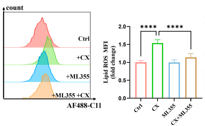
CX-5461 ameliorates disease in lupus-prone mice by triggering B-cell ferroptosis via p53-SLC7A11-ALOX12 pathway. [Abstract]2024 Oct:223:325-340. PMID: 39111584
Onvoloxastat purchased from MedChemExpress. Usage Cited in: Free Radic Biol Med. 2024 Oct:223:325-340. [Abstract]
Selected Raji cells were treated with 1 μM CX-5461, with or without 10 μM ML355, and lipid ROS levels were analyzed by flow cytometry. Representative results and quantitative analyses are presented.
-
Int Immunopharmacol
Palmitic acid inhibits macrophage-mediated chemotherapy resistance in multiple myeloma via ALOX12 signaling. [Abstract]2024 Oct 7;143(Pt 1):113320. PMID: 39378653 -
Inflammation
MAGL Inhibition Relieves Psoriasiform Inflammation and Pruritus Via Modulation of ALOX12-12-HETE Axis in Mice. [Abstract]2026 Mar 3;49(1):92. PMID: 41774275


Solvent & Solubility
In Vitro:
DMSO : ≥ 42 mg/mL (95.13 mM; Hygroscopic DMSO has a significant impact on the solubility of product, please use newly opened DMSO)
* "≥" means soluble, but saturation unknown.
Please refer to the solubility information to select the appropriate solvent. Once prepared, please aliquot and store the solution to prevent product inactivation from repeated freeze-thaw cycles.
Storage method and period of stock solution: -80°C, 1 year; -20°C, 6 months. When stored at -80°C, please use it within 1 year. When stored at -20°C, please use it within 6 months.
Please refer to the solubility information to select the appropriate solvent. Once prepared, please aliquot and store the solution to prevent product inactivation from repeated freeze-thaw cycles.
Storage method and period of stock solution: -80°C, 1 year; -20°C, 6 months. When stored at -80°C, please use it within 1 year. When stored at -20°C, please use it within 6 months.
Concentration (start) × Volume (start) = Concentration (final) × Volume (final)
In Vivo:
Select the appropriate dissolution method based on your experimental animal and administration route.
- For the following dissolution methods, please ensure to first prepare a clear stock solution using an In Vitro approach and then sequentially add co-solvents:
- To ensure reliable experimental results, the clarified stock solution can be appropriately stored based on storage conditions. As for the working solution for In Vivo experiments, it is recommended to prepare freshly and use it on the same day.
- The percentages shown for the solvents indicate their volumetric ratio in the final prepared solution. If precipitation or phase separation occurs during preparation, heat and/or sonication can be used to aid dissolution.
Add each solvent one by one: 10% DMSO 40% PEG300 5% Tween-80 45% Saline
Solubility: ≥ 2.5 mg/mL (5.66 mM); Clear solution
This protocol yields a clear solution of ≥ 2.5 mg/mL (saturation unknown).
Taking 1 mL working solution as an example, add 100 μL DMSO stock solution (25.0 mg/mL) to 400 μL PEG300, and mix evenly; then add 50 μL Tween-80 and mix evenly; then add 450 μL Saline to adjust the volume to 1 mL.
Preparation of Saline: Dissolve 0.9 g sodium chloride in ddH₂O and dilute to 100 mL to obtain a clear Saline solution.
Add each solvent one by one: 10% DMSO 90% (20% SBE-β-CD in Saline)
Solubility: ≥ 2.5 mg/mL (5.66 mM); Clear solution
This protocol yields a clear solution of ≥ 2.5 mg/mL (saturation unknown).
Taking 1 mL working solution as an example, add 100 μL DMSO stock solution (25.0 mg/mL) to 900 μL 20% SBE-β-CD in Saline, and mix evenly.
Preparation of 20% SBE-β-CD in Saline (4°C, storage for one week): 2 g SBE-β-CD powder is dissolved in 10 mL Saline, completely dissolve until clear.
In Vivo Dissolution Calculator
Please enter the basic information of animal experiments:
-
-
-
-
Recommended: Prepare an additional quantity of animals to account for potential losses during experiments.
Please enter your animal formula composition:
-
%DMSO +
Recommended: Keep the proportion of DMSO in working solution below 2% if your animal is weak.
-
%+
-
+%Tween-80 + +
-
%Saline +
The co-solvents required include: DMSO, . All of co-solvents are available by MedChemExpress (MCE). , Tween 80. All of co-solvents are available by MedChemExpress (MCE).
Working solution concentration: 0.22 mg/mL
Method for preparing stock solution: mg drug dissolved in μL DMSO. Stock solution concentration: mg/mL.
1. Take μL DMSO stock solution;
2. Add μL .
μL , mix evenly;
3. Then add μL Tween 80, mix evenly;
4. Then add μL
Please ensure that the stock solution in the first step is dissolved to a clear state, and add co-solvents in sequence. You can use ultrasonic heating (ultrasonic cleaner, recommended frequency 20-40 kHz), vortexing, etc. to assist dissolution.

Protocols
-
Kinase activity and phosphorylation assays
Kinase activity assays measure the ability of kinases to transfer phosphate groups from ATP to specific substrates, while phosphorylation assays detect the presence and levels of phosphorylated proteins. Common methods include radiolabeled ATP incorporation (e. g. ,), ADP release detection via bioluminescence (e. g. ,[3]), enzyme-linked immunosorbent assays (ELISA) for phospho-specific epitopes (e. g. ,[6]), and microtiter-based formats for high-throughput screening (e. g. ,[8]). The ADP-Glo assay quantifies kinase activity by measuring ADP produced during phosphorylation using a luciferase-based system. Radiometric assays involve autoradiography or scintillation counting after incorporation of 32P-labeled ATP into substrate proteins. ELISA-based approaches rely on phospho-specific antibodies to detect activated kinases in cell lysates or purified samples.
-
Western Blot
Western blotting (WB) is a commonly used experimental method in molecular biology, biochemistry, and immunogenetics for identifying and quantifying target proteins. It combines gel electrophoresis with immunoassay, enabling researchers to analyze protein expression, post-translational modifications, and molecular weight.
-
Cell Cytotoxicity Assay
Cytotoxicity assays are usually based on the assessment of cell membrane damage, which can also be indirectly detected by measuring cell viability. Detection methods include MTT assay, CKK-8 assay, LDH assay and ATP assay, etc.
-
ROS/oxidative-stress fluorescent staining
ROS/oxidative-stress fluorescent staining uses cell-permeant fluorogenic probes that become fluorescent after oxidation inside cells or tissues; commonly used examples include DCFH-DA/DCFDA for broad cellular oxidant detection, DHE for superoxide-related signal detection, MitoSOX for mitochondrial superoxide-related signal detection, and CellROX probes for oxidative-stress-associated fluorescence readouts. The assay detects probe oxidation rather than a single ROS species unless the probe and analysis method have been chemically validated for that species. DCFH-DA enters cells, is deacetylated by intracellular esterases to DCFH, and produces fluorescent DCF after oxidation, so the readout is used as an operational measure of total cellular oxidative stress rather than a species-specific ROS measurement. DHE and MitoSOX can report superoxide-related oxidation, but red fluorescence alone can include non-specific ethidium-like oxidation products; HPLC or optimized spectral approaches are
-
Protocol for Kinase activity and phosphorylation assays
Kinase activity assays measure transfer of phosphate from ATP to a protein or peptide substrate, generating phosphorylated substrate, ADP, or incorporated radiolabeled phosphate as the readout; phosphorylation assays measure site-specific phosphorylation in cells or tissues as a proxy for kinase-pathway activation, inhibition, or substrate regulation. Phosphorylation can be detected by phospho-specific Western blot, immunoprecipitation kinase assay, phospho-immunofluorescence, phospho-flow cytometry, luminescent ADP detection, radiolabeled ATP incorporation, or reporter-based pathway assays, and these readouts can be applied to cancer cells, primary neurons, mouse tumors, organoids, inflammatory macrophages, ferroptosis studies, and mitophagy studies when the kinase target is biologically relevant.
-
Research Protocol for Metabolic Diseases
AMP-activated protein kinase, AMPK, is a conserved cellular energy sensor that responds to reduced cellular energy status and coordinates metabolism by increasing ATP-generating catabolic pathways while suppressing ATP-consuming anabolic processes. In metabolic disease research, the AMPK pathway is experimentally relevant because it regulates hepatic lipid synthesis, fatty acid oxidation, glucose production, skeletal-muscle glucose disposal, mTORC1-linked biosynthesis, autophagy, mitochondrial homeostasis, and whole-body energy balance. The central pathway logic is that energy stress, metformin, exercise-like stimulation, or direct AMPK activators increase AMPKα Thr172 phosphorylation and downstream substrate phosphorylation, including ACC and RAPTOR. Phosphorylation of ACC suppresses lipogenesis and supports fatty acid oxidation, whereas phosphorylation of RAPTOR suppresses mTORC1 signaling and links cellular energy status to growth and protein synthesis control. The pathway is linked


Purity & Documentation
-
Data Sheet (306 KB)
-
SDS (393 KB)
- English - EN (393 KB)
- Français - FR (393 KB)
- Deutsch - DE (393 KB)
- Norwegian - NO (393 KB)
- Español - ES (393 KB)
- Swedish - SV (393 KB)
- Italian - IT (393 KB)
- Korean - KR (393 KB)
- Portuguese - PT (393 KB)
-
Handling Instructions (2659 KB)
References
[2]. Adili R, et al. First Selective 12-LOX Inhibitor, ML355, Impairs Thrombus Formation and Vessel Occlusion In Vivo With Minimal Effects on Hemostasis. Arteriosclerosis, thrombosis, and vascular biology. 2017 Oct;37(10):1828-1839. [Content Brief]
[4]. Pizzorno J, et al. Health Medicine. Integrative medicine (Encinitas, Calif.). 2022 Dec;21(6):8-14. [Content Brief]


Complete Stock Solution Preparation Table
Please refer to the solubility information to select the appropriate solvent. Once prepared, please aliquot and store the solution to prevent product inactivation from repeated freeze-thaw cycles.
Storage method and period of stock solution: -80°C, 1 year; -20°C, 6 months. When stored at -80°C, please use it within 1 year. When stored at -20°C, please use it within 6 months.
| Optional Solvent | Concentration Solvent Mass | 1 mg | 5 mg | 10 mg | 25 mg |
|---|---|---|---|---|---|
| DMSO | 1 mM | 2.2649 mL | 11.3245 mL | 22.6490 mL | 56.6226 mL |
| 5 mM | 0.4530 mL | 2.2649 mL | 4.5298 mL | 11.3245 mL | |
| 10 mM | 0.2265 mL | 1.1325 mL | 2.2649 mL | 5.6623 mL | |
| 15 mM | 0.1510 mL | 0.7550 mL | 1.5099 mL | 3.7748 mL | |
| 20 mM | 0.1132 mL | 0.5662 mL | 1.1325 mL | 2.8311 mL | |
| 25 mM | 0.0906 mL | 0.4530 mL | 0.9060 mL | 2.2649 mL | |
| 30 mM | 0.0755 mL | 0.3775 mL | 0.7550 mL | 1.8874 mL | |
| 40 mM | 0.0566 mL | 0.2831 mL | 0.5662 mL | 1.4156 mL | |
| 50 mM | 0.0453 mL | 0.2265 mL | 0.4530 mL | 1.1325 mL | |
| 60 mM | 0.0377 mL | 0.1887 mL | 0.3775 mL | 0.9437 mL | |
| 80 mM | 0.0283 mL | 0.1416 mL | 0.2831 mL | 0.7078 mL |


Onvoloxastat Related Classifications
HY-12341 Related Classifications
 Powered by Bioz