



FSL-1 TFA
Based on 5 publication(s) in Google Scholar
FSL-1 TFA, a bacterial-derived toll-like receptor 2/6 (TLR2/6) agonist, enhances resistance to experimental HSV-2 infection. FSL-1 TFA induces MMP-9 production through TLR2 and NF-κB/AP-1 signaling pathways in monocytic THP-1 cells.
For research use only. We do not sell to patients.
- Purity : 99.81%
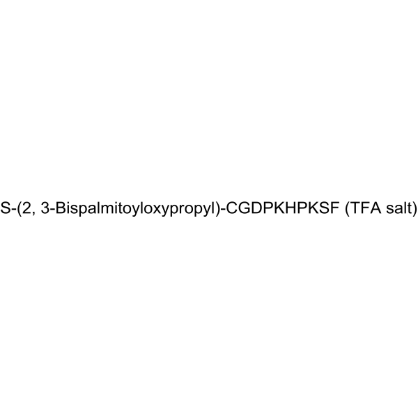
- Formula: C84H140N14O18S.xC2HF3O2
- Molecular Weight:1666.16 (free acid)
-
Storage:
-80°C

To place orders, for customer services and technical support, please contact: MedChemExpress USA
Tel: 609-228-6898 E-mail: [email protected] [email protected]


-
 Biological Activity
-
Chemical Information
-
Solvent & Solubility
- Protocols
- Purity & Documentation
- References
-
 Help & FAQs


-
HY-P1180A
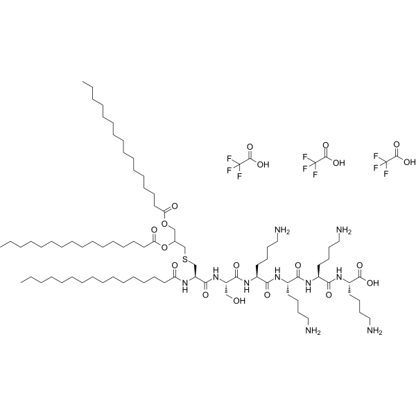
Pam3CSK4 TFATLR1/2 Agonist -
HY-13740
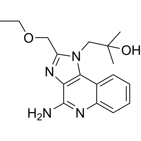
ResiquimodTLR7/8 Agonist -
HY-107202
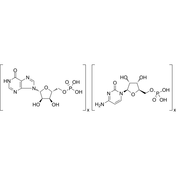
Polyinosinic-polycytidylic acidTLR-3 Agonist -

HY-10071
Y-27632ROCK Inhibitor -

HY-D1056
Lipopolysaccharides, from E. coli O55:B5Endotoxin -
HY-150218
Agatolimod sodiumTLR9 Agonist -
HY-110353
CU-T12-9TLR1/2 Agonist -

HY-107202A
Poly (I:C):Kanamycin (1:1)TLR3 Agonist
Publications Citing Use of MedChemExpress (MCE) FSL-1 TFA
MoreAll Antibiotic Isoforms
More

Biological Activity
Description
|
TLR2 |
HSV-2 |
TLR6 |
MMP-9 |
In Vitro
FSL-1 TFA significantly reduces HSV-2 replication in human vaginal epithelial cells (EC)[1].
FSL-1 TFA induces significant resistance to experimental genital HSV-2 infection through elaboration of a specific cytokine response profile[1].
FSL-1 TFA (50 ng/mL, 24 hours) induces MMP-9 expression at both mRNA and protein levels in human monocytic THP-1 cells[2].
FSL-1 TFA activates the MAP kinase/NF-κB signaling pathway[2].
MedChemExpress (MCE) has not independently confirmed the accuracy of these methods. They are for reference only. Further protocols information, click here.
-
Cell Line:V11I, V12I or V19I immortalized human vaginal EC
-
Concentration:6 μg or 0.1 μg
-
Incubation Time:Added at 24, 6 or just prior to HSV-2 inoculation (104pfu/well)
-
Result:The 6 μg does produced significant reductions when delivered at 24 or 6 h prior to HSV-2 inoculation. The 0.1 μg dose produced reduced HSV-2 replication at 24 or 6 h prior to viral challenge.
In Vivo
MedChemExpress (MCE) has not independently confirmed the accuracy of these methods. They are for reference only.
-
Animal Model:Female Swiss-Webster mice (weighing 20-25 g)[1]
-
Dosage:2 or 6 μg
-
Administration:Delivered vaginally using a positive displacement pipet, prior to or following viral challenge as specified for each experiment.
-
Result:The 2 μg does delivered 6 h prior to HSV-2 challenge increased the ID50 (260 pfu) and LD50 (660 pfu) by 10-fold compared to DPBS vehicle control.
The single 6 μg dose produced significantly improved outcomes compared to DPBS vehicle application.


Chemical Information
-
Appearance Solid
-
Molecular Weight 1666.16 (free acid)
-
Formula C84H140N14O18S.xC2HF3O2
-
Color White to off-white
-
Sequence Shortening
S-(2, 3-Bispalmitoyloxypropyl)-CGDPKHPKSF
-
Shipping
Shipping with dry ice.
-
Storage
-80°C


Publications (5)
-
Journal Impact Factor
-
Most Recent
-
Adv Sci (Weinh)
Calhm6 Governs Macrophage Polarization Through Chp1-Camk4-Creb1 Axis and Ectosomal Delivery in Inflammatory Responses. [Abstract]2025 Sep 26:e02395. PMID: 40999918 -
Food Res Int
Walnut-derived peptides ameliorate d-galactose-induced memory impairments in a mouse model via inhibition of MMP-9-mediated blood-brain barrier disruption. [Abstract]2022 Dec;162(Pt A):112029. PMID: 36461249 -
J Ethnopharmacol
Cornus officinalis var. koreana Kitam extracts alleviate cadmium-induced renal fibrosis by targeting matrix metallopeptidase 9. [Abstract]2024 May 10:325:117824. PMID: 38278375 -
Biomedicines
Integrated Network Pharmacology, Molecular Docking and Experimental Validation Reveal That Quercetin Suppresses Clear Cell Renal Cell Carcinoma via MMP9-Associated Macrophage Polarization. [Abstract]2026 Apr 16;14(4):904. PMID: 42072446 -
Int J Med Sci
SJMHE1 protects against excessive iodine-induced pyroptosis in human thyroid follicular epithelial cells through a toll-like receptor 2-dependent pathway. [Abstract]2022 Mar 21;19(4):631-639. PMID: 35582426


Solvent & Solubility
In Vitro:
H2O : 50 mg/mL (Need ultrasonic)
DMSO : 25 mg/mL (Need ultrasonic; Hygroscopic DMSO has a significant impact on the solubility of product, please use newly opened DMSO)

Protocols
-
Research Protocol for Infectious Diseases
Infectious-disease experiments test how pathogens interact with host barriers, innate immune receptors, inflammatory signaling, pathogen replication, and tissue injury; pattern-recognition receptors such as TLRs, RIG-I-like receptors, NOD-like receptors, and inflammasomes detect microbial molecules and activate NF-κB, interferon, and cytokine responses. The central hypothesis is that infection severity reflects the balance between pathogen burden and host response: protective inflammation restricts pathogen growth, whereas excessive or mislocalized inflammation contributes to tissue damage and disease phenotype. Unresolved questions include which host pathways are protective versus pathogenic, why some infection models fail to translate to human disease, and which combined readouts best predict clinically relevant infection outcomes.
-
Mitochondrial membrane-potential fluorescent assay
Mitochondrial membrane potential fluorescent assays estimate ΔΨm in living cells using lipophilic cationic dyes such as TMRM, TMRE, rhodamine 123, and JC-1, which accumulate in mitochondria according to membrane polarization; loss of signal after FCCP or CCCP treatment is interpreted as mitochondrial depolarization. TMRM/TMRE and rhodamine 123 are commonly used for semi-quantitative live-cell microscopy or flow cytometry, while JC-1 can report a shift from red aggregate fluorescence to green monomer fluorescence during depolarization; interpretation requires controls because dye concentration, quenching mode, cell type, dye efflux, and mitochondrial mass can affect fluorescence independently of ΔΨm.
-
Mitochondrial membrane-potential and mitochondrial mass staining
Mitochondrial membrane potential staining measures the electrochemical polarization across the mitochondrial inner membrane in live cells using lipophilic cationic fluorescent probes; early rhodamine-based work showed that selective mitochondrial dye accumulation is lost when the mitochondrial transmembrane potential is dissipated. JC-1 reports mitochondrial polarization by shifting from green monomer fluorescence to red J-aggregate fluorescence as dye concentration increases within energized mitochondria; therefore, the red/green fluorescence ratio is used as a relative readout of mitochondrial membrane potential. TMRE or TMRM staining provides a single-channel relative readout because these cationic rhodamine esters accumulate in polarized mitochondria, and lower fluorescence indicates reduced mitochondrial polarization when acquisition and dye-loading conditions are controlled. Mitochondrial mass staining is commonly performed with MitoTracker Green FM or related MitoTracker dyes as
-
Research Protocol for Inflammation-related Diseases
The NLRP3 inflammasome is a cytosolic innate immune signaling platform that integrates priming signals and danger-signal activation to promote caspase-1 activation, maturation of IL-1β and IL-18, and gasdermin D-mediated pyroptotic cell death. The core experimental logic is to determine whether inflammatory disease phenotypes are driven by increased NLRP3 expression, ASC-containing inflammasome assembly, caspase-1 cleavage, GSDMD cleavage, and extracellular release of IL-1β/IL-18 rather than by nonspecific cell injury alone. The pathway is strongly linked to inflammation-related disease phenotypes because monosodium urate crystals activate NALP3/NLRP3 inflammasome signaling in gout-like crystal inflammation, cholesterol crystals activate NLRP3 inflammasomes in atherogenesis models, and DSS-induced intestinal inflammation has been reported to involve NLRP3 inflammasome activity. However, experimental colitis studies also show context-dependent protective effects of NLRP3 inflammasome co


Purity & Documentation
-
Data Sheet (270 KB)
-
SDS (252 KB)
- English - EN (252 KB)
- Français - FR (252 KB)
- Deutsch - DE (252 KB)
- Norwegian - NO (252 KB)
- Español - ES (252 KB)
- Swedish - SV (252 KB)
- Italian - IT (252 KB)
- Korean - KR (252 KB)
- Portuguese - PT (252 KB)
-
Handling Instructions (2659 KB)
References
[1]. William A Rose 2nd, et al. FSL-1, a bacterial-derived toll-like receptor 2/6 agonist, enhances resistance to experimental HSV-2 infection. Virol J. 2009 Nov 10;6:195. [Content Brief]
[2]. Cathryn J Kurkjian,et al. The Toll-Like Receptor 2/6 Agonist, FSL-1 Lipopeptide, Therapeutically Mitigates Acute Radiation Syndrome. Sci Rep. 2017 Dec 11;7(1):17355. [Content Brief]

Calculators
Concentration (start) × Volume (start) = Concentration (final) × Volume (final)


FSL-1 TFA Related Classifications
HY-P2036A Related Classifications
 Powered by Bioz