



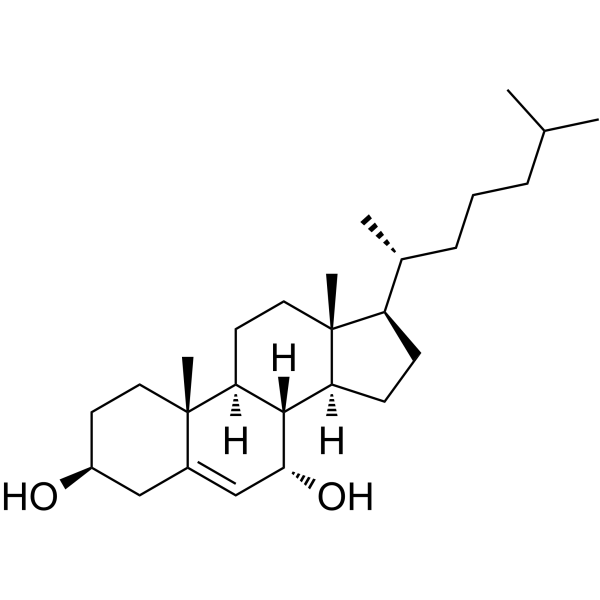
7α-Hydroxycholesterol
Based on 3 publication(s) in Google Scholar
7α-Hydroxycholesterol is a cholesterol oxide and can serve as a biomarker for oxidative stress and lipid peroxidation. 7α-Hydroxycholesterol has cytotoxic and pro-inflammatory activities. 7α-Hydroxycholesterol can also inhibit sterol synthesis and reduce the activity of HMG-CoA reductase. 7α-Hydroxycholesterol can be used in the research of diseases such as diabetes and atherosclerosis.
Nos produits utilisent uniquement pour la recherche. Nous ne vendons pas aux patients.
- Pureté : 99.99%
- CAS No.: 566-26-7
- Formule: C27H46O2
- Masse moléculaire:402.65
-
Stockage:
4°C, protect from light
* In solvent : -80°C, 6 months; -20°C, 1 month (protect from light)

To place orders, for customer services and technical support, please contact: MedChemExpress USA
Tel: 609-228-6898 E-mail: [email protected] [email protected]


-
 Activité biologique
-
Chemical Information
-
Solvant et solubilité
- Protocole
- Pureté et documentation
- Références
-
 Help & FAQs


-
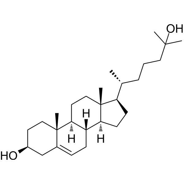
HY-113134
25-HydroxycholesterolCholesterol Metabolite -
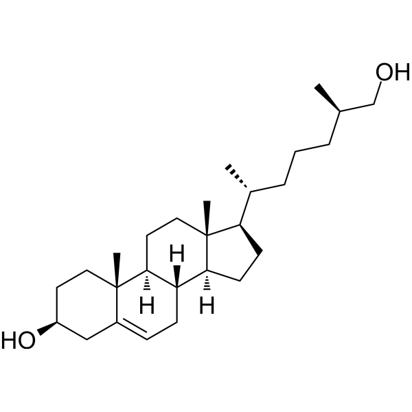
HY-N2371
27-HydroxycholesterolLXR Agonist -
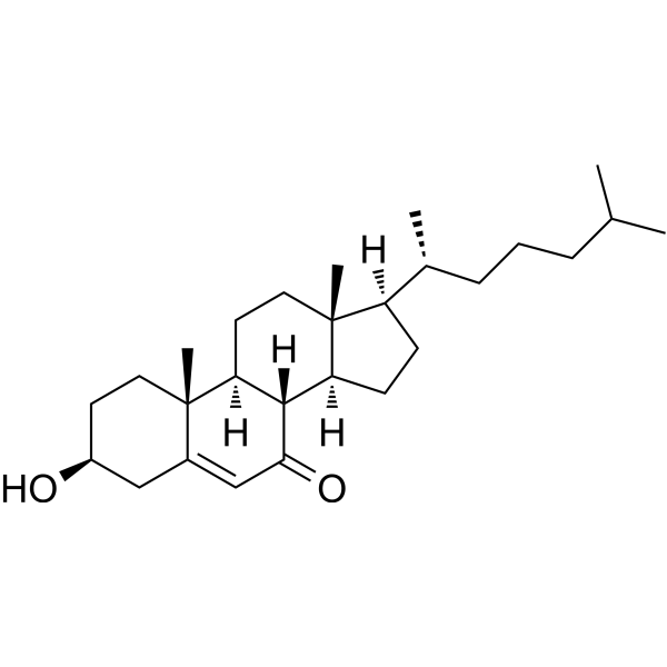
HY-113342
7-KetocholesterolCholesterol 7 Alpha-Hydroxylase Inhibitor -
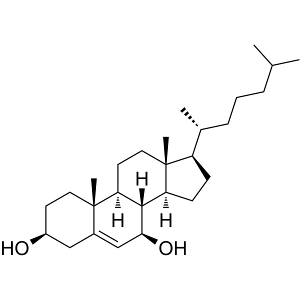
HY-113341
7β-HydroxycholesterolOxidative Stress Inducer -
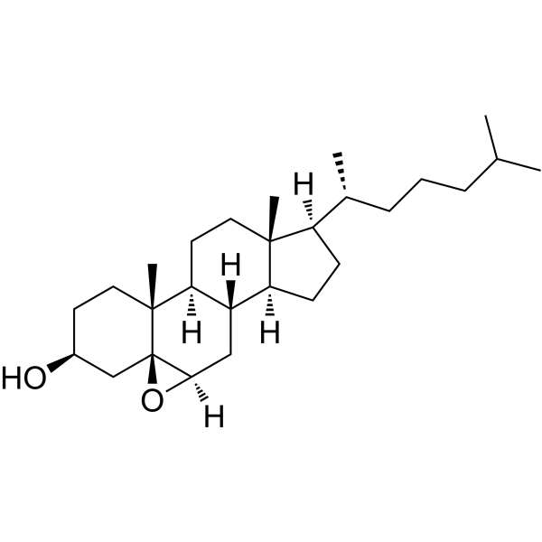
HY-130502
5β,6β-EpoxycholestanolOxysterol -
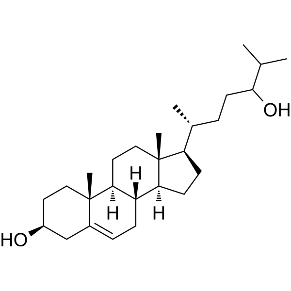
HY-N2370
24-HydroxycholesterolLXR Activator -
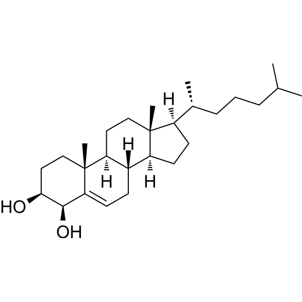
HY-124265
4β-HydroxycholesterolDrug Metabolite -
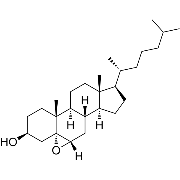
HY-133971
Cholesterol 5α,6α-epoxideBiochemical Assay Reagent -
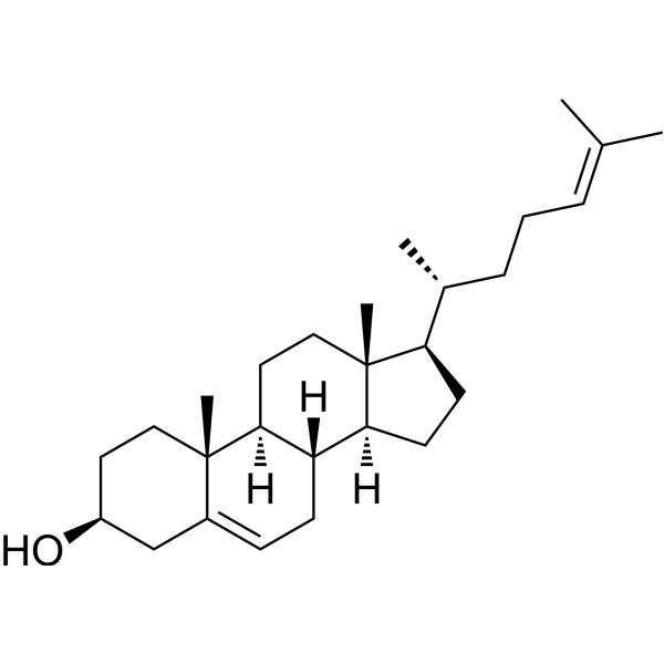
HY-113224
DesmosterolEndogenous Metabolite -
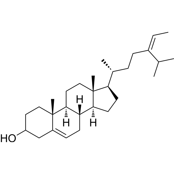
HY-N4103
FucosterolBioactive Sterol -
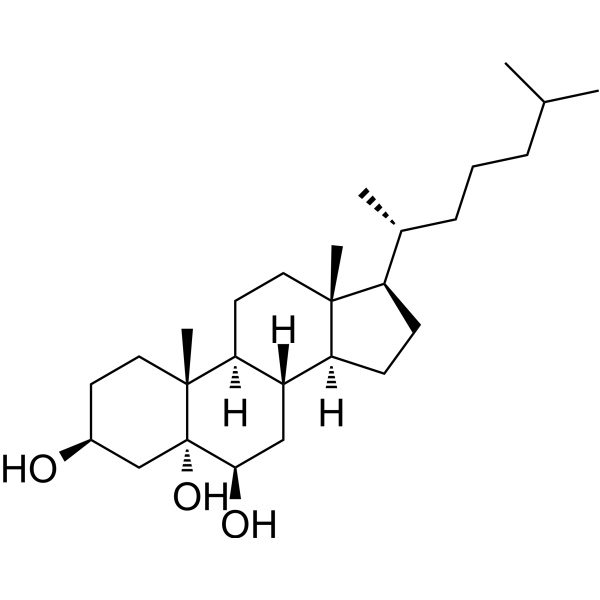
HY-W010934
3β,5α,6β-TrihydroxycholestaneBiochemical Assay Reagent -
HY-N0322
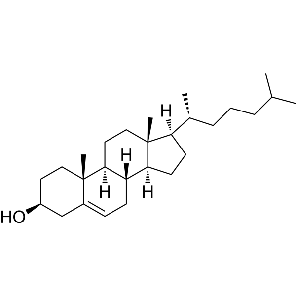
Cholesterol (from animal)ERRα Agonist
Publications Citing Use of MedChemExpress (MCE) 7α-Hydroxycholesterol
More Customer Validation & Images

-
Others

-
WB

-
Cell Imaging/Staining


Activité biologique
Description
Cellular Effect
|
Cell Line

|
Type | Value | Description | References |
|---|---|---|---|---|
| C6 | IC50 |
40 μM
Compound: 4, 7alpha-OHC
|
Growth inhibition of rat C6 cells after 24 hrs by trypan blue-based cell counting method
Growth inhibition of rat C6 cells after 24 hrs by trypan blue-based cell counting method
|
[PMID: 24211631] |
| HT-29 | IC50 |
15.5 μM
Compound: 5
|
Cytotoxicity against human HT-29 cells after 48 hrs by Alamar Blue assay
Cytotoxicity against human HT-29 cells after 48 hrs by Alamar Blue assay
|
[PMID: 19473028] |
| HT-29 | IC50 |
15.5 μM
Compound: 5
|
Cytotoxicity against human HT-29 cells assessed as cell viability after 48 hrs by alamar blue assay
Cytotoxicity against human HT-29 cells assessed as cell viability after 48 hrs by alamar blue assay
|
[PMID: 21797280] |
| HT-29 | IC50 |
15.5 μM
Compound: 7
|
Cytotoxicity against human HT-29 cells after 48 hrs by Alamar blue assay
Cytotoxicity against human HT-29 cells after 48 hrs by Alamar blue assay
|
[PMID: 20931970] |
In Vitro
7α-Hydroxycholesterol (2.5-160 μM; 24 h) has cytotoxicity against 158N and C6 cells, with IC50 values of approximately 15 μM and 40 μM, respectively[1].
7α-Hydroxycholesterol (8 μg/mL; 2-4 h) significantly reduces the sterol synthesis rate and HMG-CoA reductase activity in primary hepatocytes or L-cell cultures[2].
7α-Hydroxycholesterol (5 µg/mL; 48 h) increases the transcriptional level of TLR6, promotes the expression of CD14, and induces monocytes to produce chemokines such as CCL2, CCL3, and CCL4 in monocytes/macrophages. The mechanism involves the phosphorylation of Akt, Src, ERK1/2, and the p65 subunit[3].
MedChemExpress (MCE) has not independently confirmed the accuracy of these methods. They are for reference only.
-
Cell Line:THP-1 cells
-
Concentration:5 µg/mL
-
Incubation Time:48 h
-
Result:Significantly increased the mRNA levels of TLR6 and CD14.


Chemical Information
-
CAS No. 566-26-7
-
Appearance Solid
-
Masse moléculaire 402.65
-
Formule C27H46O2
-
Color White to off-white
-
SMILES
CC(C)CCC[C@@H](C)[C@H]1CC[C@@]2([H])[C@]3([H])[C@H](O)C=C4C[C@@H](O)CC[C@]4(C)[C@@]3([H])CC[C@]12C
-
Structure Classification
-
Initial Source
fish oil, serum
-
Livraison
Room temperature in continental US; may vary elsewhere.
-
Stockage
4°C, protect from light
* In solvent : -80°C, 6 months; -20°C, 1 month (protect from light)


Publications (3)
-
Journal Impact Factor
-
Most Recent
-
ACS Nano
Immune Modulatory Oxysterols Produced from Cholesterol-Containing Lipid Nanoparticles Regulate Tumor Growth. [Abstract]2026 Mar 13. PMID: 41825961 -
Cell Chem Biol
Transient hydroxycholesterol treatment restrains TCR signaling to promote long-term immunity. [Abstract]2024 May 16;31(5):920-931.e6. PMID: 38759618
7α-Hydroxycholesterol purchased from MedChemExpress. Usage Cited in: Cell Chem Biol. 2024 May 16;31(5):920-931.e6. [Abstract]
Both 2 ng/μL of 7α-Hydroxycholesterol (7a-HC) and 20S-HC significantly inhibited CD69 levels, with 7a-HC showing a more significant effect.
7α-Hydroxycholesterol purchased from MedChemExpress. Usage Cited in: Cell Chem Biol. 2024 May 16;31(5):920-931.e6. [Abstract]
Effects of transient 7α-Hydroxycholesterol (7a-HC) treatment on the phosphorylation levels of TCR and downstream molecules in mouse T cells. Mouse naive CD4+ T cells were transiently treated with 2 ng/μL 7a-HC for 20 min and then stimulated with 5 ng/μL α-CD3ε and 5 ng/μL α-CD28.
7α-Hydroxycholesterol purchased from MedChemExpress. Usage Cited in: Cell Chem Biol. 2024 May 16;31(5):920-931.e6. [Abstract]
Naive mouse CD8+ T cells were subjected to transient treatments with 10 ng/μL cholesterol, 2 ng/μL 7α-Hydroxycholesterol (7a-HC) for 20 min, or left untreated (n = 7). Then the plasma membrane was labeled by octadecyl rhodamine B (R18). Data were shown by the representative images during recovery (left panel) and kinetics curves (right panel). Statistical analysis was performed with the 20 s time points of the three conditions.
-
Vet Microbiol
Identification and evaluation of Nordihydroguaiaretic acid (NDGA) as an active traditional Chinese medicine compound inhibiting the 3C-like protease of feline infectious peritonitis virus. [Abstract]2025 Sep 15:310:110730. PMID: 40976146


Solvant et solubilité
In Vitro:
DMSO : 50 mg/mL (124.18 mM; Need ultrasonic; Hygroscopic DMSO has a significant impact on the solubility of product, please use newly opened DMSO)
Please refer to the solubility information to select the appropriate solvent. Once prepared, please aliquot and store the solution to prevent product inactivation from repeated freeze-thaw cycles.
Storage method and period of stock solution: -80°C, 6 months; -20°C, 1 month (protect from light). When stored at -80°C, please use it within 6 months. When stored at -20°C, please use it within 1 month.
Please refer to the solubility information to select the appropriate solvent. Once prepared, please aliquot and store the solution to prevent product inactivation from repeated freeze-thaw cycles.
Storage method and period of stock solution: -80°C, 6 months; -20°C, 1 month (protect from light). When stored at -80°C, please use it within 6 months. When stored at -20°C, please use it within 1 month.
Concentration (start) × Volume (start) = Concentration (final) × Volume (final)
In Vivo:
Select the appropriate dissolution method based on your experimental animal and administration route.
- For the following dissolution methods, please ensure to first prepare a clear stock solution using an In Vitro approach and then sequentially add co-solvents:
- To ensure reliable experimental results, the clarified stock solution can be appropriately stored based on storage conditions. As for the working solution for In Vivo experiments, it is recommended to prepare freshly and use it on the same day.
- The percentages shown for the solvents indicate their volumetric ratio in the final prepared solution. If precipitation or phase separation occurs during preparation, heat and/or sonication can be used to aid dissolution.
Add each solvent one by one: 10% DMSO 40% PEG300 5% Tween-80 45% Saline
Solubility: 10 mg/mL (24.84 mM); Suspended solution; Need ultrasonic
This protocol yields a suspended solution of 10 mg/mL. Suspended solution can be used for oral and intraperitoneal injection.
Taking 1 mL working solution as an example, add 100 μL DMSO stock solution (100.0 mg/mL) to 400 μL PEG300, and mix evenly; then add 50 μL Tween-80 and mix evenly; then add 450 μL Saline to adjust the volume to 1 mL.
Preparation of Saline: Dissolve 0.9 g sodium chloride in ddH₂O and dilute to 100 mL to obtain a clear Saline solution.
Add each solvent one by one: 10% DMSO 90% (20% SBE-β-CD in Saline)
Solubility: 10 mg/mL (24.84 mM); Suspended solution; Need ultrasonic
This protocol yields a suspended solution of 10 mg/mL. Suspended solution can be used for oral and intraperitoneal injection.
Taking 1 mL working solution as an example, add 100 μL DMSO stock solution (100.0 mg/mL) to 900 μL 20% SBE-β-CD in Saline, and mix evenly.
Preparation of 20% SBE-β-CD in Saline (4°C, storage for one week): 2 g SBE-β-CD powder is dissolved in 10 mL Saline, completely dissolve until clear.
In Vivo Dissolution Calculator
Please enter the basic information of animal experiments:
-
-
-
-
Recommended: Prepare an additional quantity of animals to account for potential losses during experiments.
Please enter your animal formula composition:
-
%DMSO +
Recommended: Keep the proportion of DMSO in working solution below 2% if your animal is weak.
-
%+
-
+%Tween-80 + +
-
%Saline +
The co-solvents required include: DMSO, . All of co-solvents are available by MedChemExpress (MCE). , Tween 80. All of co-solvents are available by MedChemExpress (MCE).
Working solution concentration: 0.22 mg/mL
Method for preparing stock solution: mg drug dissolved in μL DMSO. Stock solution concentration: mg/mL. * In solvent : -80°C, 6 months; -20°C, 1 month (protect from light)
1. Take μL DMSO stock solution;
2. Add μL .
μL , mix evenly;
3. Then add μL Tween 80, mix evenly;
4. Then add μL
Please ensure that the stock solution in the first step is dissolved to a clear state, and add co-solvents in sequence. You can use ultrasonic heating (ultrasonic cleaner, recommended frequency 20-40 kHz), vortexing, etc. to assist dissolution.


Pureté et documentation
-
Fiche technique (277 KB)
-
SDS (252 KB)
- English - EN (252 KB)
- Français - FR (252 KB)
- Deutsch - DE (252 KB)
- Norwegian - NO (252 KB)
- Español - ES (252 KB)
- Swedish - SV (252 KB)
- Italian - IT (252 KB)
- Korean - KR (252 KB)
- Portuguese - PT (252 KB)
-
Instruction de manipulation (2659 KB)
Références
[1]. Nury T, et al. Improved synthesis and in vitro evaluation of the cytotoxic profile of oxysterols oxidized at C4 (4α- and 4β-hydroxycholesterol) and C7 (7-ketocholesterol, 7α- and 7β-hydroxycholesterol) on cells of the central nervous system. Eur J Med Chem. 2013;70:558-67. [Content Brief]
[2]. Kandutsch AA, et al. Inhibition of sterol synthesis in cultured mouse cells by 7alpha-hydroxycholesterol, 7beta-hydroxycholesterol, and 7-ketocholesterol. J Biol Chem. 1973 Dec 25;248(24):8408-17. [Content Brief]
[3]. Son Y, et al. Glucocorticoids Impair the 7α-Hydroxycholesterol-Enhanced Innate Immune Response. Immune Netw. 2023 Oct 19;23(5):e40. [Content Brief]
[4]. Ferderbar S, et al. Cholesterol oxides as biomarkers of oxidative stress in type 1 and type 2 diabetes mellitus. Diabetes Metab Res Rev. 2007 Jan;23(1):35-42. [Content Brief]
[5]. Kitano S, et al. Oxidative status of human low density lipoprotein isolated by anion-exchange high-performance liquid chromatography--assessment by total hydroxyoctadecadienoic acid, 7-hydroxycholesterol, and 8-iso-prostaglandin F(2alpha). Anal Chim Acta. 2007 Feb 28;585(1):86-93. [Content Brief]


Complete Stock Solution Preparation Table
Please refer to the solubility information to select the appropriate solvent. Once prepared, please aliquot and store the solution to prevent product inactivation from repeated freeze-thaw cycles.
Storage method and period of stock solution: -80°C, 6 months; -20°C, 1 month (protect from light). When stored at -80°C, please use it within 6 months. When stored at -20°C, please use it within 1 month.
| Optional Solvent | Concentration Solvent Mass | 1 mg | 5 mg | 10 mg | 25 mg |
|---|---|---|---|---|---|
| DMSO | 1 mM | 2.4835 mL | 12.4177 mL | 24.8355 mL | 62.0887 mL |
| 5 mM | 0.4967 mL | 2.4835 mL | 4.9671 mL | 12.4177 mL | |
| 10 mM | 0.2484 mL | 1.2418 mL | 2.4835 mL | 6.2089 mL | |
| 15 mM | 0.1656 mL | 0.8278 mL | 1.6557 mL | 4.1392 mL | |
| 20 mM | 0.1242 mL | 0.6209 mL | 1.2418 mL | 3.1044 mL | |
| 25 mM | 0.0993 mL | 0.4967 mL | 0.9934 mL | 2.4835 mL | |
| 30 mM | 0.0828 mL | 0.4139 mL | 0.8278 mL | 2.0696 mL | |
| 40 mM | 0.0621 mL | 0.3104 mL | 0.6209 mL | 1.5522 mL | |
| 50 mM | 0.0497 mL | 0.2484 mL | 0.4967 mL | 1.2418 mL | |
| 60 mM | 0.0414 mL | 0.2070 mL | 0.4139 mL | 1.0348 mL | |
| 80 mM | 0.0310 mL | 0.1552 mL | 0.3104 mL | 0.7761 mL | |
| 100 mM | 0.0248 mL | 0.1242 mL | 0.2484 mL | 0.6209 mL |


7α-Hydroxycholesterol Related Classifications
HY-N7264 Related Classifications
 Powered by Bioz