



Chymotrypsin
Based on 3 publication(s) in Google Scholar
Chymotrypsin (EC 3.4.21.1; Chymotrypsin A) is an orally effective inhibitor targeting molecules such as TLR4, NF-κB, MMP-1, TNF-α, IL-1β, and IL-6. Chymotrypsin downregulates the TLR4/NF-κB signaling pathway, inhibiting the release of inflammatory factors, reducing cell infiltration and tissue damage. It also reduces the expression of tumor cell adhesion molecules (such as CD44 and CD54) and can be specifically detected by fluorescent probes (such as NBD-3). Chymotrypsin has anti-inflammatory, hepatoprotective, joint damage-reducing, liver protection against lipotoxicity, and anti-tumor metastasis functions. It can be used in research on diseases such as rheumatoid arthritis, non-alcoholic fatty liver disease, and melanoma metastasis. Chymotrypsin can be used in studies of inflammation, edema, and expectoration.
Nos produits utilisent uniquement pour la recherche. Nous ne vendons pas aux patients.
- CAS No.: 9004-07-3
-
Stockage:
Please store the product under the recommended conditions in the Certificate of Analysis.

To place orders, for customer services and technical support, please contact: MedChemExpress USA
Tel: 609-228-6898 E-mail: [email protected] [email protected]


-
 Activité biologique
-
Technical Parameters
-
Chemical Information
-
Solvant et solubilité
- Protocole
- Pureté et documentation
- Références
-
 Help & FAQs


-

HY-129047
TrypsinSerine Protease -

HY-P1635
PepsinGastric Protease -

HY-P2974
Elastase, Porcine pancreasSerine Protease -

HY-108910B
TLCK-treated ChymotrypsinChymotrypsin -
HY-108910A
Chymotrypsin (MS grade)Serine Protease -
HY-108717
Proteinase KSerine Protease -

HY-P1645
PapainPapain -
HY-148905
p-Aminophenylmercuric acetateMatrix Metalloproteinases Activator -

HY-18739
Phorbol 12-myristate 13-acetatePKC Activator -
HY-E70198
Pepsin (MS Grade)Gastric Protease -
HY-P3208
Endoproteinase Lys-CLys-C Protease
Publications Citing Use of MedChemExpress (MCE) Chymotrypsin
More Customer Validation & Images

-
WB


Activité biologique
Description
IC50 & Target
|
MMP-1 |
TLR4 |
In Vitro
Chymotrypsin mixed with Trypsin (HY-129047) and Papain (HY-P1645) was used as a treatment group in in vitro studies (with a Chymotrypsin content of 0.2 mg/0.1 mL), causing a decrease in CD44 expression in SMMU-2, SK-MEL 28, B16F10 melanoma cells, MOLT 4/8 leukemia cells, and breast cancer cells[3].
Chymotrypsin selectively reduces the density of CD4, CD44, and CD80 molecules in peripheral lymphocytes[3].
Chymotrypsin can be detected by the probe NBD-3 (2 mM), with a detection concentration of 0-6 g/mL and a reaction time of 20 minutes, producing red fluorescence at 634 nm under an excitation wavelength of 550 nm, with a detection limit of 15. ng/mL. Furthermore, its activity can be inhibited by the inhibitor phenylmethylsulfonyl fluoride (PMSF)[4];
MedChemExpress (MCE) has not independently confirmed the accuracy of these methods. They are for reference only.
-
Cell Line:SMMU-2 melanoma cells, SK-MEL 28 melanoma cells, MOLT 4/8 leukemia cells, mammary carcinoma cells, peripheral lymphocytes, B16F10 mouse melanoma cells
-
Concentration:Mixture containing chymotrypsin (0.2 mg/0.1 ml), trypsin (0.2 mg/0.1 ml), and papain (0.5 mg/0.1 ml)
-
Incubation Time:
-
Result:Exposure to the mixture of chymotrypsin, trypsin, and papain reduced the expression of CD44 in SMMU-2 melanoma cells, SK-MEL 28 melanoma cells, MOLT 4/8 leukemia cells, mammary carcinoma cells, and B16F10 mouse melanoma cells.
Additionally, it selectively decreased the density of CD4, CD44, and CD80 molecules on peripheral lymphocytes in vitro.
In Vivo
Chymotrypsin (in combination with trypsin, 3 EAU/kg; oral gavage; once daily; 5 weeks) protectes the liver from damage in an olanzapine-induced non-alcoholic steatohepatitis (NASH) model in young adult male albino rats, significantly reducing olanzapine-induced elevated ALT, AST, ALP, total cholesterol, and LDL-C, reducing steatosis scores and hepatocyte necrosis, increasing Ki67 expression, and not affecting the memory function of the rats[2].
Chymotrypsin (mixed with trypsin and papain, 0.2 mg/0.1 mL mixture; rectal administration; twice daily; 100 days) inhibits primary tumor growth, reduces tumor recurrence and metastasis (including lung and distant metastasis), prolongs mouse survival time, and reduced the expression of CD44 and CD54 molecules in tumor cells in a B16 melanoma metastasis model in female inbred C57Bl6 mice[3].
The NBD-3 probe detects endogenous chymotrypsin in mouse liver[4].
MedChemExpress (MCE) has not independently confirmed the accuracy of these methods. They are for reference only.
-
Animal Model:Sprague-Dawley rats adjuvant-induced arthritis (AIA) model[1]
-
Dosage:Low dose 0.53 mg/kg, high dose 1.06 mg/kg
-
Administration:
-
Result:Attenuated joint damage of AIA rats, suppressed paw swelling and arthritic scores, and reduced synovial hyperplasia, inflammatory cell infiltration, pannus formation, and bone destruction.
The overproduction of MMP-1, TNF-α, IL-1β, and IL-6 in serum was remarkably attenuated, and the protein levels of TLR4 and NF-κB in synovial tissue as well as the mRNA expression of TLR4, NF-κB, TNF-α, IL-1β, and IL-6 in synovial tissue were substantially declined.
-
Animal Model:Female inbred C57Bl6 mice (average body weight 18-20 g) syngeneic B16 melanoma metastatic model[3]
-
Dosage:0.2 mg chymotrypsin combined with 0.2 mg trypsin and 0.5 mg papain per 0.1 mL
-
Administration:
-
Result:Inhibited the growth of primary tumors, with the mean tumor size in the treated group (E1) being statistically significantly smaller (47.2 mm3) than that in all control groups. Reduced the number of tumor recurrences (6% in E1 and 2% in E2 vs 30% in C1 and C2) and considerably curtailed metastasis (46% pulmonary metastases in E1 and 68% in E2 vs 95% in C1 and 90% in C2). Extended the survival time of the mice, with the mean survival time of 72.06 days in E1 and 54.92 days in E2, compared to 27.25 days in C1 and 28.65 days in C2, and 46% of mice in E1 and 30% in E2 survived until the end of the 100-day study.
Correlated with a decreased expression of CD44 and CD54 molecules in ascitic tumor cells.
- RNA extraction experimental
- Collagen-Induced Arthritis
- Mitochondrial membrane-potential fluorescent assay
- Mitochondrial membrane-potential and mitochondrial mass staining
- Research Protocol for Inflammation-related Diseases
- Research Protocol for Metabolic Diseases
- Orthotopic Cell-Line Xenograft
- Pyroptosis Solutions
Essai clinique
| NCT Number | Sponsor | Condition | Start Date |
Phase

|
|---|---|---|---|---|
| NCT01329991 | Plexxikon| | 2011-05 | PHASE1 |
EC Number
3.4.21.1
Enzyme Activity
≥1000 U/mg soild
Unit Definition
One unit is defined as the amount of enzyme that causes a decrease in the absorbance at 237 nm of 0.001 in per minute at 25°C, pH 7.0.

Technical Parameters
-
Biological Source
Pancreas


Chemical Information
-
CAS No. 9004-07-3
-
Appearance Solid
-
Color White to off-white
-
SMILES
[Chymotrypsin]
-
Synonyms
EC 3.4.21.1; Chymotrypsin A
-
Livraison
Room temperature in continental US; may vary elsewhere.
-
Stockage
Please store the product under the recommended conditions in the Certificate of Analysis.


Publications (3)
-
Journal Impact Factor
-
Most Recent
-
Nat Protoc
Design, performance, processing, and validation of a pooled CRISPR perturbation screen for bacterial toxins. [Abstract]2025 May;20(5):1158-1195. PMID: 39487259
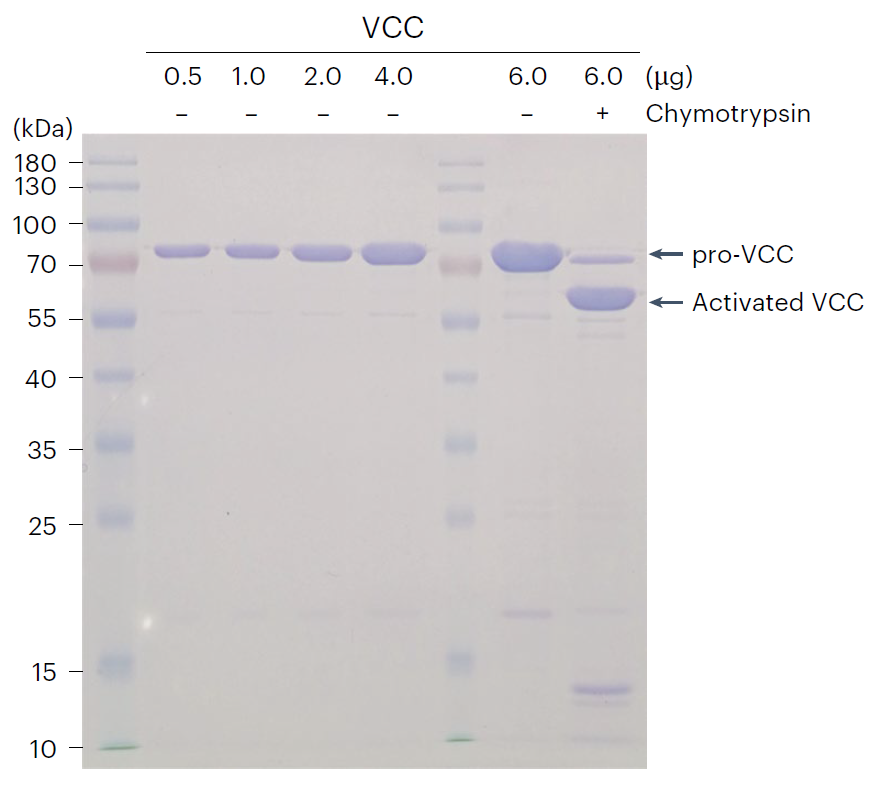
Chymotrypsin purchased from MedChemExpress. Usage Cited in: Nat Protoc. 2025 May;20(5):1158-1195. [Abstract]
Vibrio cholerae cytolysin (VCC) was produced as an inactive pro-toxin (80 kDa), and the mature toxin (65 kDa) was generated through proteolysis by Chymotrypsin (incubated with VCC at 1000:1 molar ratio; room temperature; 2 h). A representative gel for in-house produced His-tagged pro-VCC and its proteolysis activation was shown. Both the toxin purity (the major band was toxin, with few degradation bands) and activation efficiency were satisfactory.
-
J Med Chem
Peptidomimetic Analogues Act as Effective Inhibitors against SARS-CoV-2 by Blocking the Function of Cathepsin L. [Abstract]2024 Oct 10;67(19):17124-17143. PMID: 39292661 -
PLoS Pathog
Serine protease-driven entry and S2 ' cleavage flexibility of feline coronavirus during feline enterocyte infections. [Abstract]2026 Jan 8;22(1):e1013854. PMID: 41505483


Solvant et solubilité
In Vitro:
H2O : ≥ 33.33 mg/mL
* "≥" means soluble, but saturation unknown.


Pureté et documentation
-
Fiche technique (279 KB)
-
SDS (702 KB)
- English - EN (702 KB)
- Français - FR (702 KB)
- Deutsch - DE (702 KB)
- Norwegian - NO (702 KB)
- Español - ES (702 KB)
- Swedish - SV (702 KB)
- Italian - IT (702 KB)
- Korean - KR (702 KB)
- Portuguese - PT (702 KB)
-
Instruction de manipulation (2659 KB)
Références
[1]. Li J, et al. Chymotrypsin attenuates adjuvant-induced arthritis by downregulating TLR4, NF-κB, MMP-1, TNF-α, IL-1β, and IL-6 expression in Sprague-Dawley rats. Immunopharmacol Immunotoxicol. 2022 Dec;44(6):959-969. [Content Brief]
[2]. Mahmoud GS, et al. Hepatoprotective effect of trypsin/chymotrypsin against olanzapine-induced non-alcoholic steatohepatitis in rats. Can J Physiol Pharmacol. 2021 Oct;99(10):1088-1096. [Content Brief]
[3]. Wald M, et al. Mixture of trypsin, chymotrypsin and papain reduces formation of metastases and extends survival time of C57Bl6 mice with syngeneic melanoma B16. Cancer Chemother Pharmacol. 2001 Jul;47 Suppl:S16-22. [Content Brief]
[4]. Jancsó Z, et al. Chymotrypsin Reduces the Severity of Secretagogue-Induced Pancreatitis in Mice. Gastroenterology. 2018 Oct;155(4):1017-1021. [Content Brief]

Calculators
Concentration (start) × Volume (start) = Concentration (final) × Volume (final)


Chymotrypsin Related Classifications
HY-108910 Related Classifications
 Powered by Bioz