



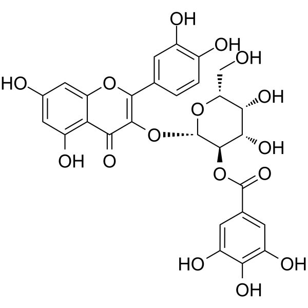
2"-O-Galloylhyperin
Based on 2 publication(s) in Google Scholar
2''-O-Galloylhyperin is an active natural compound with anti‑inflammatory, antioxidant, anti‑adipogenic, antifibrotic, and cytostatic activities. 2''-O-Galloylhyperin upregulates SIRT1/Nrf2 signaling, inhibits NF-κB and MAPK (ERK1/2, p38, JNK) phosphorylation, suppresses TSHR activation, reduces ROS accumulation, and enhances SOD and GSH-Px activities. 2''-O-Galloylhyperin protects against LPS-induced tissue injury, enhances survival, and inhibits adipogenesis and fibrosis. 2"-O-Galloylhyperin can be used for the research of sepsis, acute lung injury, and thyroid eye disease.
Nur für Forschungszwecke. Wir verkaufen nicht an Patienten.
- Reinheit : 99.57%
- CAS. Nr.: 53209-27-1
- Formel: C28H24O16
- Molecular Weight:616.48
-
Speicherung:
4°C, protect from light
* In solvent : -80°C, 6 months; -20°C, 1 month (protect from light)

To place orders, for customer services and technical support, please contact: MedChemExpress USA
Tel: 609-228-6898 E-mail: [email protected] [email protected]


-
 Biologische Aktivität
-
Chemical Information
-
Lösungsmittel & Löslichkeit
- Protokoll
- Reinheit & Dokumentation
- Verweise
-
 Help & FAQs


-
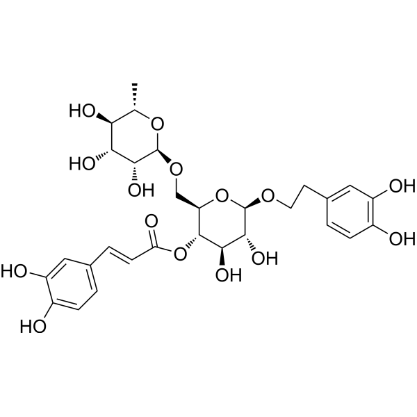
HY-N0028
Forsythiaside AAnti-Inflammatory Agent -
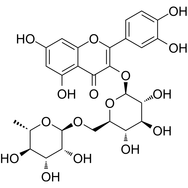
HY-N0148
RutinCBR1 Inhibitor -
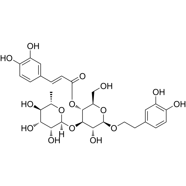
HY-N0021
VerbascosideBacterial Inhibitor -
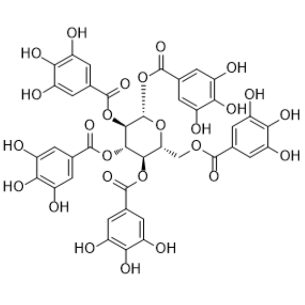
HY-N0527
PentagalloylglucoseAntioxidant -
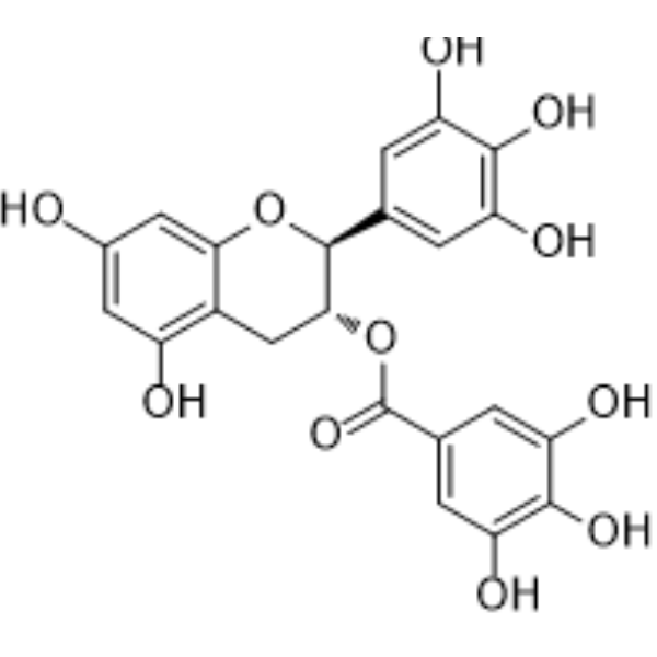
HY-N0522
(-)-Gallocatechin gallateAnticancer Agent -
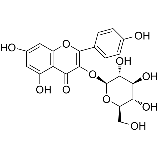
HY-N0015
AstragalinNF-κB Inhibitor -
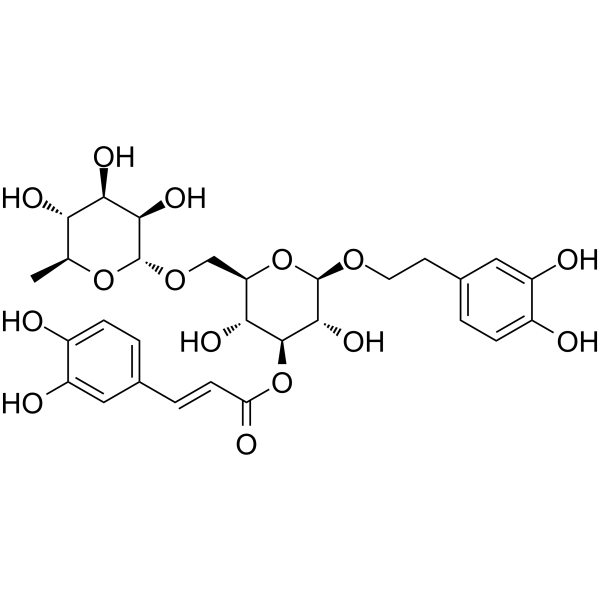
HY-N5042
Forsythoside IAnti-inflammatory Agent -
HY-N4089
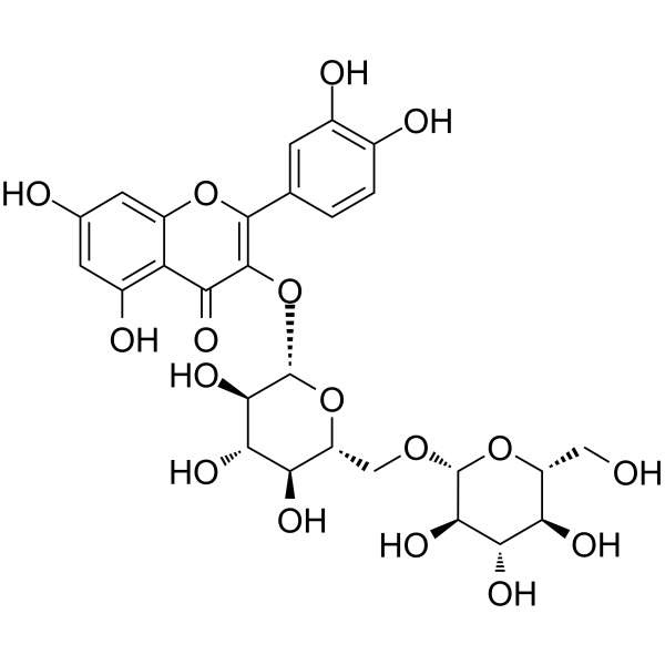
Quercetin 3-gentiobiosideAromatase Inhibitor -
HY-N1996
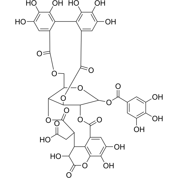
Chebulagic acidCOX Inhibitor -
HY-113529
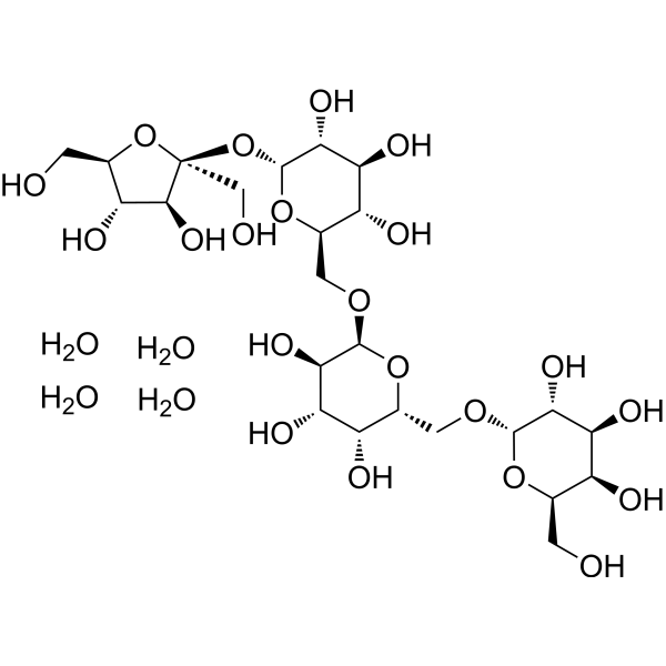
Stachyose tetrahydratePrebiotic -
HY-N0356
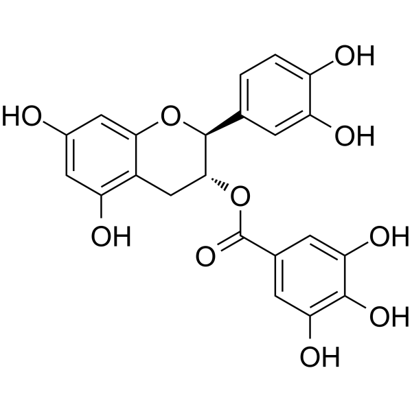
(-)-Catechin gallateCOX Inhibitor
Publications Citing Use of MedChemExpress (MCE) 2"-O-Galloylhyperin
More

Biologische Aktivität
Beschreibung
IC50 & Target
[1]|
SIRT1 |
NFκB |
ERK1 |
ERK2 |
p38 |
In Vivo
MedChemExpress (MCE) has not independently confirmed the accuracy of these methods. They are for reference only.
-
Animal Model:ICR (male, 6 weeks old, 20-22 g, intranasal LPS-induced)[2]
-
Dosage:10 mg/kg; 50 mg/kg
-
Administration:i.p.; single dose; 1 hour prior to LPS challenge
-
Result:Reduced lung W/D ratio, MPO activity, inflammatory cell infiltration, pro‑inflammatory cytokine levels, and MDA content, enhanced SOD and GSH‑Px activities, alleviated histopathological damage, inhibited MAPK and NF‑κB activation, promoted AMPK phosphorylation, and upregulated Nrf2/HO‑1 expression at 10 mg/kg.
Achieved stronger protective effects on lung injury, more potent suppression of inflammation and oxidative stress, and greater regulation of AMPK/Nrf2/NF‑κB/MAPK signaling at 50 mg/kg.
- Western Blot
- Collagen: Sirius Red Staining
- ROS/oxidative-stress fluorescent staining
- Connective Tissue: Masson's Trichrome/Collagen Trichrome Staining
- Research Protocol for Inflammation-related Diseases
- Kinase activity and phosphorylation assays
- Fibrosis/Collagen Morphometry
- 3T3-L1 preadipocyte-to-adipocyte differentiation
- LPS-Induced Endotoxemia/Systemic Inflammation
- Research Protocol for Endocrine Diseases
- Protocol for Kinase activity and phosphorylation assays
- Inhalation Toxicity Study
- Lipid Droplets: Oil Red O/Sudan Dye Lipid Staining


Chemical Information
-
CAS. Nr. 53209-27-1
-
Appearance Solid
-
Molecular Weight 616.48
-
Formel C28H24O16
-
Color Light yellow to yellow
-
SMILES
O=C1C(O[C@H](O[C@H](CO)[C@H](O)[C@@H]2O)[C@@H]2OC(C3=CC(O)=C(O)C(O)=C3)=O)=C(C4=CC(O)=C(O)C=C4)OC5=CC(O)=CC(O)=C15
-
Structure Classification
-
Versand
Room temperature in continental US; may vary elsewhere.
-
Speicherung
4°C, protect from light
* In solvent : -80°C, 6 months; -20°C, 1 month (protect from light)


Publications (2)
-
Journal Impact Factor
-
Most Recent
-
Food Chem
Effects of sun drying combined with baking processes on the flavor quality of Chongqing Tuocha raw tea. [Abstract]2025 Dec 30:497:146992. PMID: 41285060 -
Food Chem
Flavonoid-mediated metabolic underpinning quality variation in red bud-sport pear mutants. [Abstract]2025 Oct 15:489:144992. PMID: 40466530


Lösungsmittel & Löslichkeit
In Vitro:
DMSO : 100 mg/mL (162.21 mM; Need ultrasonic; Hygroscopic DMSO has a significant impact on the solubility of product, please use newly opened DMSO)
Please refer to the solubility information to select the appropriate solvent. Once prepared, please aliquot and store the solution to prevent product inactivation from repeated freeze-thaw cycles.
Storage method and period of stock solution: -80°C, 6 months; -20°C, 1 month (protect from light). When stored at -80°C, please use it within 6 months. When stored at -20°C, please use it within 1 month.
Please refer to the solubility information to select the appropriate solvent. Once prepared, please aliquot and store the solution to prevent product inactivation from repeated freeze-thaw cycles.
Storage method and period of stock solution: -80°C, 6 months; -20°C, 1 month (protect from light). When stored at -80°C, please use it within 6 months. When stored at -20°C, please use it within 1 month.
Konzentration (Stammlösung) × Volumen (Stammlösung) = Konzentration (Ziellösung) × Volumen (Ziellösung)
In Vivo:
Select the appropriate dissolution method based on your experimental animal and administration route.
- For the following dissolution methods, please ensure to first prepare a clear stock solution using an In Vitro approach and then sequentially add co-solvents:
- To ensure reliable experimental results, the clarified stock solution can be appropriately stored based on storage conditions. As for the working solution for In Vivo experiments, it is recommended to prepare freshly and use it on the same day.
- The percentages shown for the solvents indicate their volumetric ratio in the final prepared solution. If precipitation or phase separation occurs during preparation, heat and/or sonication can be used to aid dissolution.
Add each solvent one by one: 10% DMSO 40% PEG300 5% Tween-80 45% Saline
Solubility: ≥ 0.83 mg/mL (1.35 mM); Clear solution
This protocol yields a clear solution of ≥ 0.83 mg/mL (saturation unknown).
Taking 1 mL working solution as an example, add 100 μL DMSO stock solution (8.3 mg/mL) to 400 μL PEG300, and mix evenly; then add 50 μL Tween-80 and mix evenly; then add 450 μL Saline to adjust the volume to 1 mL.
Preparation of Saline: Dissolve 0.9 g sodium chloride in ddH₂O and dilute to 100 mL to obtain a clear Saline solution.
Add each solvent one by one: 10% DMSO 90% (20% SBE-β-CD in Saline)
Solubility: ≥ 0.83 mg/mL (1.35 mM); Clear solution
This protocol yields a clear solution of ≥ 0.83 mg/mL (saturation unknown).
Taking 1 mL working solution as an example, add 100 μL DMSO stock solution (8.3 mg/mL) to 900 μL 20% SBE-β-CD in Saline, and mix evenly.
Preparation of 20% SBE-β-CD in Saline (4°C, storage for one week): 2 g SBE-β-CD powder is dissolved in 10 mL Saline, completely dissolve until clear.
In Vivo Dissolution Calculator
Please enter the basic information of animal experiments:
-
-
-
-
Recommended: Prepare an additional quantity of animals to account for potential losses during experiments.
Please enter your animal formula composition:
-
%DMSO +
Recommended: Keep the proportion of DMSO in working solution below 2% if your animal is weak.
-
%+
-
+%Tween-80 + +
-
%Saline +
The co-solvents required include: DMSO, . All of co-solvents are available by MedChemExpress (MCE). , Tween 80. All of co-solvents are available by MedChemExpress (MCE).
Working solution concentration: 0.22 mg/mL
Method for preparing stock solution: mg drug dissolved in μL DMSO. Stock solution concentration: mg/mL. * In solvent : -80°C, 6 months; -20°C, 1 month (protect from light)
1. Take μL DMSO stock solution;
2. Add μL .
μL , mix evenly;
3. Then add μL Tween 80, mix evenly;
4. Then add μL
Please ensure that the stock solution in the first step is dissolved to a clear state, and add co-solvents in sequence. You can use ultrasonic heating (ultrasonic cleaner, recommended frequency 20-40 kHz), vortexing, etc. to assist dissolution.


Reinheit & Dokumentation
-
Data Sheet (284 KB)
-
SDS (394 KB)
- English - EN (394 KB)
- Français - FR (394 KB)
- Deutsch - DE (394 KB)
- Norwegian - NO (394 KB)
- Español - ES (394 KB)
- Swedish - SV (394 KB)
- Italian - IT (394 KB)
- Korean - KR (394 KB)
- Portuguese - PT (394 KB)
-
Handling Instructions (2659 KB)
Verweise
[1]. Wang P, et al. 2'-O-Galloylhyperin Isolated From Pyrola incarnata Fisch. Attenuates LPS-Induced Inflammatory Response by Activation of SIRT1/Nrf2 and Inhibition of the NF-κB Pathways in Vitro and Vivo. Front Pharmacol. 2018 Jun 27;9:679. [Content Brief]
[2]. Zhang SD, et al. 2'O-galloylhyperin attenuates LPS-induced acute lung injury via up-regulation antioxidation and inhibition of inflammatory responses in vivo. Chem Biol Interact. 2019;304:20-27. [Content Brief]
[3]. Guo Y, et al. 2'-O-Galloylhyperin Prevents Tissue Remodeling in Thyroid Eye Disease: Prospects as a Thyrotropin Receptor Antagonist. J Clin Endocrinol Metab. 2025;110(8):e2711-e2722. [Content Brief]


Complete Stock Solution Preparation Table
Please refer to the solubility information to select the appropriate solvent. Once prepared, please aliquot and store the solution to prevent product inactivation from repeated freeze-thaw cycles.
Storage method and period of stock solution: -80°C, 6 months; -20°C, 1 month (protect from light). When stored at -80°C, please use it within 6 months. When stored at -20°C, please use it within 1 month.
| Optional Solvent | Concentration Solvent Mass | 1 mg | 5 mg | 10 mg | 25 mg |
|---|---|---|---|---|---|
| DMSO | 1 mM | 1.6221 mL | 8.1106 mL | 16.2211 mL | 40.5528 mL |
| 5 mM | 0.3244 mL | 1.6221 mL | 3.2442 mL | 8.1106 mL | |
| 10 mM | 0.1622 mL | 0.8111 mL | 1.6221 mL | 4.0553 mL | |
| 15 mM | 0.1081 mL | 0.5407 mL | 1.0814 mL | 2.7035 mL | |
| 20 mM | 0.0811 mL | 0.4055 mL | 0.8111 mL | 2.0276 mL | |
| 25 mM | 0.0649 mL | 0.3244 mL | 0.6488 mL | 1.6221 mL | |
| 30 mM | 0.0541 mL | 0.2704 mL | 0.5407 mL | 1.3518 mL | |
| 40 mM | 0.0406 mL | 0.2028 mL | 0.4055 mL | 1.0138 mL | |
| 50 mM | 0.0324 mL | 0.1622 mL | 0.3244 mL | 0.8111 mL | |
| 60 mM | 0.0270 mL | 0.1352 mL | 0.2704 mL | 0.6759 mL | |
| 80 mM | 0.0203 mL | 0.1014 mL | 0.2028 mL | 0.5069 mL | |
| 100 mM | 0.0162 mL | 0.0811 mL | 0.1622 mL | 0.4055 mL |


2"-O-Galloylhyperin Related Classifications
HY-N0526 Related Classifications
 Powered by Bioz