



Amitriptyline hydrochloride
Based on 9 publication(s) in Google Scholar
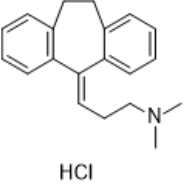
Amitriptyline hydrochloride is an orally active tricyclic antidepressant (TCA). Amitriptyline hydrochloride mainly exerts its antidepressant effect by blocking SERT (Ki = 3.45 nM) and NET (Ki = 13.3 nM), thereby increasing the concentrations of 5-hydroxytryptamine (5-HT) and norepinephrine (NE) in the synaptic cleft. Amitriptyline hydrochloride is also an agonist at α2A and TrkA/TrkB receptors, thereby exerting analgesic and neurotrophic activities (inhibiting cell apoptosis). Amitriptyline hydrochloride can reduce inflammation, angiogenesis and fibrosis. Amitriptyline hydrochloride binds to DAT (with Ki = 2.58 μM). Amitriptyline hydrochloride has high affinity for a series of receptors and can antagonize muscarinic cholinergic receptors (M1/M2/M3/M4/M5 receptors) (Ki = 11-24 nM), H1 receptors (Ki = 0.5-1.1 nM), adrenergic α1 receptors (Ki = 4.4 nM), etc., resulting in a series of side effects. Amitriptyline hydrochloride can block sodium channels and hERG potassium channel (IC50 = 4.78 μM) and it has cardiotoxicity.
Nur für Forschungszwecke. Wir verkaufen nicht an Patienten.
- Reinheit : 99.94%
- CAS. Nr.: 549-18-8
- Formel: C20H24ClN
- Molecular Weight:313.86
-
Speicherung:
4°C, sealed storage, away from moisture
* In solvent : -80°C, 6 months; -20°C, 1 month (sealed storage, away from moisture)

To place orders, for customer services and technical support, please contact: MedChemExpress USA
Tel: 609-228-6898 E-mail: [email protected] [email protected]


-
 Biologische Aktivität
-
Chemical Information
-
Lösungsmittel & Löslichkeit
- Protokoll
- Reinheit & Dokumentation
- Verweise
-
 Help & FAQs


-
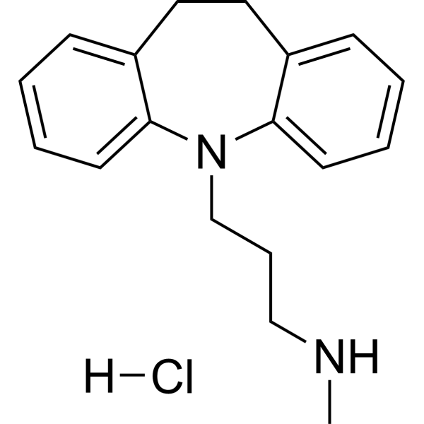
HY-B1272
Desipramine hydrochlorideSerotonin Transporter Inhibitor -
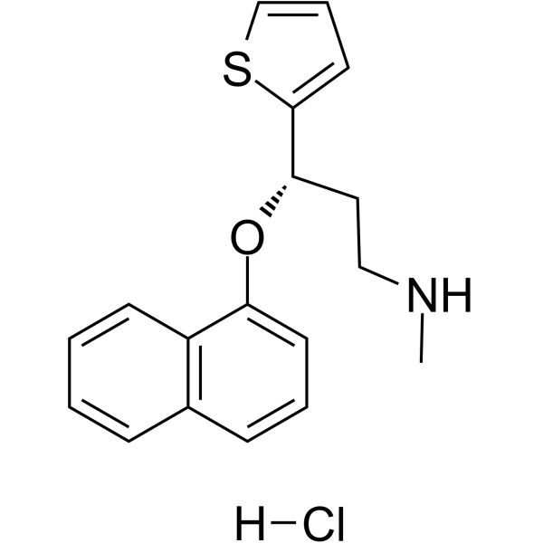
HY-B0161A
Duloxetine hydrochlorideSerotonin-norepinephrine Reuptake Inhibitor -
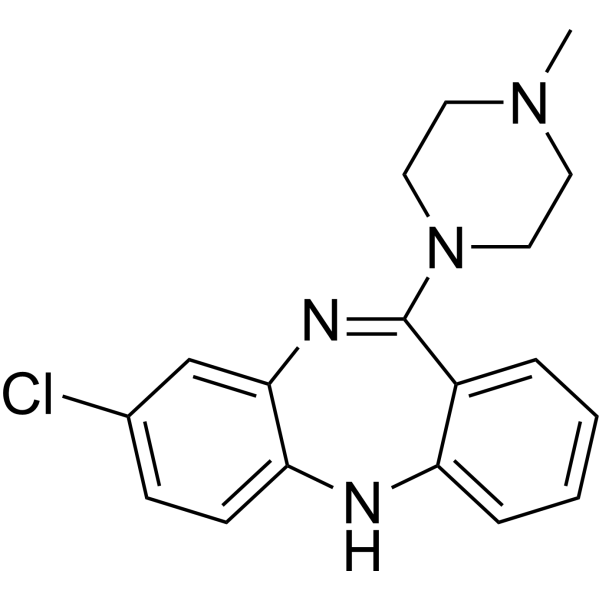
HY-14539
ClozapineAntipsychotic Agent -
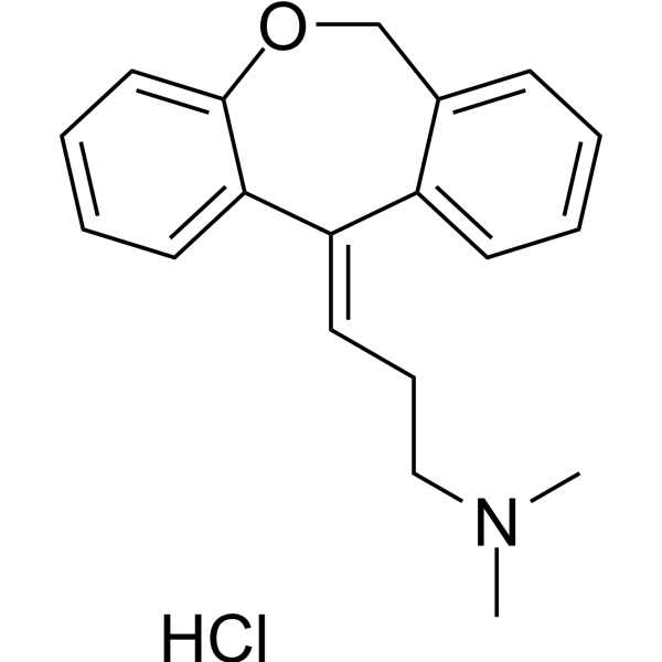
HY-B0725
Doxepin HydrochlorideAntidepressant -
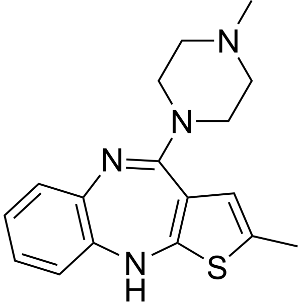
HY-14541
Olanzapine5-HT2/D1/D2 Antagonist -
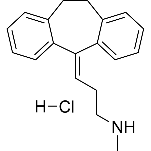
HY-B1417
Nortriptyline hydrochlorideAntidepressant -
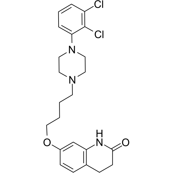
HY-14546
AripiprazoleD2/5-HT1A Agonist -
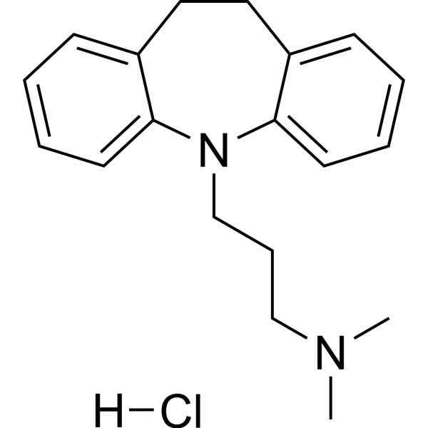
HY-B1490
Imipramine hydrochlorideAntidepressant Agent -
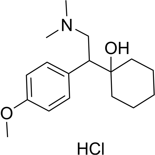
HY-B0196A
Venlafaxine hydrochlorideSerotonin/Norepinephrine Reuptake Inhibitor -
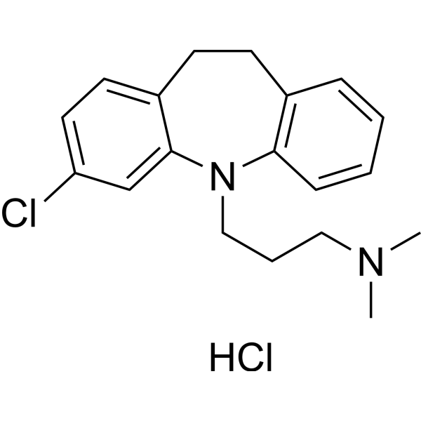
HY-B0457
Clomipramine hydrochloride5-HT Reuptake Inhibitor -
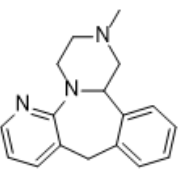
HY-B0352
MirtazapineNaSSA Agent -
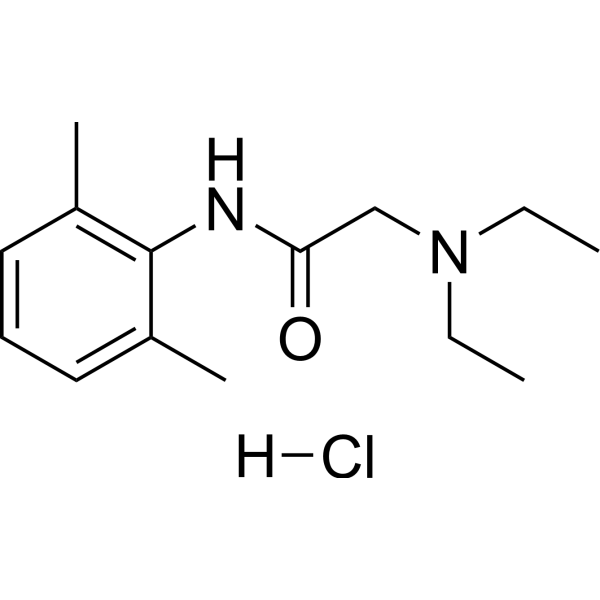
HY-B0185A
Lidocaine hydrochlorideNa+ Channel Blocker
Publications Citing Use of MedChemExpress (MCE) Amitriptyline hydrochloride
More- NPJ Digit Med. 2025 Nov 17;8(1):663. [Abstract]
- J Am Chem Soc. 2025 Mar 26. [Abstract]
- Cell Death Dis. 2026 Jul 10.
- Cell Commun Signal. 2023 May 25;21(1):123. [Abstract]
- Neurosci Bull. 2025 Mar 17. [Abstract]
- Int Immunopharmacol. 2025 Feb 6:147:113969. [Abstract]
- Skelet Muscle. 2024 Jul 18;14(1):16. [Abstract]
- J Med Virol. 2023 Jan;95(1):e28266. [Abstract]
- PLoS Negl Trop Dis. 2019 Aug 20;13(8):e0007681. [Abstract]
Alle 5-HT Receptor Isoform-spezifische Produkte anzeigen
MoreAlle Histamine Receptor Isoform-spezifische Produkte anzeigen
MoreAlle Adrenergic Receptor Isoform-spezifische Produkte anzeigen
More

Biologische Aktivität
Beschreibung
|
5-HT1A Receptor 450 nM (IC50) |
5-HT1B Receptor 40 nM (IC50) |
5-HT2A Receptor 4 nM (IC50) |
5-HT2B Receptor 40 nM (IC50) |
5-HT2C Receptor 6 nM (IC50) |
H1 Receptor 1.1 nM (IC50) |
H3 receptor 1 μM (IC50) |
H4 receptor 33.6 nM (IC50) |
SERT 3.45 nM (Ki) |
NET 13.3 nM (Ki) |
DAT 2.58 μM (Ki) |
Adrenergic receptor 24 nM (IC50) |
muscarinic receptor 7.2 nM (IC50) |
TrkA |
TrkB |
Cellular Effect
|
Cell Line

|
Type | Value | Description | References |
|---|---|---|---|---|
| HEK293 | IC50 |
0.2 μM
Compound: Amitriptyline
|
Inhibition of the Norepinephrine transporter (NET, SLC6A2) as assessed by GPCR-mediated changes in cell morphology using the impedance-based transporter activity through receptor activation (TRACT) assay in HEK-293 JumpIN-SLC6A2 cells (PubChem AID: 174586
Inhibition of the Norepinephrine transporter (NET, SLC6A2) as assessed by GPCR-mediated changes in cell morphology using the impedance-based transporter activity through receptor activation (TRACT) assay in HEK-293 JumpIN-SLC6A2 cells (PubChem AID: 174586
|
[PMID: 34112854] |
In Vitro
Amitriptyline hydrochloride (0.5-10 μM, 3.5-16.5 h) effectively resists cell apotosis (EC50 = 50 nM) in T17 cells and shows no protective effect on the SN56 cells, and in primary rat hippocampal neurons stimulated by glutamate and subjected to oxygen-glucose deprivation (OGD), the neuronal apoptosis is significantly reduced[3].
Amitriptyline hydrochloride (0.5 μM, 30 min) induces TrkA and TrkB receptor phosphorylation and activation in hippocampal neurons[3].
Amitriptyline hydrochloride (0.5 μM, 5 days) induces neurite outgrowth in PC12 cells[3].
MedChemExpress (MCE) has not independently confirmed the accuracy of these methods. They are for reference only. Further protocols information, click here.
-
Cell Line:Primary rat hippocampal neurons
-
Concentration:0.5 μM
-
Incubation Time:30 min
-
Result:Strongly induced phosphorylation of TrkA, TrkB, Akt, and ERK.
Induced TrkA-TrkA homodimerization, TrkB-TrkB homodimerization, and TrkA-TrkB heterodimerization.
In Vivo
Amitriptyline hydrochloride (15 mg/kg, i.p., single dose) produces antinociception in a mouse model by activating α2A-adrenoceptor receptors in the central nervous system[4].
Amitriptyline hydrochloride (5 mg/kg, p.o., once daily for 7 days) is able to down-regulate angiogenesis and foreign body reaction (FBR) in 14-day-old implants[5].
MedChemExpress (MCE) has not independently confirmed the accuracy of these methods. They are for reference only.
-
Animal Model:Kainic acid induced excitatory epilepsy model established in male C57BL/6 mice[3]
-
Dosage:15 mg/kg
-
Administration:Intraperitoneal injection (i.p.), once daily for 5 days
-
Result:Effectively activated TrkA, TrkB and their downstream signaling pathways in the brain. Significantly reduced KA-induced hippocampal (70%).
-
Animal Model:Hot-plate test and abdominal constriction test established in male Swiss albino mice (23-30 g)[4]
-
Dosage:15 mg/kg
-
Administration:Intraperitoneal injection (i.p.), single dose
-
Result:Significantly increased pain threshold in both models (increased hot plate latency and reduced writhing times).
Required the presence of endogenous monoamine neurotransmitters (such as NE).
Exhibited the analgesic effect be completely blocked by BRL 44408 (HY-12716).
-
Animal Model:Polyether-polyurethane sponge disks induced FBR model established in male C57BL/6 mice[5]
-
Dosage:5 mg/kg
-
Administration:Oral administration (p.o.), once daily for 7 days
-
Result:Had no effect on 7-day implants.
Significantly reduced all key parameters of the 14-day implants, reducing angiogenesis, fibrosis markers, and FBR markers.
Clinical Trial
| NCT Number | Sponsor | Condition | Start Date |
Phase
|
|---|---|---|---|---|
| NCT01329991 | Plexxikon| | 2011-05 | PHASE1 |


Chemical Information
-
CAS. Nr. 549-18-8
-
Appearance Solid
-
Molecular Weight 313.86
-
Formel C20H24ClN
-
Color White to off-white
-
SMILES
CN(C)CC/C=C1C2=CC=CC=C2CCC3=C\1C=CC=C3.Cl
-
Versand
Room temperature in continental US; may vary elsewhere.
-
Speicherung
4°C, sealed storage, away from moisture
* In solvent : -80°C, 6 months; -20°C, 1 month (sealed storage, away from moisture)


Publications (9)
-
Journal Impact Factor
-
Most Recent
-
NPJ Digit Med
Tandospirone augments cisplatin treatment by lowering cholesterol and managing distress in NSCLC patients. [Abstract]2025 Nov 17;8(1):663. PMID: 41249389 -
J Am Chem Soc
Fluorinated Ribonucleocarbohydrate Nanoparticles Allow Ultraefficient mRNA Delivery and Protein Expression in Tumor-Associated Myeloid Cells. [Abstract]2025 Mar 26. PMID: 40135499 -
-
Cell Commun Signal
Tricyclic antidepressants induce liver inflammation by targeting NLRP3 inflammasome activation. [Abstract]2023 May 25;21(1):123. PMID: 37231437 -
Neurosci Bull
Electrophysiological Abnormalities and Pharmacological Corrections of Pathogenic Missense Variants in KCNQ3. [Abstract]2025 Mar 17. PMID: 40095209 -
Int Immunopharmacol
Innovative role of the antidepressant imipramine in esophageal squamous cell carcinoma treatment: Promoting apoptosis and protective autophagy. [Abstract]2025 Feb 6:147:113969. PMID: 39764996 -
Skelet Muscle
ASM is a therapeutic target in dermatomyositis by regulating the differentiation of naive CD4 + T cells into Th17 and Treg subsets. [Abstract]2024 Jul 18;14(1):16. PMID: 39026344 -
J Med Virol
2023 Jan;95(1):e28266. PMID: 36319186 -
PLoS Negl Trop Dis
Identification of anti-flaviviral drugs with mosquitocidal and anti-Zika virus activity in Aedes aegypti. [Abstract]2019 Aug 20;13(8):e0007681. PMID: 31430351


Lösungsmittel & Löslichkeit
In Vitro:
DMSO : ≥ 100 mg/mL (318.61 mM; Hygroscopic DMSO has a significant impact on the solubility of product, please use newly opened DMSO)
H2O : ≥ 50 mg/mL (159.31 mM)
* "≥" means soluble, but saturation unknown.
Please refer to the solubility information to select the appropriate solvent. Once prepared, please aliquot and store the solution to prevent product inactivation from repeated freeze-thaw cycles.
Storage method and period of stock solution: -80°C, 6 months; -20°C, 1 month (sealed storage, away from moisture). When stored at -80°C, please use it within 6 months. When stored at -20°C, please use it within 1 month.
* Note: If you choose water as the stock solution, please dilute it to the working solution, then filter and sterilize it with a 0.22 μm filter before use.
Please refer to the solubility information to select the appropriate solvent. Once prepared, please aliquot and store the solution to prevent product inactivation from repeated freeze-thaw cycles.
Storage method and period of stock solution: -80°C, 6 months; -20°C, 1 month (sealed storage, away from moisture). When stored at -80°C, please use it within 6 months. When stored at -20°C, please use it within 1 month.
* Note: If you choose water as the stock solution, please dilute it to the working solution, then filter and sterilize it with a 0.22 μm filter before use.
Konzentration (Stammlösung) × Volumen (Stammlösung) = Konzentration (Ziellösung) × Volumen (Ziellösung)
In Vivo:
For the following dissolution methods, please prepare the working solution directly:
It is recommended to prepare fresh solutions and use them promptly within a short period of time.
The percentages shown for the solvents indicate their volumetric ratio in the final prepared solution. If precipitation or phase separation occurs during preparation, heat and/or sonication can be used to aid dissolution.
Add each solvent one by one: PBS
Solubility: 120 mg/mL (382.34 mM); Clear solution; Need ultrasonic

Protokoll
-
Collagen: Sirius Red Staining
Sirius Red or picrosirius red staining is a histochemical method for visualizing collagen-rich extracellular matrix in tissue sections, and collagen fibers are detected as red-stained structures under bright-field microscopy with enhanced birefringence under polarized light. Picrosirius red is useful for assessing total collagen organization, distribution, and fibrosis burden, but polarized color should not be interpreted as a definitive collagen type I versus type III readout because color is affected by fiber orientation, thickness, and packing.
-
Human pluripotent stem cell endothelial-cell differentiation
Human pluripotent stem cell endothelial differentiation is based on stepwise developmental patterning: early activation of WNT/GSK3β inhibition promotes mesodermal or vascular progenitor entry, followed by endothelial specification using VEGF-related signaling, BMP4, FGF2, Notch modulation, or cAMP depending on the published protocol. Endothelial differentiation is read out by acquisition of CD31, CD34, VE-cadherin/CD144, KDR/VEGFR2, vWF, Tie2, NOS3, acetylated LDL uptake, tube/network formation, barrier function, and in vivo vessel-forming capacity where tested.
-
Vascular/Branching Fractal Analysis
Vascular/branching fractal analysis quantifies the geometric complexity of vessel trees or vascular networks from segmented 2D images, commonly by converting vessels into binary and/or skeletonized maps and estimating fractal dimension using box-counting or related approaches. Fractal dimension is interpreted as an image-derived readout of vascular branching complexity, space filling, or density, and has been applied to retinal photographs, fluorescein angiography, OCT angiography, capillary perfusion maps, and in vitro Matrigel angiogenesis networks. The assay readout is generated from vessel-positive pixels after image preprocessing, vessel segmentation, binarization, and optional skeletonization; reported outputs include fractal dimension, vessel density, branchpoint density, endpoint density, vessel length density, tortuosity, and generation-based branching metrics when VESGEN-style analysis is used. The biological interpretation is limited to quantitative vascular patterning and s
-
Annexin V plus membrane-impermeant dye apoptosis staining
Annexin V-based apoptosis assays rely on the detection of phosphatidylserine (PS) externalization from the inner leaflet of the plasma membrane to the outer leaflet, an early biochemical hallmark of apoptosis. Fluorescently labeled Annexin V binds PS in a calcium-dependent manner, enabling identification of early apoptotic cells by flow cytometry or fluorescence microscopy. When combined with a membrane-impermeant DNA-binding dye (e. g. , propidium iodide), this approach allows discrimination between viable (Annexin V−/dye−), early apoptotic (Annexin V+/dye−), and late apoptotic or necrotic (Annexin V+/dye+) cell populations by assessing membrane integrity and PS exposure.
-
Connective Tissue: Masson's Trichrome/Collagen Trichrome Staining
Masson’s Trichrome (collagen/trichrome staining) is a histological technique that differentially stains tissue compartments using sequential acidic dyes to distinguish collagen from muscle and cytoplasmic components based on dye affinity and tissue permeability differences, enabling visualization of fibrosis and connective tissue architecture in histological sections. The classical formulation typically uses Weigert's iron hematoxylin for nuclear staining, Biebrich scarlet-acid fuchsin for cytoplasm and muscle, and aniline blue (or light green variants) for collagen, producing a characteristic blue/green collagen signal contrasted against red cytoplasm and dark nuclei. The staining principle relies on selective displacement of smaller dye molecules by larger anionic dyes in collagen-rich regions under controlled acidified conditions, which enhances collagen-specific dye retention. This property makes the method widely used for fibrosis assessment in organs such as heart, liver, lung, a
-
Research Protocol for Inflammation-related Diseases
The NLRP3 inflammasome is a cytosolic innate immune signaling platform that integrates priming signals and danger-signal activation to promote caspase-1 activation, maturation of IL-1β and IL-18, and gasdermin D-mediated pyroptotic cell death. The core experimental logic is to determine whether inflammatory disease phenotypes are driven by increased NLRP3 expression, ASC-containing inflammasome assembly, caspase-1 cleavage, GSDMD cleavage, and extracellular release of IL-1β/IL-18 rather than by nonspecific cell injury alone. The pathway is strongly linked to inflammation-related disease phenotypes because monosodium urate crystals activate NALP3/NLRP3 inflammasome signaling in gout-like crystal inflammation, cholesterol crystals activate NLRP3 inflammasomes in atherogenesis models, and DSS-induced intestinal inflammation has been reported to involve NLRP3 inflammasome activity. However, experimental colitis studies also show context-dependent protective effects of NLRP3 inflammasome co
-
Fibrosis/Collagen Morphometry
Fibrosis and collagen morphometry is based on the quantitative visualization of fibrillar collagen deposition in tissue sections using histochemical stains such as Sirius Red (Picrosirius Red) or Masson's trichrome, followed by image-based or polarization-enhanced analysis to estimate collagen proportional area as a surrogate of extracellular matrix accumulation during fibrotic remodeling. Sirius Red combined with polarized light microscopy enhances detection of collagen fibers due to birefringence properties, enabling more specific visualization of collagen type I and III fibrils compared to conventional bright-field histology, while whole-section or region-restricted digital morphometry reduces field-selection bias in fibrosis assessment. Alternative quantitative approaches include second harmonic generation (SHG) and two-photon excited fluorescence microscopy, which enable label-free detection of fibrillar collagen and have been validated against histological staining and biochemica
-
Apoptosis
Apoptosis, also called programmed cell death, is generally characterized by distinct morphological characteristics.
-
TUNEL staining for apoptotic DNA fragmentation
TUNEL staining detects DNA strand breaks by using terminal deoxynucleotidyl transferase to add labeled nucleotides to exposed 3′-OH DNA termini, generating either microscopic staining in fixed cells or tissue sections, or fluorescence/cytometric signal in cell suspensions. TUNEL positivity reflects DNA fragmentation but should not be interpreted alone as definitive apoptosis, because TUNEL can also label necrotic, autolytic, mechanically damaged, or DNA-repair-associated DNA breaks.
-
Cell Cytotoxicity Assay
Cytotoxicity assays are usually based on the assessment of cell membrane damage, which can also be indirectly detected by measuring cell viability. Detection methods include MTT assay, CKK-8 assay, LDH assay and ATP assay, etc.
-
Endothelial Tube Formation Assay
Endothelial tube formation assay evaluates the ability of endothelial cells to attach, migrate, align, and organize into capillary-like networks when cultured on gelled basement membrane extract or Matrigel; the readout is the morphology and quantity of tube-like networks, which reflects an in vitro endothelial morphogenesis step related to angiogenesis. Basement membrane extract/Matrigel provides laminin-rich extracellular matrix cues that support endothelial differentiation into capillary-like structures, but it can contain biologically active growth factors, so growth-factor-reduced matrix is preferred when testing defined angiogenic stimulators or inhibitors.
-
Apoptosis Solutions
Apoptosis is a regulated, generally non-lytic cell-death pathway that removes unwanted, damaged, infected, or abnormal cells through coordinated morphological changes, caspase activation, DNA fragmentation, and membrane remodeling. The intrinsic apoptosis pathway is controlled mainly by mitochondrial outer membrane permeabilization, BCL-2 family proteins, cytochrome c release, apoptosome formation, caspase-9 activation, and downstream executioner caspase-3/7 activation. The extrinsic apoptosis pathway is initiated by death receptors such as Fas, TNFR, and TRAIL receptors, which recruit adaptor proteins and activate caspase-8 before engaging executioner caspases or mitochondrial amplification through BID cleavage. Apoptosis is linked to many phenotypes, including cancer cell killing, tissue homeostasis, immune regulation, neurodegeneration, infection response, and treatment-induced cytotoxicity; unresolved questions include how apoptosis interacts with necroptosis, pyroptosis, ferroptos
-
Endothelial Cell Migration/Angiogenic Sprouting Assay
Endothelial cell migration and angiogenic sprouting assays are in vitro (and partially ex vivo-adapted) functional models that quantify the ability of endothelial cells to undergo coordinated migration, extracellular matrix invasion, and multicellular organization into capillary-like sprouts in response to pro-angiogenic stimuli such as VEGF, bFGF, or conditioned microenvironments. These assays are used to model early angiogenic events including tip-cell formation, directional migration, and lumen-like sprout extension, which collectively reflect angiogenic activation and vascular morphogenesis processes observed in vivo.


Reinheit & Dokumentation
-
Data Sheet (286 KB)
-
SDS (480 KB)
- English - EN (480 KB)
- Français - FR (480 KB)
- Deutsch - DE (480 KB)
- Norwegian - NO (480 KB)
- Español - ES (480 KB)
- Swedish - SV (480 KB)
- Italian - IT (480 KB)
- Korean - KR (480 KB)
- Portuguese - PT (480 KB)
-
Handling Instructions (2659 KB)
Verweise
[1]. Kim Lawson. A Brief Review of the Pharmacology of Amitriptyline and Clinical Outcomes in Treating Fibromyalgia. Biomedicines. 2017 Jun; 5(2): 24. [Content Brief]
[2]. S Neil Vaishnavi , et al. Milnacipran: a comparative analysis of human monoamine uptake and transporter binding affinity. Biol Psychiatry. 2004 Feb 1;55(3):320-2. [Content Brief]
[3]. Jang, S.W., et al., Amitriptyline is a TrkA and TrkB receptor agonist that promotes TrkA/TrkB heterodimerization and has potent neurotrophic activity. Chem Biol, 2009. 16(6): p. 644-56. [Content Brief]
[4]. Ghelardini C, et al. Antinociception induced by amitriptyline and imipramine is mediated by alpha2A-adrenoceptors. Jpn J Pharmacol. 2000 Feb;82(2):130-7. [Content Brief]
[5]. Scheuermann K, et al. Amitriptyline efficacy in decreasing implant-induced foreign body reaction. IUBMB Life. 2023 Sep;75(9):732-742. [Content Brief]


Complete Stock Solution Preparation Table
Please refer to the solubility information to select the appropriate solvent. Once prepared, please aliquot and store the solution to prevent product inactivation from repeated freeze-thaw cycles.
Storage method and period of stock solution: -80°C, 6 months; -20°C, 1 month (sealed storage, away from moisture). When stored at -80°C, please use it within 6 months. When stored at -20°C, please use it within 1 month.
| Optional Solvent | Concentration Solvent Mass | 1 mg | 5 mg | 10 mg | 25 mg |
|---|---|---|---|---|---|
| H2O / DMSO | 1 mM | 3.1861 mL | 15.9307 mL | 31.8613 mL | 79.6534 mL |
| 5 mM | 0.6372 mL | 3.1861 mL | 6.3723 mL | 15.9307 mL | |
| 10 mM | 0.3186 mL | 1.5931 mL | 3.1861 mL | 7.9653 mL | |
| 15 mM | 0.2124 mL | 1.0620 mL | 2.1241 mL | 5.3102 mL | |
| 20 mM | 0.1593 mL | 0.7965 mL | 1.5931 mL | 3.9827 mL | |
| 25 mM | 0.1274 mL | 0.6372 mL | 1.2745 mL | 3.1861 mL | |
| 30 mM | 0.1062 mL | 0.5310 mL | 1.0620 mL | 2.6551 mL | |
| 40 mM | 0.0797 mL | 0.3983 mL | 0.7965 mL | 1.9913 mL | |
| 50 mM | 0.0637 mL | 0.3186 mL | 0.6372 mL | 1.5931 mL | |
| 60 mM | 0.0531 mL | 0.2655 mL | 0.5310 mL | 1.3276 mL | |
| 80 mM | 0.0398 mL | 0.1991 mL | 0.3983 mL | 0.9957 mL | |
| 100 mM | 0.0319 mL | 0.1593 mL | 0.3186 mL | 0.7965 mL |
* Note: If you choose water as the stock solution, please dilute it to the working solution, then filter and sterilize it with a 0.22 μm filter before use.


Amitriptyline hydrochloride Related Classifications
HY-B0527A Related Classifications
 Powered by Bioz