



Tenidap

Based on 1 Customer Validation
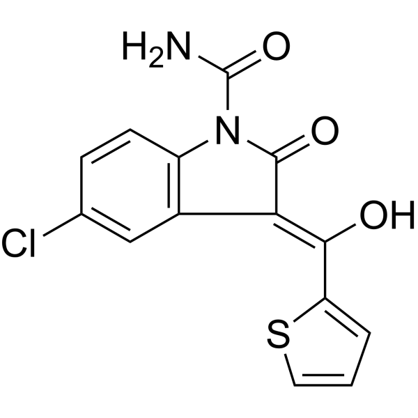
Tenidap (CP-66248) is an orally active dual inhibitor of 5-LOX and COX with anti-inflammatory and immunomodulatory properties. Tenidap downregulates the expression of IL-1 receptors in chondrocytes, reduces the release of pro-inflammatory cytokines such as IL-1, IL-6 and TNF-α, and inhibits MMP production and cartilage degradation. Tenidap also blocks bone resorption and leukocyte adhesion to vascular endothelium, interferes with ion and pH changes associated with mouse sperm capacitation, and selectively enhances the activity of hKir2.3 channels (EC50=1.3 μM). Tenidap is applicable to research related to rheumatoid arthritis.
Nur für Forschungszwecke. Wir verkaufen nicht an Patienten.
- Reinheit : 99.93%
- CAS. Nr.: 120210-48-2
- Formel: C14H9ClN2O3S
- Molecular Weight:320.75
-
Speicherung:Powder -20°C, 3 years , 4°C, 2 years ; In solvent -80°C, 6 months , -20°C, 1 month

To place orders, for customer services and technical support, please contact: MedChemExpress USA
Tel: 609-228-6898 E-mail: [email protected] [email protected]


-
 Biologische Aktivität
-
Chemical Information
-
Lösungsmittel & Löslichkeit
- Protokoll
- Reinheit & Dokumentation
- Verweise
-
 Help & FAQs


-

HY-105028R
Tenidap (Standard)COX-1 Inhibitor -
HY-13568
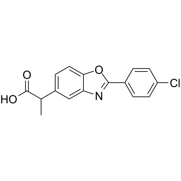
BenoxaprofenNonsteroidal Anti-inflammatory Agent -
HY-B0253
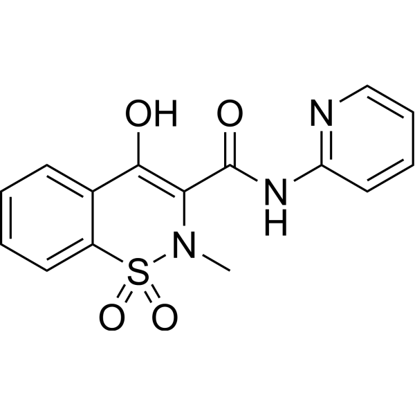
PiroxicamCOX Inhibitor -
HY-B0138
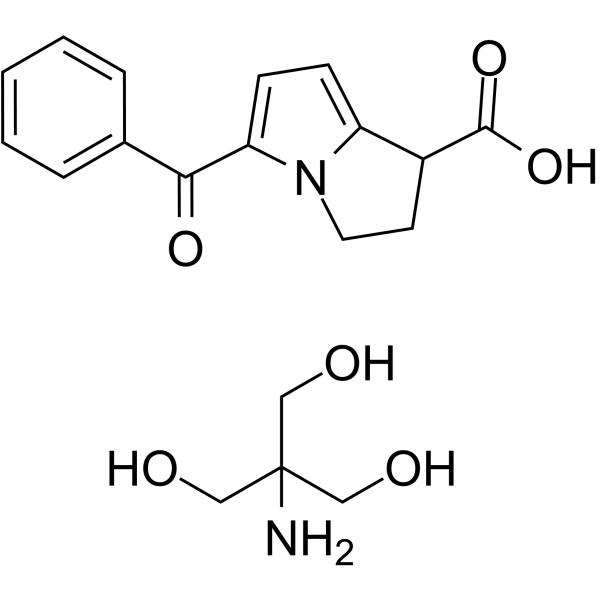
Ketorolac tromethamine saltCOX Inhibitor -
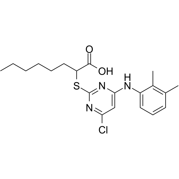
HY-111140
YS121mPGES-1/5-LOX Inhibitor -
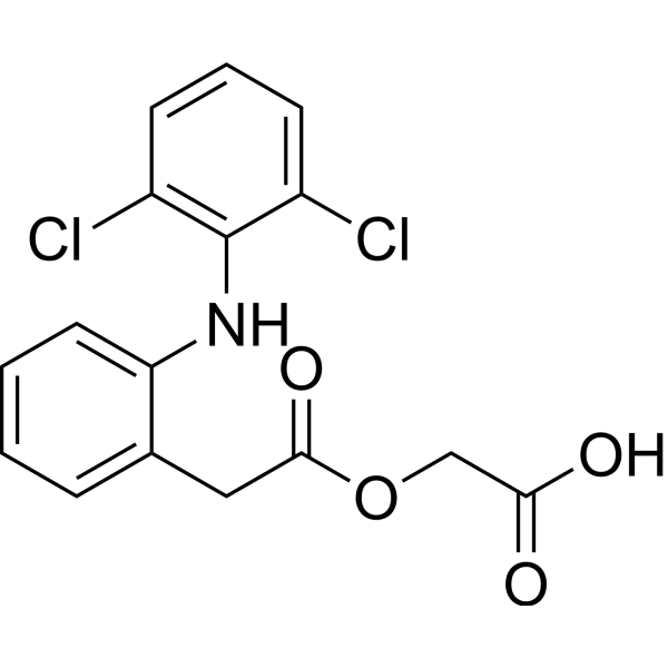
HY-B0634
AceclofenacNSAID -
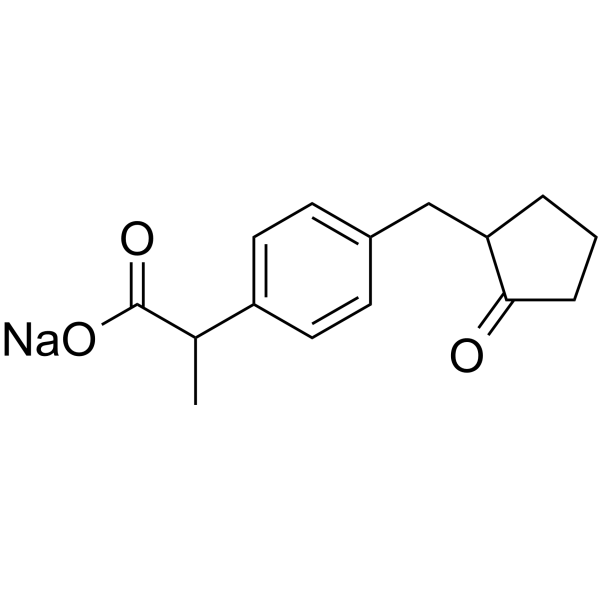
HY-B0578A
Loxoprofen sodiumAnti-Inflammatory Agent -

HY-B0138R
Ketorolac tromethamine salt (Standard)COX Inhibitor -
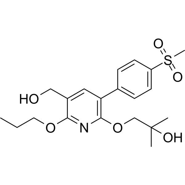
HY-115866
COX-2-IN-6COX Inhibitor -

HY-13568R
Benoxaprofen (Standard)Anti-inflammatory/Antipyretic Compound -

HY-B0253R
Piroxicam (Standard)COX Inhibitor -
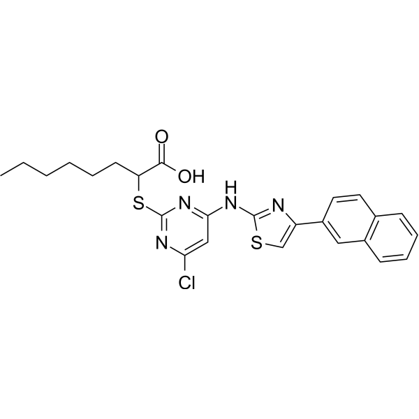
HY-126898
5-LO/mPGES1-IN-15-LO/mPGES-1 Inhibitor


Biologische Aktivität
Beschreibung
IC50 & Target
|
COX-1 0.03 μM (IC50) |
COX-2 1.2 μM (IC50) |
15-LOX |
MMP-8 |
Cellular Effect
|
Cell Line

|
Type | Value | Description | References |
|---|---|---|---|---|
| RBL-1 | IC50 |
0.01 μM
Compound: 2 (tenidap)
|
In vitro inhibition of Prostaglandin G/H synthase pathway in rat basophilic leukemia (RBL-1) cells
In vitro inhibition of Prostaglandin G/H synthase pathway in rat basophilic leukemia (RBL-1) cells
|
[PMID: 2113951] |
| RBL-1 | IC50 |
9 μM
Compound: 2 (tenidap)
|
In vitro inhibition of 5-lipoxygenase pathway in rat basophilic leukemia (RBL-1) cells
In vitro inhibition of 5-lipoxygenase pathway in rat basophilic leukemia (RBL-1) cells
|
[PMID: 2113951] |
| THP-1 | IC50 |
16 μM
Compound: Tenidap
|
In vitro effect on production of lipopolysaccharide (LPS) -induced IL1-beta in human THP-1 cells
In vitro effect on production of lipopolysaccharide (LPS) -induced IL1-beta in human THP-1 cells
|
[PMID: 10821716] |
In Vitro
Tenidap potently inhibits leukotriene B4 and prostanoid synthesis in human polymorphonuclear leukocytes in vitro[1].
Tenidap (20 μg/mL; therapeutic concentrations) reduces IL-1β receptor expression in normal and osteoarthritic human chondrocytes, and potently suppresses IL-1β-induced collagenase and stromelysin synthesis and mRNA levels in these cells[2].
Tenidap potently inhibits bone resorption induced by parathyroid hormone, vitamin D3, IL-1α, TNF-α, and prostaglandin E2 in in vitro bone cultures, with effects independent of prostaglandin synthesis suppression[2].
Tenidap inhibits vitamin D3-induced CD14 expression, partially reverses reduced 3H-thymidine uptake, and suppresses early-stage IFN-γ-stimulated complement C2 production in HL-60 myelomonocytic cells, with no effect on C2 production in differentiated cells[2].
Tenidap potently inhibits adhesion of neutrophils, eosinophils, monocytes, and T cells to endothelial cells in in vitro adhesion assays, including IL-1-stimulated models[2].
Tenidap (0.01-10 μM; 50 min) potently and dose-dependently enhances Ba2+-sensitive 86Rb+ efflux through hKir2.3 channels in CHO cells with an EC50 of 402 nM[4].
Tenidap (30 μM; 5 min, not specified) is selective for hKir2.3 channels, as it has little or no effect on hKv1.5, μ1 Na+, or hKir2.1 channels in stably transfected cells[4].
MedChemExpress (MCE) has not independently confirmed the accuracy of these methods. They are for reference only.
- RNA extraction experimental
- Collagen-Induced Arthritis
- Mitochondrial membrane-potential fluorescent assay
- LPS-Induced Endotoxemia/Systemic Inflammation
- Mitochondrial membrane-potential and mitochondrial mass staining
- Research Protocol for Inflammation-related Diseases
- Osteoclast differentiation from monocyte/macrophage precursors


Chemical Information
-
CAS. Nr. 120210-48-2
-
Appearance Solid
-
Molecular Weight 320.75
-
Formel C14H9ClN2O3S
-
Color White to yellow
-
SMILES
O/C(C1=CC=CS1)=C2C3=C(C=CC(Cl)=C3)N(C(N)=O)C/2=O
-
Synonyms
CP-66248
-
Versand
Room temperature in continental US; may vary elsewhere.
-
Speicherung
Powder -20°C 3 years 4°C 2 years In solvent -80°C 6 months -20°C 1 month


Lösungsmittel & Löslichkeit
In Vitro:
DMSO : 25 mg/mL (77.94 mM; Need ultrasonic; Hygroscopic DMSO has a significant impact on the solubility of product, please use newly opened DMSO)
Please refer to the solubility information to select the appropriate solvent. Once prepared, please aliquot and store the solution to prevent product inactivation from repeated freeze-thaw cycles.
Storage method and period of stock solution: -80°C, 6 months; -20°C, 1 month. When stored at -80°C, please use it within 6 months. When stored at -20°C, please use it within 1 month.
Please refer to the solubility information to select the appropriate solvent. Once prepared, please aliquot and store the solution to prevent product inactivation from repeated freeze-thaw cycles.
Storage method and period of stock solution: -80°C, 6 months; -20°C, 1 month. When stored at -80°C, please use it within 6 months. When stored at -20°C, please use it within 1 month.
Konzentration (Stammlösung) × Volumen (Stammlösung) = Konzentration (Ziellösung) × Volumen (Ziellösung)
In Vivo:
Select the appropriate dissolution method based on your experimental animal and administration route.
- For the following dissolution methods, please ensure to first prepare a clear stock solution using an In Vitro approach and then sequentially add co-solvents:
- To ensure reliable experimental results, the clarified stock solution can be appropriately stored based on storage conditions. As for the working solution for In Vivo experiments, it is recommended to prepare freshly and use it on the same day.
- The percentages shown for the solvents indicate their volumetric ratio in the final prepared solution. If precipitation or phase separation occurs during preparation, heat and/or sonication can be used to aid dissolution.
Add each solvent one by one: 10% DMSO 40% PEG300 5% Tween-80 45% Saline
Solubility: 2.5 mg/mL (7.79 mM); Suspended solution; Need ultrasonic
This protocol yields a suspended solution of 2.5 mg/mL. Suspended solution can be used for oral and intraperitoneal injection.
Taking 1 mL working solution as an example, add 100 μL DMSO stock solution (25.0 mg/mL) to 400 μL PEG300, and mix evenly; then add 50 μL Tween-80 and mix evenly; then add 450 μL Saline to adjust the volume to 1 mL.
Preparation of Saline: Dissolve 0.9 g sodium chloride in ddH₂O and dilute to 100 mL to obtain a clear Saline solution.
In Vivo Dissolution Calculator
Please enter the basic information of animal experiments:
-
-
-
-
Recommended: Prepare an additional quantity of animals to account for potential losses during experiments.
Please enter your animal formula composition:
-
%DMSO +
Recommended: Keep the proportion of DMSO in working solution below 2% if your animal is weak.
-
%+
-
+%Tween-80 + +
-
%Saline +
The co-solvents required include: DMSO, . All of co-solvents are available by MedChemExpress (MCE). , Tween 80. All of co-solvents are available by MedChemExpress (MCE).
Working solution concentration: 0.22 mg/mL
Method for preparing stock solution: mg drug dissolved in μL DMSO. Stock solution concentration: mg/mL.
1. Take μL DMSO stock solution;
2. Add μL .
μL , mix evenly;
3. Then add μL Tween 80, mix evenly;
4. Then add μL
Please ensure that the stock solution in the first step is dissolved to a clear state, and add co-solvents in sequence. You can use ultrasonic heating (ultrasonic cleaner, recommended frequency 20-40 kHz), vortexing, etc. to assist dissolution.


Reinheit & Dokumentation
-
Data Sheet (280 KB)
-
SDS (393 KB)
- English - EN (393 KB)
- Français - FR (393 KB)
- Deutsch - DE (393 KB)
- Norwegian - NO (393 KB)
- Español - ES (393 KB)
- Swedish - SV (393 KB)
- Italian - IT (393 KB)
- Korean - KR (393 KB)
- Portuguese - PT (393 KB)
-
Handling Instructions (2659 KB)
Verweise
[2]. Breedveld F, et al. Tenidap: a novel cytokine-modulating antirheumatic drug for the treatment of rheumatoid arthritis. Scand J Rheumatol Suppl. 1994;100:31-44. [Content Brief]
[3]. Chávez JC, et al. Participation of the Cl-/HCO(3)- exchangers SLC26A3 and SLC26A6, the Cl- channel CFTR, and the regulatory factor SLC9A3R1 in mouse sperm capacitation. Biol Reprod. 2012;86(1):1-14. Published 2012 Jan 19. [Content Brief]
[4]. Liu Y, et al. Tenidap, a novel anti-inflammatory agent, is an opener of the inwardly rectifying K+ channel hKir2.3. Eur J Pharmacol. 2002;435(2-3):153-160. [Content Brief]


Complete Stock Solution Preparation Table
Please refer to the solubility information to select the appropriate solvent. Once prepared, please aliquot and store the solution to prevent product inactivation from repeated freeze-thaw cycles.
Storage method and period of stock solution: -80°C, 6 months; -20°C, 1 month. When stored at -80°C, please use it within 6 months. When stored at -20°C, please use it within 1 month.
| Optional Solvent | Concentration Solvent Mass | 1 mg | 5 mg | 10 mg | 25 mg |
|---|---|---|---|---|---|
| DMSO | 1 mM | 3.1177 mL | 15.5885 mL | 31.1769 mL | 77.9423 mL |
| 5 mM | 0.6235 mL | 3.1177 mL | 6.2354 mL | 15.5885 mL | |
| 10 mM | 0.3118 mL | 1.5588 mL | 3.1177 mL | 7.7942 mL | |
| 15 mM | 0.2078 mL | 1.0392 mL | 2.0785 mL | 5.1962 mL | |
| 20 mM | 0.1559 mL | 0.7794 mL | 1.5588 mL | 3.8971 mL | |
| 25 mM | 0.1247 mL | 0.6235 mL | 1.2471 mL | 3.1177 mL | |
| 30 mM | 0.1039 mL | 0.5196 mL | 1.0392 mL | 2.5981 mL | |
| 40 mM | 0.0779 mL | 0.3897 mL | 0.7794 mL | 1.9486 mL | |
| 50 mM | 0.0624 mL | 0.3118 mL | 0.6235 mL | 1.5588 mL | |
| 60 mM | 0.0520 mL | 0.2598 mL | 0.5196 mL | 1.2990 mL |


Tenidap Related Classifications
HY-105028 Related Classifications
 Powered by Bioz