



Dexpramipexole
Based on 2 publication(s) in Google Scholar
Dexpramipexole ((R)-Pramipexole) is an orally active, blood-brain barrier permeable mitochondrial protective agent. Dexpramipexole upregulates the expression of Parkin, PINK1, GPX4 and FSP1; binds to mitochondrial F1/Fo-ATP synthase; blocks the Nav1.8 sodium channel; and inhibits the activation of the NLRP3 inflammasome. Dexpramipexole induces mitophagy, inhibits ferroptosis, pyroptosis, apoptosis, neuroinflammation and eosinophilopoiesis; maintains mitochondrial function and redox homeostasis; reduces reactive oxygen species production; and decreases myocardial infarct size. Dexpramipexole is applicable to studies on eosinophilic asthma, myocardial ischemia/reperfusion injury, sepsis-associated encephalopathy, analgesia, and more.
Para uso exclusivo en investigación. No vendemos a pacientes.
- Pureza : 99.69%
- No. CAS: 104632-28-2
- Fòrmula: C10H17N3S
- Peso molecular:211.33
-
Almacenamiento:Powder -20°C, 3 years ; In solvent -80°C, 6 months , -20°C, 1 month

To place orders, for customer services and technical support, please contact: MedChemExpress USA
Tel: 609-228-6898 E-mail: [email protected] [email protected]


-
 Actividad biológica
-
Chemical Information
-
Solvente y solubilidad
- Protocolo
- Pureza y Documentación
- Referencias
-
 Help & FAQs


Publications Citing Use of MedChemExpress (MCE) Dexpramipexole
More Customer Validation & Images

-
IF
-
IF


Actividad biológica
Descripciòn
|
Nav1.8 |
GPX4 |
PINK1 |
NLRP3 inflammasome |
In Vitro
Dexpramipexole (5-200 μM; 24 h) shows no cytotoxicity to primary cardiomyocytes isolated from neonatal rats at concentrations below 100 μM, while the compound at 200 μM reduces cell viability[1].
Dexpramipexole (2-50 μM) protects primary neonatal rat cardiomyocytes against hypoxia/reoxygenation (H/R)-induced injury by enhancing cell viability and reducing LDH release[1].
Dexpramipexole (2-50 μM) upregulates the expression level of PINK1 protein in primary cardiomyocytes from neonatal rats treated with H/R[1].
Dexpramipexole protects primary cardiomyocytes from neonatal rats against H/R-induced mitochondrial dysfunction by reducing mPTP opening, decreasing ROS production and restoring mitochondrial membrane potential[1].
Dexpramipexole enhances ATG7-dependent mitophagy in primary neonatal rat cardiomyocytes subjected to H/R, which is characterized by increased formation of mitophagosomes, enhanced autophagic mitochondrial sequestration, and improved mitochondrial clearance[1].
Dexpramipexole activates PINK1 and Parkin and promotes mitochondrial fission in primary neonatal rat cardiomyocytes exposed to H/R[1].
Dexpramipexole protects primary cardiomyocytes from neonatal rats against H/R injury by upregulating PINK1- and Parkin-dependent mitophagy[1].
Dexpramipexole (10 μM; 5 min) inhibits tetrodotoxin-resistant sodium currents in primary cultured rat dorsal root ganglion neurons with an IC50 of 294.4 nM, and does not alter the voltage-dependent activation or inactivation properties of the channels[5].
Dexpramipexole (10 μM) selectively inhibits Nav1.8-mediated sodium currents in primary cultured dorsal root ganglion neurons from wild-type mice[5].
MedChemExpress (MCE) has not independently confirmed the accuracy of these methods. They are for reference only. Further protocols information, click here.
In Vivo
Dexpramipexole (5-20 mg/kg; i.p., once daily for 14 consecutive days) improves survival rate, body weight gain, spontaneous activity, motor coordination and forelimb muscle strength, reduces hematoma volume, alleviates white matter injury, decreases iron and ROS accumulation, and inhibits ferroptosis by upregulating GPX4 and FSP1 in mice with intracerebral hemorrhage[2].
Dexpramipexole (3 mg/kg; i.p.; once daily for 6 consecutive days) improves lipopolysaccharide (LPS)-induced cognitive impairment in sepsis-associated encephalopathy (SAE), preserves the morphology and function of hippocampal mitochondria in mice, and inhibits mitochondria-mediated pyroptosis and apoptosis[3].
Dexpramipexole (3-20 mg/kg; 20-140 μg/20 μL; p.o.; i.p.; hind paw s.c.; single administration) exerts effective analgesic effects in various mouse models of nociceptive pain (e.g., inflammatory pain, visceral pain) and neuropathic pain (e.g., chemotherapy-induced pain, nerve compression pain, diabetic neuropathic pain)[5].
MedChemExpress (MCE) has not independently confirmed the accuracy of these methods. They are for reference only.
-
Animal Model:C57BL/6 (male, 9-10 weeks old, 24-26 g; intracerebral hemorrhage induced by stereotactic injection of autologous tail vein blood)[2]
-
Dosage:5 mg/kg; 10 mg/kg; 20 mg/kg
-
Administration:i.p., once daily for 14 days
-
Result:Significantly increased the survival rate of mice after cerebral hemorrhage.
Slowed down the postoperative weight loss and promoted the recovery of motor function, motor coordination ability, muscle strength and comprehensive neurological function (BMS).
Reduced iron deposition around the hematoma and the accumulation of reactive oxygen species.
Increased the expression of GPX4 and FSP1.
-
Animal Model:C57BL/6 (male, 3 months old, 20-28 g, LPS-induced sepsis-associated encephalopathy)[3]
-
Dosage:3 mg/kg
-
Administration:i.p.; daily; 6 consecutive days
-
Result:Improved the long-term cognitive dysfunction caused by LPS, particularly spatial working memory and hippocampus-dependent memory.
Protected the mitochondria of hippocampal neurons, improved their morphology (reduced swelling) and function (maintained ATP levels, reduced ROS).
Inhibited pyroptosis and apoptosis of cells
-
Animal Model:CD-1 (male, 20-25 g, intraplantar formalin injection-induced inflammatory pain)[5]
-
Dosage:3 mg/kg; 10 mg/kg; 20 μg/20 μL; 70 μg/20 μL; 140 μg/20 μL
-
Administration:p.o.; single dose 1 h before test; i.p.; single dose 1 h before test; s.c. in hind paw; single dose 10 min before test
-
Result:Reduced time spent in pain-like behaviors during the early formalin phase (10 mg/kg p.o.).
Reduced time spent in pain-like behaviors dose-dependently during the late formalin phase (3 mg/kg, 10 mg/kg p.o.).
Reduced time spent in pain-like behaviors dose-dependently during the late formalin phase (3 mg/kg, 10 mg/kg i.p.).
Reduced time spent in pain-like behaviors during the late formalin phase (70 μg/20 μL, 140 μg/20 μL s.c.).
Caused no effect on pain-like behaviors in the early phase at any tested subdermal dose.
-
Animal Model:CD-1 (male, 20-25 g; female, intraperitoneal acetic acid injection-induced visceral pain)[5]
-
Dosage:10 mg/kg; 20 mg/kg
-
Administration:i.p.; single dose 1 h before test
-
Result:Reduced the number of abdominal writhings dose-dependently in male mice
. Reduced the number of abdominal writhings in female mice.
-
Animal Model:CD-1 (male, 20-25 g, ankle complete Freund adjuvant injection-induced chronic inflammatory arthritis pain)[5]
-
Dosage:70 μg/20 μL
-
Administration:s.c. in hind paw; single dose 10 min before each test on day 7, 14, 21
-
Result:Increased mechanical thresholds at day 7, 14, and 21 post CFA injection.
-
Animal Model:CD-1 (subcutaneous oxaliplatin injection-induced acute chemotherapy-induced neuropathic pain)[5]
-
Dosage:10 mg/kg; 70 μg/20 μL
-
Administration:i.p.; single dose 30 min post oxaliplatin; s.c. in hind paw; single dose 50 min post oxaliplatin
-
Result:Reduced cold pain-related behaviors at 1, 2, 3, 4, and 5 h post oxaliplatin injection (10 mg/kg i.p.).
Reduced cold pain-related behaviors at 1 hour post oxaliplatin injection (70 μg/20 μL, s.c.).
-
Animal Model:CD-1 (intraperitoneal oxaliplatin injection-induced chronic chemotherapy-induced neuropathic pain)[5]
-
Dosage:10 mg/kg
-
Administration:i.p.; single dose 1 h before test; p.o.; single dose 1 h before test
-
Result:Reduced cold allodynia at day 21 post initial oxaliplatin injection (10 mg/kg i.p.).
Reduced cold allodynia at 1, 3, and 6 hours post dosing, with effects diminishing by 9 hours (10 mg/kg p.o.).
-
Animal Model:CD-1 (male, 20-25 g, chronic constriction injury of right sciatic nerve-induced neuropathic pain)[5]
-
Dosage:10 mg/kg
-
Administration:i.p.; single dose 1 h before test
-
Result:Reduced mechanical allodynia in the injured hind paw.
-
Animal Model:CD-1 (intraperitoneal streptozotocin injection-induced diabetic neuropathic pain, blood glucose > 250 mg/dl confirmed 3 weeks post-injection)[5]
-
Dosage:10 mg/kg
-
Administration:i.p.; single dose 1 h before test
-
Result:Increased latency to pain-related behaviors in diabetic mice.


Chemical Information
-
No. CAS 104632-28-2
-
Appearance Solid
-
Peso molecular 211.33
-
Fòrmula C10H17N3S
-
Color White to off-white
-
SMILES
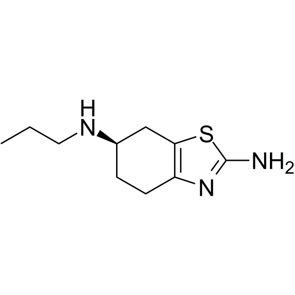
NC1=NC(CC[C@@H](NCCC)C2)=C2S1
-
Synonyms
(R)-Pramipexole; R-(+)-Pramipexole; KNS-760704
-
Envío
Room temperature in continental US; may vary elsewhere.
-
Almacenamiento
Powder -20°C 3 years In solvent -80°C 6 months -20°C 1 month


Publications (2)
-
Journal Impact Factor
-
Most Recent
-
Neuroreport
Dexpramipexole ameliorates cognitive deficits in sepsis-associated encephalopathy through suppressing mitochondria-mediated pyroptosis and apoptosis. [Abstract]2023 Mar 1;34(4):220-231. PMID: 36719835
Dexpramipexole purchased from MedChemExpress. Usage Cited in: Neuroreport. 2023 Mar 1;34(4):220-231. [Abstract]
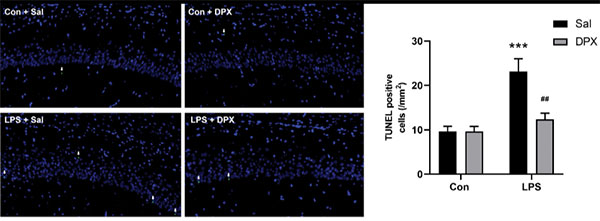
Dexpramipexole (DPX; 3 mg/kg; i.p.; once daily for 6 consecutive days) attenuates the LPS-induced increase in the number of TUNEL-positive cells in the hippocampus in SAE mice.
Dexpramipexole purchased from MedChemExpress. Usage Cited in: Neuroreport. 2023 Mar 1;34(4):220-231. [Abstract]
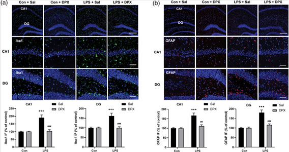
Dexpramipexole (DPX; 3 mg/kg; i.p.; once daily for 6 consecutive days) attenuates LPS-induced activation of microglia and astrocytes in the hippocampus in SAE mice. (a) Representative images of Iba-1 staining and quantification of Iba-1 (green) fluorescence intensity in the hippocampal CA1 and DG regions. (b) Representative images of GFAP staining and quantification of GFAP (red) fluorescence intensity in the hippocampal CA1 and DG regions. DAPI staining is shown in blue.
-
Oxid Med Cell Longev
Dexpramipexole Attenuates White Matter Injury to Facilitate Locomotion and Motor Coordination Recovery via Reducing Ferroptosis after Intracerebral Hemorrhage. [Abstract]2022 Aug 4;2022:6160701. PMID: 35965685


Solvente y solubilidad
In Vitro:
DMSO : 10 mg/mL (47.32 mM; ultrasonic and warming and heat to 60°C; Hygroscopic DMSO has a significant impact on the solubility of product, please use newly opened DMSO)
Please refer to the solubility information to select the appropriate solvent. Once prepared, please aliquot and store the solution to prevent product inactivation from repeated freeze-thaw cycles.
Storage method and period of stock solution: -80°C, 6 months; -20°C, 1 month. When stored at -80°C, please use it within 6 months. When stored at -20°C, please use it within 1 month.
Please refer to the solubility information to select the appropriate solvent. Once prepared, please aliquot and store the solution to prevent product inactivation from repeated freeze-thaw cycles.
Storage method and period of stock solution: -80°C, 6 months; -20°C, 1 month. When stored at -80°C, please use it within 6 months. When stored at -20°C, please use it within 1 month.
Concentration (start) × Volume (start) = Concentration (final) × Volume (final)
In Vivo:
Select the appropriate dissolution method based on your experimental animal and administration route.
- For the following dissolution methods, please ensure to first prepare a clear stock solution using an In Vitro approach and then sequentially add co-solvents:
- To ensure reliable experimental results, the clarified stock solution can be appropriately stored based on storage conditions. As for the working solution for In Vivo experiments, it is recommended to prepare freshly and use it on the same day.
- The percentages shown for the solvents indicate their volumetric ratio in the final prepared solution. If precipitation or phase separation occurs during preparation, heat and/or sonication can be used to aid dissolution.
Add each solvent one by one: 10% DMSO 40% PEG300 5% Tween-80 45% Saline
Solubility: ≥ 1 mg/mL (4.73 mM); Clear solution
This protocol yields a clear solution of ≥ 1 mg/mL (saturation unknown).
Taking 1 mL working solution as an example, add 100 μL DMSO stock solution (10.0 mg/mL) to 400 μL PEG300, and mix evenly; then add 50 μL Tween-80 and mix evenly; then add 450 μL Saline to adjust the volume to 1 mL.
Preparation of Saline: Dissolve 0.9 g sodium chloride in ddH₂O and dilute to 100 mL to obtain a clear Saline solution.
Add each solvent one by one: 10% DMSO 90% (20% SBE-β-CD in Saline)
Solubility: ≥ 1 mg/mL (4.73 mM); Clear solution
This protocol yields a clear solution of ≥ 1 mg/mL (saturation unknown).
Taking 1 mL working solution as an example, add 100 μL DMSO stock solution (10.0 mg/mL) to 900 μL 20% SBE-β-CD in Saline, and mix evenly.
Preparation of 20% SBE-β-CD in Saline (4°C, storage for one week): 2 g SBE-β-CD powder is dissolved in 10 mL Saline, completely dissolve until clear.
In Vivo Dissolution Calculator
Please enter the basic information of animal experiments:
-
-
-
-
Recommended: Prepare an additional quantity of animals to account for potential losses during experiments.
Please enter your animal formula composition:
-
%DMSO +
Recommended: Keep the proportion of DMSO in working solution below 2% if your animal is weak.
-
%+
-
+%Tween-80 + +
-
%Saline +
The co-solvents required include: DMSO, . All of co-solvents are available by MedChemExpress (MCE). , Tween 80. All of co-solvents are available by MedChemExpress (MCE).
Working solution concentration: 0.22 mg/mL
Method for preparing stock solution: mg drug dissolved in μL DMSO. Stock solution concentration: mg/mL.
1. Take μL DMSO stock solution;
2. Add μL .
μL , mix evenly;
3. Then add μL Tween 80, mix evenly;
4. Then add μL
Please ensure that the stock solution in the first step is dissolved to a clear state, and add co-solvents in sequence. You can use ultrasonic heating (ultrasonic cleaner, recommended frequency 20-40 kHz), vortexing, etc. to assist dissolution.

Protocolo
-
RNA extraction experimental
By lysing cells, releasing RNA, and removing impurities such as proteins and DNA, high-purity RNA products are finally obtained. The commonly used traditional method is the guanidine isothiocyanate/phenol/chloroform method (Trizol), which is suitable for a variety of animal materials including animal tissues, microorganisms, cultured cells, etc., and most plant materials.
-
Mitochondrial membrane-potential fluorescent assay
Mitochondrial membrane potential fluorescent assays estimate ΔΨm in living cells using lipophilic cationic dyes such as TMRM, TMRE, rhodamine 123, and JC-1, which accumulate in mitochondria according to membrane polarization; loss of signal after FCCP or CCCP treatment is interpreted as mitochondrial depolarization. TMRM/TMRE and rhodamine 123 are commonly used for semi-quantitative live-cell microscopy or flow cytometry, while JC-1 can report a shift from red aggregate fluorescence to green monomer fluorescence during depolarization; interpretation requires controls because dye concentration, quenching mode, cell type, dye efflux, and mitochondrial mass can affect fluorescence independently of ΔΨm.
-
Annexin V plus membrane-impermeant dye apoptosis staining
Annexin V-based apoptosis assays rely on the detection of phosphatidylserine (PS) externalization from the inner leaflet of the plasma membrane to the outer leaflet, an early biochemical hallmark of apoptosis. Fluorescently labeled Annexin V binds PS in a calcium-dependent manner, enabling identification of early apoptotic cells by flow cytometry or fluorescence microscopy. When combined with a membrane-impermeant DNA-binding dye (e. g. , propidium iodide), this approach allows discrimination between viable (Annexin V−/dye−), early apoptotic (Annexin V+/dye−), and late apoptotic or necrotic (Annexin V+/dye+) cell populations by assessing membrane integrity and PS exposure.
-
ROS/oxidative-stress fluorescent staining
ROS/oxidative-stress fluorescent staining uses cell-permeant fluorogenic probes that become fluorescent after oxidation inside cells or tissues; commonly used examples include DCFH-DA/DCFDA for broad cellular oxidant detection, DHE for superoxide-related signal detection, MitoSOX for mitochondrial superoxide-related signal detection, and CellROX probes for oxidative-stress-associated fluorescence readouts. The assay detects probe oxidation rather than a single ROS species unless the probe and analysis method have been chemically validated for that species. DCFH-DA enters cells, is deacetylated by intracellular esterases to DCFH, and produces fluorescent DCF after oxidation, so the readout is used as an operational measure of total cellular oxidative stress rather than a species-specific ROS measurement. DHE and MitoSOX can report superoxide-related oxidation, but red fluorescence alone can include non-specific ethidium-like oxidation products; HPLC or optimized spectral approaches are
-
Research Protocol for Neurological Diseases
PINK1/Parkin-mediated mitophagy pathway is a mitochondrial quality-control signaling axis in which mitochondrial depolarization stabilizes PINK1 on damaged mitochondria, activates Parkin recruitment and E3 ubiquitin ligase activity, promotes ubiquitination of outer mitochondrial membrane proteins, recruits selective autophagy adaptors, and drives lysosomal degradation of damaged mitochondria. In neurological disease research, this pathway is experimentally important because neurons, especially dopaminergic neurons, are highly dependent on mitochondrial integrity, and defective mitochondrial turnover can lead to mitochondrial dysfunction, oxidative stress, impaired neuronal survival, α-synuclein accumulation, and neuroinflammatory damage-associated signals. The genetic disease link is strongest in Parkinson’s disease because mutations in PRKN/parkin cause autosomal recessive juvenile parkinsonism, mutations in PINK1 cause hereditary early-onset Parkinson’s disease, and Drosophila studie
-
Apoptosis
Apoptosis, also called programmed cell death, is generally characterized by distinct morphological characteristics.
-
TUNEL staining for apoptotic DNA fragmentation
TUNEL staining detects DNA strand breaks by using terminal deoxynucleotidyl transferase to add labeled nucleotides to exposed 3′-OH DNA termini, generating either microscopic staining in fixed cells or tissue sections, or fluorescence/cytometric signal in cell suspensions. TUNEL positivity reflects DNA fragmentation but should not be interpreted alone as definitive apoptosis, because TUNEL can also label necrotic, autolytic, mechanically damaged, or DNA-repair-associated DNA breaks.
-
LPS-Induced Endotoxemia/Systemic Inflammation
Lipopolysaccharide (LPS)-induced endotoxemia is a widely used in vivo model of acute systemic inflammation in which LPS, a Gram-negative bacterial endotoxin, activates innate immune signaling primarily through TLR4, leading to rapid and transient induction of pro-inflammatory cytokines such as TNF-α, IL-6, and IL-1β in circulation and tissues. This cytokine surge is commonly used as a measurable readout of systemic inflammatory activation and immune dysregulation, and is typically assessed within hours after intraperitoneal LPS administration in mouse models of endotoxemia. The model captures key features of systemic inflammatory response syndrome, including cytokine release, immune cell activation, and downstream tissue responses, and has been used to evaluate anti-inflammatory interventions such as cytokine modulation, lipid mediators, and immune cell-targeting therapies.
-
Mitochondrial membrane-potential and mitochondrial mass staining
Mitochondrial membrane potential staining measures the electrochemical polarization across the mitochondrial inner membrane in live cells using lipophilic cationic fluorescent probes; early rhodamine-based work showed that selective mitochondrial dye accumulation is lost when the mitochondrial transmembrane potential is dissipated. JC-1 reports mitochondrial polarization by shifting from green monomer fluorescence to red J-aggregate fluorescence as dye concentration increases within energized mitochondria; therefore, the red/green fluorescence ratio is used as a relative readout of mitochondrial membrane potential. TMRE or TMRM staining provides a single-channel relative readout because these cationic rhodamine esters accumulate in polarized mitochondria, and lower fluorescence indicates reduced mitochondrial polarization when acquisition and dye-loading conditions are controlled. Mitochondrial mass staining is commonly performed with MitoTracker Green FM or related MitoTracker dyes as
-
Apoptosis Solutions
Apoptosis is a regulated, generally non-lytic cell-death pathway that removes unwanted, damaged, infected, or abnormal cells through coordinated morphological changes, caspase activation, DNA fragmentation, and membrane remodeling. The intrinsic apoptosis pathway is controlled mainly by mitochondrial outer membrane permeabilization, BCL-2 family proteins, cytochrome c release, apoptosome formation, caspase-9 activation, and downstream executioner caspase-3/7 activation. The extrinsic apoptosis pathway is initiated by death receptors such as Fas, TNFR, and TRAIL receptors, which recruit adaptor proteins and activate caspase-8 before engaging executioner caspases or mitochondrial amplification through BID cleavage. Apoptosis is linked to many phenotypes, including cancer cell killing, tissue homeostasis, immune regulation, neurodegeneration, infection response, and treatment-induced cytotoxicity; unresolved questions include how apoptosis interacts with necroptosis, pyroptosis, ferroptos
-
Ferroptosis Solutions
Ferroptosis is an iron-dependent, non-apoptotic form of regulated cell death characterized by lethal lipid peroxidation and sensitivity to suppression by iron chelators or lipophilic radical-trapping antioxidants. The core pathway links cystine uptake through system Xc−, glutathione availability, GPX4-dependent detoxification of phospholipid hydroperoxides, iron-dependent oxidative reactions, and polyunsaturated-phospholipid metabolism into a cell-death program that is biochemically and morphologically distinct from apoptosis, necrosis, and autophagy. The ferroptosis pathway is experimentally linked to phenotype through chemical and genetic perturbation. Erastin induces ferroptosis by inhibiting cystine uptake through system Xc− and weakening antioxidant defenses, while GPX4 inhibition or depletion causes lipid peroxide accumulation and ferroptotic cancer-cell death. ACSL4 and oxidizable arachidonoyl- or adrenoyl-containing phosphatidylethanolamines shape ferroptosis sensitivity by con
-
Mitophagy Solutions
Mitophagy is the selective autophagic degradation of mitochondria and functions as a mitochondrial quality-control pathway that removes damaged, depolarized, excess, or developmentally programmed mitochondria. The pathway links mitochondrial damage recognition, autophagosome recruitment, lysosomal delivery, and mitochondrial turnover to phenotypes such as mitochondrial homeostasis, oxidative-stress control, metabolic remodeling, differentiation, and neurodegeneration-related mitochondrial fidelity. The best-characterized damage-induced pathway is the PINK1-Parkin axis. Parkin is recruited selectively to impaired mitochondria and promotes their autophagic elimination, while mitochondrial depolarization stabilizes PINK1 on damaged mitochondria, recruits Parkin, and activates Parkin-dependent mitophagy. PINK1 also phosphorylates ubiquitin to activate Parkin E3 ubiquitin ligase activity, and PINK1-driven ubiquitin phosphorylation creates a feed-forward signal for recruiting autophagy machi
-
Pyroptosis Solutions
Pyroptosis is a lytic inflammatory cell-death pathway executed by gasdermin pores, most classically through inflammasome-mediated activation of caspase-1, cleavage of gasdermin D, membrane pore formation, LDH release, and secretion of IL-1β and IL-18. The canonical pathway is commonly modeled by priming cells with an inflammatory signal such as LPS to induce pro-IL-1β and inflammasome components, followed by an activation signal such as ATP or nigericin to activate NLRP3, ASC speck formation, caspase-1 cleavage, GSDMD cleavage, cytokine release, and pyroptotic membrane rupture. The non-canonical pathway is triggered when cytosolic LPS activates mouse caspase-11 or human caspase-4/5, leading to GSDMD cleavage and pyroptosis, and this can secondarily activate NLRP3-dependent IL-1β release. Pyroptosis is linked to inflammatory injury, infection, cancer, liver disease, ocular disease, placental inflammation, and other disease phenotypes, but unresolved questions include which gasdermin fam


Pureza y Documentación
-
Ficha de datos (292 KB)
-
SDS (396 KB)
- English - EN (396 KB)
- Français - FR (396 KB)
- Deutsch - DE (396 KB)
- Norwegian - NO (396 KB)
- Español - ES (396 KB)
- Swedish - SV (396 KB)
- Italian - IT (396 KB)
- Korean - KR (396 KB)
- Portuguese - PT (396 KB)
-
Instrucciones de manejo (2659 KB)
Referencias
[1]. Tang L, et al. Dexpramipexole attenuates myocardial ischemia/reperfusion injury through upregulation of mitophagy. Eur J Pharmacol. 2021;899:173962. [Content Brief]
[2]. Wang B, et al. Dexpramipexole Attenuates White Matter Injury to Facilitate Locomotion and Motor Coordination Recovery via Reducing Ferroptosis after Intracerebral Hemorrhage. Oxid Med Cell Longev. 2022;2022:6160701. Published 2022 Aug 4. [Content Brief]
[3]. Zhang Y, et al. Dexpramipexole ameliorates cognitive deficits in sepsis-associated encephalopathy through suppressing mitochondria-mediated pyroptosis and apoptosis. Neuroreport. 2023;34(4):220-231. [Content Brief]
[4].
Cusack RP, Sulaiman I, Gauvreau GM. Refashioning dexpramipexole: A new horizon in eosinophilic asthma? J Allergy Clin Immunol. 2023 Nov;152(5):1092-1094.
[Content Brief]
[5].
Urru M, et al. Dexpramipexole blocks Nav1.8 sodium channels and provides analgesia in multiple nociceptive and neuropathic pain models. Pain. 2020 Apr;161(4):831-841.
[Content Brief]
[6]. Panch SR, et al. Dexpramipexole as an oral steroid-sparing agent in hypereosinophilic syndromes. Blood. 2018;132(5):501-509. [Content Brief]


Complete Stock Solution Preparation Table
Please refer to the solubility information to select the appropriate solvent. Once prepared, please aliquot and store the solution to prevent product inactivation from repeated freeze-thaw cycles.
Storage method and period of stock solution: -80°C, 6 months; -20°C, 1 month. When stored at -80°C, please use it within 6 months. When stored at -20°C, please use it within 1 month.
| Optional Solvent | Concentration Solvent Mass | 1 mg | 5 mg | 10 mg | 25 mg |
|---|---|---|---|---|---|
| DMSO | 1 mM | 4.7319 mL | 23.6597 mL | 47.3194 mL | 118.2984 mL |
| 5 mM | 0.9464 mL | 4.7319 mL | 9.4639 mL | 23.6597 mL | |
| 10 mM | 0.4732 mL | 2.3660 mL | 4.7319 mL | 11.8298 mL | |
| 15 mM | 0.3155 mL | 1.5773 mL | 3.1546 mL | 7.8866 mL | |
| 20 mM | 0.2366 mL | 1.1830 mL | 2.3660 mL | 5.9149 mL | |
| 25 mM | 0.1893 mL | 0.9464 mL | 1.8928 mL | 4.7319 mL | |
| 30 mM | 0.1577 mL | 0.7887 mL | 1.5773 mL | 3.9433 mL | |
| 40 mM | 0.1183 mL | 0.5915 mL | 1.1830 mL | 2.9575 mL |


Dexpramipexole Related Classifications
HY-17355B Related Classifications
 Powered by Bioz
Keywords
- Dexpramipexole
- 104632-28-2
- (R)-Pramipexole
- R-(+)-Pramipexole
- KNS-760704
- KNS760704
- KNS 760704
- KNS-760704
- PINK1/Parkin
- Glutathione Peroxidase
- Sodium Channel
- ATP Synthase
- NOD-like Receptor (NLR)
- Mitophagy
- Ferroptosis
- Autophagy
- Apoptosis
- Reactive Oxygen Species (ROS)
- Parkin
- PINK1
- Na?1.8 sodium channels
- neonatal rat primary cardiomyocytes
- FSP1
- F1/F0 ATP synthase b-subunits
- oligomycin sensitivity-conferring protein
- NLRP3 inflammasome
- GPX4
- dorsal root ganglion neurons
- Inhibitor
- inhibitor
- inhibit