



Gomisin N

Based on 1 Customer Validation
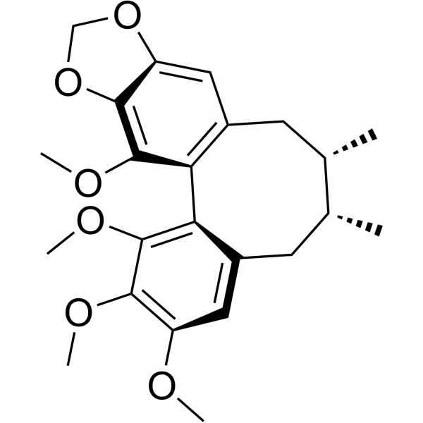
Gomisin N is an orally active lignan compound. Gomisin N can be isolated from Schisandra chinensis. Gomisin N induces Apoptosis in a variety of cells. Gomisin N activates AMPK, Akt, MAPK/ERK, Nrf2, caspase-3 and PARP-1. Gomisin N inhibits GSK3β, nitric oxide (NO), and proinflammatory cytokines (IL-1β, IL-6, TNF-α). Gomisin N has anti-inflammatory, antioxidant, anti-obesity, anti-diabetic, and anti-melanogenesis activities. Gomisin N has anti-tumor activity against cervical cancer and liver cancer. Gomisin N improves Alzheimer's disease.
商品は「研究用試薬」です。人や動物の医療用・臨床診断用・食品用の製品ではありません。
研究用途以外に使用した場合、当社は一切の責任を負いかねます。
- 純度 : 99.94%
- CAS 番号: 69176-52-9
- 分子式: C23H28O6
- 分子量:400.46
-
保管条件:
4°C, protect from light
* In solvent : -80°C, 6 months; -20°C, 1 month (protect from light)

To place orders, for customer services and technical support, please contact: MedChemExpress USA
Tel: 609-228-6898 E-mail: [email protected] [email protected]


-
 生物活性
生物活性
-
化学情報
-
溶剤 & 溶解度
- プロトコル
- 純度とドキュメンテーション
- 参考文献
-
 Help & FAQs


-
HY-N0385
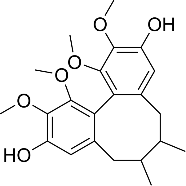
Gomisin JCalcium Channel Activator -
HY-N0694
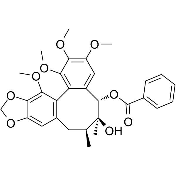
Schisantherin ANF-κB Inhibitor -
HY-N2259
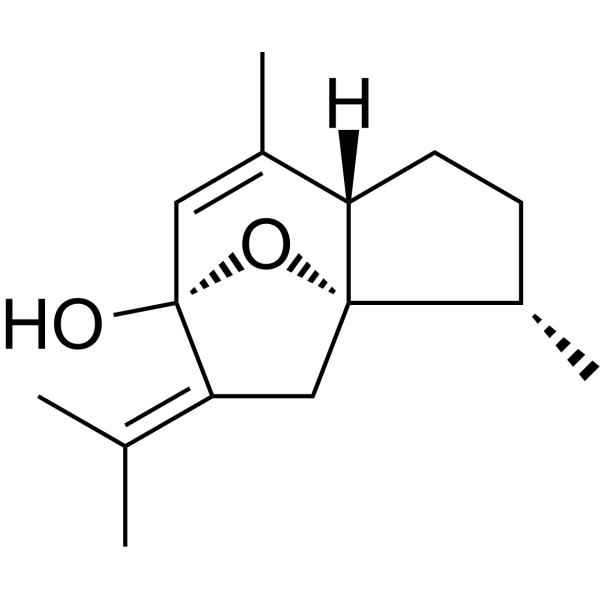
CurcumenolCYP3A4 Inhibitor -
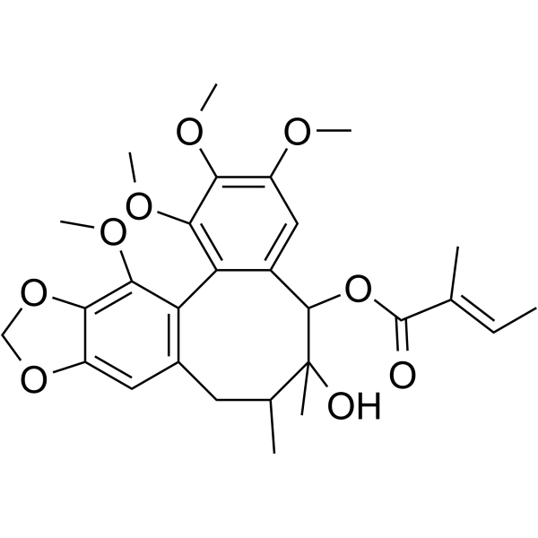
HY-N0695
Schisantherin BPI3K/AKT Activator -
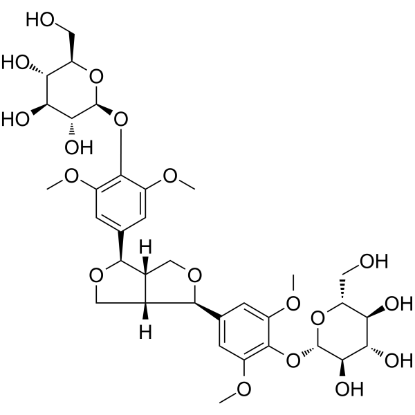
HY-N1958
Syringaresinol diglucosideNatural Product -

HY-N2259R
Curcumenol (Standard)Reference Standard -

HY-N0695R
Schisantherin B (Standard)Standard of Schisantherin B -

HY-N1958R
Syringaresinol diglucoside (Standard)Natural Product


生物活性
製品説明
|
GSK-3β |
PARP-1 |
Caspase-3 |
IL-1β |
IL-6 |
Cellular Effect
|
Cell Line

|
Type | Value | Description | References |
|---|---|---|---|---|
| BV-2 | IC50 |
>100 μM
Compound: 15
|
Antineuroinflammatory activity in mouse BV2 cells assessed as inhibition of LPS-induced NO production after 24 hrs in presence of LPS by Griess reaction based assay
Antineuroinflammatory activity in mouse BV2 cells assessed as inhibition of LPS-induced NO production after 24 hrs in presence of LPS by Griess reaction based assay
|
[PMID: 27919656] |
| HL-60 | IC50 |
52.4 μM
Compound: 9
|
Cytotoxicity against human HL60 cells by XTT assay
Cytotoxicity against human HL60 cells by XTT assay
|
[PMID: 16562834] |
| MDA-MB-231 | IC50 |
>100 μM
Compound: Gomisin N
|
Antiproliferative activity against human MDA-MB-231 cells after 72 hrs by SRB assay
Antiproliferative activity against human MDA-MB-231 cells after 72 hrs by SRB assay
|
[PMID: 18063366] |
| SK-HEP1 | IC50 |
>100 μM
Compound: Gomisin N
|
Antiproliferative activity against human SK-HEP1 cells after 72 hrs by SRB assay
Antiproliferative activity against human SK-HEP1 cells after 72 hrs by SRB assay
|
[PMID: 18063366] |
体外実験
Gomisin N (100 μM, 30 min) enhances TRAIL-induced apoptosis in Hela cells[1].
Gomisin N (10-100 μM; 30 min-6 h) activates AMPK and Akt signaling and stimulates glucose uptake in C2C12 myotubes[2].
Gomisin N (2.5-20 μM; 1 h) shows anti-inflammatory effects in LPS-stimulated Raw 264.7 cells, reducing nitric oxide (NO) production and pro-inflammatory cytokines (IL-1β, IL-6, TNF-α) expression[3].
Gomisin N (25-100 μM; 48 h) reduces viability of HepG2 and HCCLM3 cells[5].
Gomisin N (40-320 μM; 24 h) significantly induces anti-proliferative and pro-apoptotic effects in HepG2 cells[6].
Gomisin N (10-100 μM; 16 h) shows protective effect against endoplasmic reticulum stress-induced hepatic steatosis in HepG2 cells[8].
Gomisin N (1-10 μM; 72 h) inhibits melanogenesis through activating the PI3K/Akt and MAPK/ERK signaling pathways in Melan-A cells[9].
Gomisin N (50 μM; 24 h) exerts an antioxidant effect by inhibiting GSK3β, activating the Nrf2 signaling pathway in H2O2-treated SH-SY5Y/APP695swe cells[10].
MedChemExpress (MCE) has not independently confirmed the accuracy of these methods. They are for reference only.
-
Cell Line:Hela cells treated with TRAIL
-
Concentration:100 μM
-
Incubation Time:30 min
-
Result:Significantly enhanced the cleavage of caspase-3 and PARP-1.
Enhanced TRAIL-induced apoptosis.
-
Cell Line:HepG2
-
Concentration:10 μM, 50 μM, 100 μM
-
Incubation Time:Pre-incubation for 16 h
-
Result:Inhibited the expression of ER stress markers GRP78, CHOP, and XBP-1.
Inhibited the expression of inflammatory genes IL-6, TNF-α, and MCP-1 and lipogenic genes FAS, ACC1, and GPAT.
体内実験
Gomisin N (2-10 mg/kg; p.o.; 3 times per week; 8 weeks) prevents HFD-induced obesity and ameliorates hepatic steatosis in C57BL/6 mice[4].
Gomisin N (1-30 mg/kg; p.o.; 4 days) alleviates Tunicamycin (HY-A0098)-induced hepatic endoplasmic reticulum stress and triglyceride accumulation and improves liver injury in C57BL/6 mice[8].
Gomisin N (10-20 mg/kg; i.g.; 2 months) can improve the learning and memory abilities of AD rats induced by Aβ25-35, reduce the area of Aβ plaques in the hippocampus and cortex, increase the number of neurons, and improve the oxidative stress state[10].
MedChemExpress (MCE) has not independently confirmed the accuracy of these methods. They are for reference only.
-
Animal Model:C57BL/6 mice (male, 6-week-old) + high-fat diet (HFD)-induced obese model[2]
-
Dosage:20 mg/kg
-
Administration:Oral gavage (p.o.), once a day for 6 weeks
-
Result:Reduced HFD-induced fasting glucose and insulin levels.
Lowered blood glucose levels at 30-120 min after glucose loading.
Increased the phosphorylation of AMPK and Akt.
Increased the ratio of plasma membrane GLUT4 to cytoplasmic GLUT4.
Stimulated the expression of mitochondria biogenesis genes in the skeletal muscle.
-
Animal Model:C57BL/6 mice (male and female, 8-week-old), Tunicamycin-induced hepatic steatosis model[8]
-
Dosage:1 mg/kg, 30 mg/kg
-
Administration:Oral gavage (p.o.), 4 days
-
Result:Inhibited the increase in liver TG levels and the expression of ER stress markers.
Decreased serum GOT and GPT levels.
- Detection of Apoptosis
- TUNEL staining for apoptotic DNA fragmentation
- Annexin V plus membrane-impermeant dye apoptosis staining
- Apoptosis Solutions
- Research Protocol for Inflammation-related Diseases
- Liver Cancer Modeling
- Research Protocol for Metabolic Diseases
- Pyroptosis Solutions
- Alzheimer’s Disease Modeling


化学情報
-
CAS 番号 69176-52-9
-
性状 Solid
-
分子量 400.46
-
分子式 C23H28O6
-
Color White to off-white
-
SMILES
COC1=C2OCOC2=CC(C[C@@H]3C)=C1C(C(C[C@H]3C)=CC(OC)=C4OC)=C4OC
-
Structure Classification
-
Initial Source
-
輸送条件
Room temperature in continental US; may vary elsewhere.
-
保管条件
4°C, protect from light
* In solvent : -80°C, 6 months; -20°C, 1 month (protect from light)


溶剤 & 溶解度
体外:
DMSO : 100 mg/mL (249.71 mM; Need ultrasonic; Hygroscopic DMSO has a significant impact on the solubility of product, please use newly opened DMSO)
Please refer to the solubility information to select the appropriate solvent. Once prepared, please aliquot and store the solution to prevent product inactivation from repeated freeze-thaw cycles.
Storage method and period of stock solution: -80°C, 6 months; -20°C, 1 month (protect from light). When stored at -80°C, please use it within 6 months. When stored at -20°C, please use it within 1 month.
Please refer to the solubility information to select the appropriate solvent. Once prepared, please aliquot and store the solution to prevent product inactivation from repeated freeze-thaw cycles.
Storage method and period of stock solution: -80°C, 6 months; -20°C, 1 month (protect from light). When stored at -80°C, please use it within 6 months. When stored at -20°C, please use it within 1 month.
濃度 (開始) × 体積 (開始) = 濃度 (終了) × 体積 (終了)
体内:
Select the appropriate dissolution method based on your experimental animal and administration route.
- For the following dissolution methods, please ensure to first prepare a clear stock solution using an In Vitro approach and then sequentially add co-solvents:
- To ensure reliable experimental results, the clarified stock solution can be appropriately stored based on storage conditions. As for the working solution for In Vivo experiments, it is recommended to prepare freshly and use it on the same day.
- The percentages shown for the solvents indicate their volumetric ratio in the final prepared solution. If precipitation or phase separation occurs during preparation, heat and/or sonication can be used to aid dissolution.
Add each solvent one by one: 10% DMSO 40% PEG300 5% Tween-80 45% Saline
Solubility: ≥ 2.5 mg/mL (6.24 mM); Clear solution
This protocol yields a clear solution of ≥ 2.5 mg/mL (saturation unknown).
Taking 1 mL working solution as an example, add 100 μL DMSO stock solution (25.0 mg/mL) to 400 μL PEG300, and mix evenly; then add 50 μL Tween-80 and mix evenly; then add 450 μL Saline to adjust the volume to 1 mL.
Preparation of Saline: Dissolve 0.9 g sodium chloride in ddH₂O and dilute to 100 mL to obtain a clear Saline solution.
In Vivo Dissolution Calculator
Please enter the basic information of animal experiments:
-
-
-
-
Recommended: Prepare an additional quantity of animals to account for potential losses during experiments.
Please enter your animal formula composition:
-
%DMSO +
Recommended: Keep the proportion of DMSO in working solution below 2% if your animal is weak.
-
%+
-
+%Tween-80 + +
-
%Saline +
The co-solvents required include: DMSO, . All of co-solvents are available by MedChemExpress (MCE). , Tween 80. All of co-solvents are available by MedChemExpress (MCE).
Working solution concentration: 0.22 mg/mL
Method for preparing stock solution: mg drug dissolved in μL DMSO. Stock solution concentration: mg/mL. * In solvent : -80°C, 6 months; -20°C, 1 month (protect from light)
1. Take μL DMSO stock solution;
2. Add μL .
μL , mix evenly;
3. Then add μL Tween 80, mix evenly;
4. Then add μL
Please ensure that the stock solution in the first step is dissolved to a clear state, and add co-solvents in sequence. You can use ultrasonic heating (ultrasonic cleaner, recommended frequency 20-40 kHz), vortexing, etc. to assist dissolution.


純度とドキュメンテーション
-
データシート (309 KB)
-
SDS (393 KB)
- English - EN (393 KB)
- Français - FR (393 KB)
- Deutsch - DE (393 KB)
- Norwegian - NO (393 KB)
- Español - ES (393 KB)
- Swedish - SV (393 KB)
- Italian - IT (393 KB)
- Korean - KR (393 KB)
- Portuguese - PT (393 KB)
-
取扱説明書 (2659 KB)
参考文献
[1]. Inoue H, et al. Gomisin N enhances TRAIL-induced apoptosis via reactive oxygen species-mediated up-regulation of death receptors 4 and 5. Int J Oncol. 2012 Apr;40(4):1058-65. [Content Brief]
[2]. Jung DY, et al. Antidiabetic effect of gomisin N via activation of AMP-activated protein kinase. Biochem Biophys Res Commun. 2017 Dec 16;494(3-4):587-593. [Content Brief]
[3]. Oh SY, et al. Anti-inflammatory effects of gomisin N, gomisin J, and schisandrin C isolated from the fruit of Schisandra chinensis. Biosci Biotechnol Biochem. 2010;74(2):285-91. [Content Brief]
[4]. Jang MK, et al. Gomisin N inhibits adipogenesis and prevents high-fat diet-induced obesity. Sci Rep. 2017 Jan 9;7:40345. [Content Brief]
[5]. Zhu PL, et al. Gomisin N Exerts Anti-liver Cancer Effects and Regulates PI3K-Akt and mTOR-ULK1 Pathways in Vitro. Biol Pharm Bull. 2020;43(8):1267-1271. [Content Brief]
[6]. Yim SY, et al. Gomisin N isolated from Schisandra chinensis significantly induces anti-proliferative and pro-apoptotic effects in hepatic carcinoma. Mol Med Rep. 2009 Sep-Oct;2(5):725-32. [Content Brief]
[7]. Takimoto Y, et al. Gomisin N in the herbal drug gomishi (Schisandra chinensis) suppresses inducible nitric oxide synthase gene via C/EBPβ and NF-κB in rat hepatocytes. Nitric Oxide. 2013 Jan 15;28:47-56. [Content Brief]
[8]. Jang MK, et al. Protective Effect of Gomisin N against Endoplasmic Reticulum Stress-Induced Hepatic Steatosis. Biol Pharm Bull. 2016 May 1;39(5):832-8. [Content Brief]
[9]. Chae JK, et al. Gomisin N Inhibits Melanogenesis through Regulating the PI3K/Akt and MAPK/ERK Signaling Pathways in Melanocytes. Int J Mol Sci. 2017 Feb 22;18(2):471. [Content Brief]
[10]. Song M, et al. Gomisin N rescues cognitive impairment of Alzheimer's disease by targeting GSK3β and activating Nrf2 signaling pathway. Phytomedicine. 2024 Sep;132:155811. [Content Brief]


Complete Stock Solution Preparation Table
Please refer to the solubility information to select the appropriate solvent. Once prepared, please aliquot and store the solution to prevent product inactivation from repeated freeze-thaw cycles.
Storage method and period of stock solution: -80°C, 6 months; -20°C, 1 month (protect from light). When stored at -80°C, please use it within 6 months. When stored at -20°C, please use it within 1 month.
| Optional Solvent | Concentration Solvent Mass | 1 mg | 5 mg | 10 mg | 25 mg |
|---|---|---|---|---|---|
| DMSO | 1 mM | 2.4971 mL | 12.4856 mL | 24.9713 mL | 62.4282 mL |
| 5 mM | 0.4994 mL | 2.4971 mL | 4.9943 mL | 12.4856 mL | |
| 10 mM | 0.2497 mL | 1.2486 mL | 2.4971 mL | 6.2428 mL | |
| 15 mM | 0.1665 mL | 0.8324 mL | 1.6648 mL | 4.1619 mL | |
| 20 mM | 0.1249 mL | 0.6243 mL | 1.2486 mL | 3.1214 mL | |
| 25 mM | 0.0999 mL | 0.4994 mL | 0.9989 mL | 2.4971 mL | |
| 30 mM | 0.0832 mL | 0.4162 mL | 0.8324 mL | 2.0809 mL | |
| 40 mM | 0.0624 mL | 0.3121 mL | 0.6243 mL | 1.5607 mL | |
| 50 mM | 0.0499 mL | 0.2497 mL | 0.4994 mL | 1.2486 mL | |
| 60 mM | 0.0416 mL | 0.2081 mL | 0.4162 mL | 1.0405 mL | |
| 80 mM | 0.0312 mL | 0.1561 mL | 0.3121 mL | 0.7804 mL | |
| 100 mM | 0.0250 mL | 0.1249 mL | 0.2497 mL | 0.6243 mL |


Gomisin N 関連分類
HY-N6866 関連分類
 Powered by Bioz