



Chelidonic acid
Based on 1 publication(s) in Google Scholar
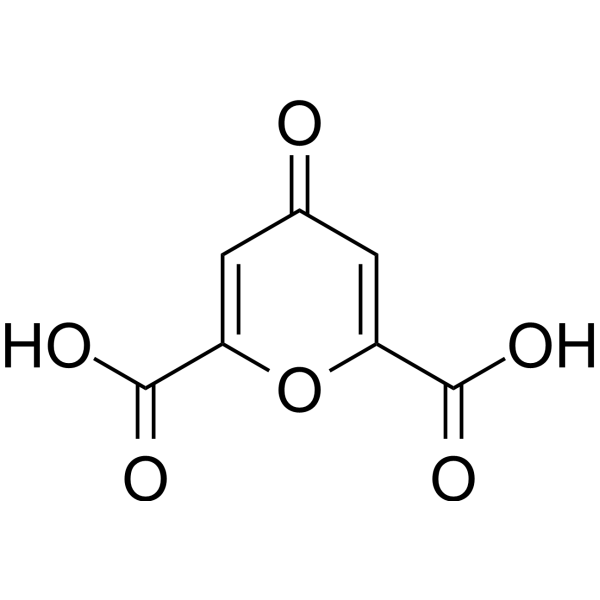
Chelidonic acid is a component of Chelidonium majus L., used as an antimicrobial. Chelidonic acid also shows anti-inflammatory activity. Chelidonic acid has potential to inhibit IL-6 production by blocking NF-κB and caspase-1. Chelidonic acid is a glutamate decarboxylase inhibitor, with a Ki of 1.2 μM.
For research use only. We do not sell to patients.
- Purity : 99.91%
- CAS No.: 99-32-1
- Formula: C7H4O6
- Molecular Weight:184.10
-
Storage:Powder -20°C, 3 years , 4°C, 2 years ; In solvent -80°C, 6 months , -20°C, 1 month

To place orders, for customer services and technical support, please contact: MedChemExpress USA
Tel: 609-228-6898 E-mail: [email protected] [email protected]


-
 Biological Activity
-
Chemical Information
-
Solvent & Solubility
- Protocols
- Purity & Documentation
- References
-
 Help & FAQs


-
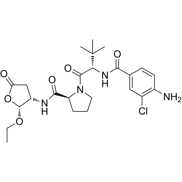
HY-13205
BelnacasanCaspase-1 Inhibitor -

HY-W041489R
Chelidonic acid (Standard) -
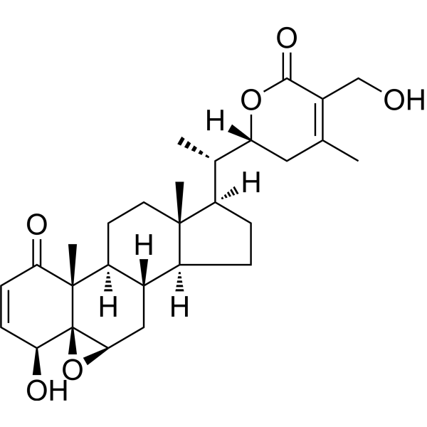
HY-N2065
Withaferin ANF-κB Inhibitor -
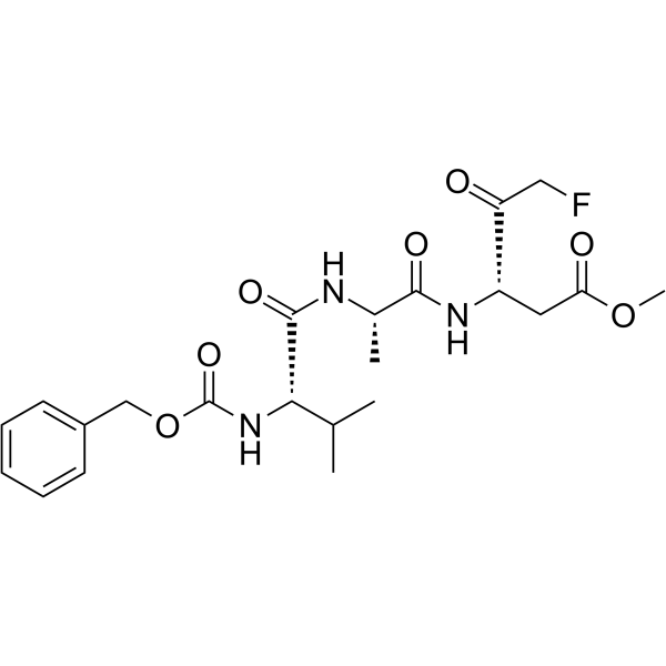
HY-16658
Z-VAD(OMe)-FMKPan-caspase Inhibitor -
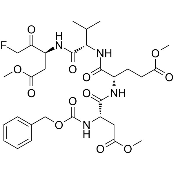
HY-12466
Z-DEVD-FMKCaspase-3 Inhibitor -
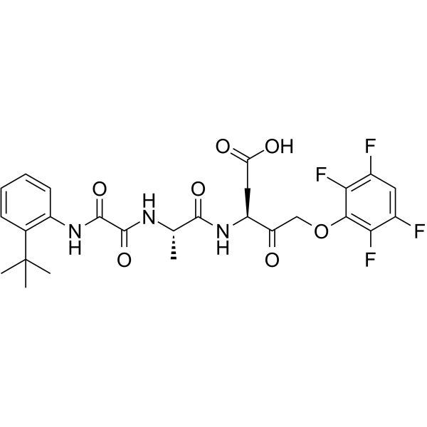
HY-10396
Emricasanpan-Caspase Inhibitor -
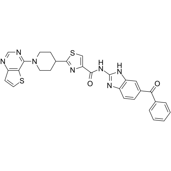
HY-10496
SC75741NF-κB Inhibitor -
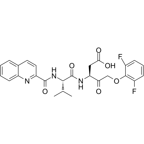
HY-12305
Q-VD-OPhPan-caspase Inhibitor -
HY-P1001
Ac-DEVD-CHOCaspase-3 Inhibitor -
HY-N1378
(E)-CardamoninhTRPA1 Antagonist -
HY-13982
JSH-23NF-κB Inhibitor -

HY-13205R
Belnacasan (Standard)Caspase-1 Inhibitor
Publications Citing Use of MedChemExpress (MCE) Chelidonic acid
MoreAll Caspase Isoforms
More

Biological Activity
Description
|
NF-κB |
Caspase-1 |
Glutamate decarboxylase 1.2 μM (Ki) |
In Vitro
Chelidonic acid dose-dependently decreases IL-6 production at 0.1-10 μM, inhibits expression of IL-6 mRNA at 1-10 μM[1].
Chelidonic acid (0.1-10 μM) decreases caspase-1 activation, nuclear NF-κB activation, and increases cytosol NF-κB activation[1].
Chelidonic acid is a glutamate decarboxylase inhibitor, with a Ki of 1.2 μM. Chelidonic acid does not promote formation of apoenzyme or react with free pyridoxal-P[2].
MedChemExpress (MCE) has not independently confirmed the accuracy of these methods. They are for reference only.
In Vivo
MedChemExpress (MCE) has not independently confirmed the accuracy of these methods. They are for reference only.


Chemical Information
-
CAS No. 99-32-1
-
Appearance Solid
-
Molecular Weight 184.10
-
Formula C7H4O6
-
Color Gray to brown
-
SMILES
O=C(C1=CC(C=C(C(O)=O)O1)=O)O
-
Structure Classification
-
Initial Source
-
Shipping
Room temperature in continental US; may vary elsewhere.
-
Storage
Powder -20°C 3 years 4°C 2 years In solvent -80°C 6 months -20°C 1 month


Publications (1)
-
Journal Impact Factor
-
Most Recent
-
Adv Healthc Mater
An Erythrocyte Membrane-Derived Nanosystem for Efficient Reversal of Endothelial Injury in Sepsis. [Abstract]2024 Jan;13(3):e2302320. PMID: 37883686


Solvent & Solubility
In Vitro:
DMSO : 10 mg/mL (54.32 mM; ultrasonic and warming and heat to 60°C; Hygroscopic DMSO has a significant impact on the solubility of product, please use newly opened DMSO)
H2O : 2.5 mg/mL (13.58 mM; Need ultrasonic)
Please refer to the solubility information to select the appropriate solvent. Once prepared, please aliquot and store the solution to prevent product inactivation from repeated freeze-thaw cycles.
Storage method and period of stock solution: -80°C, 6 months; -20°C, 1 month. When stored at -80°C, please use it within 6 months. When stored at -20°C, please use it within 1 month.
* Note: If you choose water as the stock solution, please dilute it to the working solution, then filter and sterilize it with a 0.22 μm filter before use.
Please refer to the solubility information to select the appropriate solvent. Once prepared, please aliquot and store the solution to prevent product inactivation from repeated freeze-thaw cycles.
Storage method and period of stock solution: -80°C, 6 months; -20°C, 1 month. When stored at -80°C, please use it within 6 months. When stored at -20°C, please use it within 1 month.
* Note: If you choose water as the stock solution, please dilute it to the working solution, then filter and sterilize it with a 0.22 μm filter before use.
Concentration (start) × Volume (start) = Concentration (final) × Volume (final)
In Vivo:
Select the appropriate dissolution method based on your experimental animal and administration route.
- For the following dissolution methods, please ensure to first prepare a clear stock solution using an In Vitro approach and then sequentially add co-solvents:
- To ensure reliable experimental results, the clarified stock solution can be appropriately stored based on storage conditions. As for the working solution for In Vivo experiments, it is recommended to prepare freshly and use it on the same day.
- The percentages shown for the solvents indicate their volumetric ratio in the final prepared solution. If precipitation or phase separation occurs during preparation, heat and/or sonication can be used to aid dissolution.
Add each solvent one by one: 10% DMSO 40% PEG300 5% Tween-80 45% Saline
Solubility: ≥ 2.5 mg/mL (13.58 mM); Clear solution
This protocol yields a clear solution of ≥ 2.5 mg/mL (saturation unknown).
Taking 1 mL working solution as an example, add 100 μL DMSO stock solution (25.0 mg/mL) to 400 μL PEG300, and mix evenly; then add 50 μL Tween-80 and mix evenly; then add 450 μL Saline to adjust the volume to 1 mL.
Preparation of Saline: Dissolve 0.9 g sodium chloride in ddH₂O and dilute to 100 mL to obtain a clear Saline solution.
Add each solvent one by one: 10% DMSO 90% (20% SBE-β-CD in Saline)
Solubility: ≥ 2.5 mg/mL (13.58 mM); Clear solution
This protocol yields a clear solution of ≥ 2.5 mg/mL (saturation unknown).
Taking 1 mL working solution as an example, add 100 μL DMSO stock solution (25.0 mg/mL) to 900 μL 20% SBE-β-CD in Saline, and mix evenly.
Preparation of 20% SBE-β-CD in Saline (4°C, storage for one week): 2 g SBE-β-CD powder is dissolved in 10 mL Saline, completely dissolve until clear.
For the following dissolution methods, please prepare the working solution directly:
It is recommended to prepare fresh solutions and use them promptly within a short period of time.
The percentages shown for the solvents indicate their volumetric ratio in the final prepared solution. If precipitation or phase separation occurs during preparation, heat and/or sonication can be used to aid dissolution.
Add each solvent one by one: PBS
Solubility: 2 mg/mL (10.86 mM); Clear solution; Need ultrasonic and warming and heat to 60°C
In Vivo Dissolution Calculator
Please enter the basic information of animal experiments:
-
-
-
-
Recommended: Prepare an additional quantity of animals to account for potential losses during experiments.
Please enter your animal formula composition:
-
%DMSO +
Recommended: Keep the proportion of DMSO in working solution below 2% if your animal is weak.
-
%+
-
+%Tween-80 + +
-
%Saline +
The co-solvents required include: DMSO, . All of co-solvents are available by MedChemExpress (MCE). , Tween 80. All of co-solvents are available by MedChemExpress (MCE).
Working solution concentration: 0.22 mg/mL
Method for preparing stock solution: mg drug dissolved in μL DMSO. Stock solution concentration: mg/mL.
1. Take μL DMSO stock solution;
2. Add μL .
μL , mix evenly;
3. Then add μL Tween 80, mix evenly;
4. Then add μL
Please ensure that the stock solution in the first step is dissolved to a clear state, and add co-solvents in sequence. You can use ultrasonic heating (ultrasonic cleaner, recommended frequency 20-40 kHz), vortexing, etc. to assist dissolution.


Purity & Documentation
-
Data Sheet (274 KB)
-
SDS (393 KB)
- English - EN (393 KB)
- Français - FR (393 KB)
- Deutsch - DE (393 KB)
- Norwegian - NO (393 KB)
- Español - ES (393 KB)
- Swedish - SV (393 KB)
- Italian - IT (393 KB)
- Korean - KR (393 KB)
- Portuguese - PT (393 KB)
-
Handling Instructions (2659 KB)
References
[1]. Shin HJ, et al. Inhibitory effects of chelidonic acid on IL-6 production by blocking NF-κB and caspase-1 in HMC-1 cells. Immunopharmacol Immunotoxicol. 2011 Dec;33(4):614-9. [Content Brief]
[2]. Porter TG, et al. Chelidonic acid and other conformationally restricted substrate analogues as inhibitors of rat brain glutamate decarboxylase. Biochem Pharmacol. 1985 Dec 1;34(23):4145-50. [Content Brief]
[3]. Oh HA, et al. Beneficial effects of chelidonic acid on a model of allergic rhinitis. Send to Int Immunopharmacol. 2011 Jan;11(1):39-45. [Content Brief]


Complete Stock Solution Preparation Table
Please refer to the solubility information to select the appropriate solvent. Once prepared, please aliquot and store the solution to prevent product inactivation from repeated freeze-thaw cycles.
Storage method and period of stock solution: -80°C, 6 months; -20°C, 1 month. When stored at -80°C, please use it within 6 months. When stored at -20°C, please use it within 1 month.
| Optional Solvent | Concentration Solvent Mass | 1 mg | 5 mg | 10 mg | 25 mg |
|---|---|---|---|---|---|
| H2O / DMSO | 1 mM | 5.4318 mL | 27.1592 mL | 54.3183 mL | 135.7958 mL |
| 5 mM | 1.0864 mL | 5.4318 mL | 10.8637 mL | 27.1592 mL | |
| 10 mM | 0.5432 mL | 2.7159 mL | 5.4318 mL | 13.5796 mL | |
| DMSO | 15 mM | 0.3621 mL | 1.8106 mL | 3.6212 mL | 9.0531 mL |
| 20 mM | 0.2716 mL | 1.3580 mL | 2.7159 mL | 6.7898 mL | |
| 25 mM | 0.2173 mL | 1.0864 mL | 2.1727 mL | 5.4318 mL | |
| 30 mM | 0.1811 mL | 0.9053 mL | 1.8106 mL | 4.5265 mL | |
| 40 mM | 0.1358 mL | 0.6790 mL | 1.3580 mL | 3.3949 mL | |
| 50 mM | 0.1086 mL | 0.5432 mL | 1.0864 mL | 2.7159 mL |
* Note: If you choose water as the stock solution, please dilute it to the working solution, then filter and sterilize it with a 0.22 μm filter before use.


Chelidonic acid Related Classifications
HY-W041489 Related Classifications
 Powered by Bioz