



Avotaciclib
Based on 4 publication(s) in Google Scholar
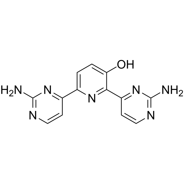
Avotaciclib (BEY1107) is an orally active cyclin-dependent kinase 1 (CDK1) inhibitor. Avotaciclib can inhibit the proliferation and induce apoptosis of tumor cells. Avotaciclib can be used in the research of cancer such as pancreatic cancer and lung cancer.
商品は「研究用試薬」です。人や動物の医療用・臨床診断用・食品用の製品ではありません。
研究用途以外に使用した場合、当社は一切の責任を負いかねます。
- 純度 : 99.72%
- CAS 番号: 1983983-41-0
- 分子式: C13H11N7O
- 分子量:281.27
-
保管条件:
-20°C, protect from light, stored under nitrogen
* In solvent : -80°C, 6 months; -20°C, 1 month (protect from light, stored under nitrogen)

To place orders, for customer services and technical support, please contact: MedChemExpress USA
Tel: 609-228-6898 E-mail: [email protected] [email protected]


-
 生物活性
生物活性
-
化学情報
-
溶剤 & 溶解度
- プロトコル
- 純度とドキュメンテーション
- 参考文献
-
 Help & FAQs


MedChemExpress(MCE)の使用を引用している文献 Avotaciclib
More

生物活性
製品説明
IC50 & Target
[1]|
CDK1 |
体外実験
Avotaciclib (0-64 μM; 48 h) can inhibit cell proliferation and promote apoptosis in radiotherapy-resistant non-small cell lung cancer cell lines, and the effect is better when combined with PV-1019 (HY-125203)[2].
MedChemExpress (MCE) has not independently confirmed the accuracy of these methods. They are for reference only. Further protocols information, click here.
-
Cell Line:Non-small cell lung cancer cells (H1437R, H1568R, H1703R and H1869R)
-
Concentration:0, 0.1, 1, 2, 4, 8, 16, 32 and 64 μM
-
Incubation Time:48 h
-
Result:Inhibited the cell viability, with EC50 values of 0.918, 0.580, 0.735 and 0.662 μM for H1437R, H1568R, H1703R and H1869R, respectively.
臨床実験
| NCT Number | Sponsor | Condition | Start Date |
Phase

|
|---|---|---|---|---|
| NCT01329991 | Plexxikon| | 2011-05 | PHASE1 |


化学情報
-
CAS 番号 1983983-41-0
-
性状 Solid
-
分子量 281.27
-
分子式 C13H11N7O
-
Color Light yellow to yellow
-
SMILES
OC1=CC=C(C2=NC(N)=NC=C2)N=C1C3=NC(N)=NC=C3
-
別名
BEY1107
-
輸送条件
Room temperature in continental US; may vary elsewhere.
-
保管条件
-20°C, protect from light, stored under nitrogen
* In solvent : -80°C, 6 months; -20°C, 1 month (protect from light, stored under nitrogen)


Publications (4)
-
Journal Impact Factor
-
Most Recent
-
Clin Sci
Glutamine alleviates immunosuppression in polymicrobial sepsis by augmenting bacterial phagocytosis through sustaining the GFAT-DRP1 dependent mitochondrial calcium dynamics. [Abstract]2025 Oct 22;139(20):1163-1185. PMID: 41082631 -
Cancer Cell Int
Proapoptotic role of CDK1 in overcoming paclitaxel resistance in ovarian cancer cells in response to combined treatment with paclitaxel and duloxetine. [Abstract]2024 Dec 19;24(1):409. PMID: 39702300 -
-
Histol Histopathol
Targeting of CDK1 and PRMT5 as a potential therapeutic combination for non-small cell lung cancer. [Abstract]2026 Jul;41(7):1275-1288. PMID: 41487038


溶剤 & 溶解度
体外:
DMSO : 100 mg/mL (355.53 mM; Need ultrasonic; Hygroscopic DMSO has a significant impact on the solubility of product, please use newly opened DMSO)
Please refer to the solubility information to select the appropriate solvent. Once prepared, please aliquot and store the solution to prevent product inactivation from repeated freeze-thaw cycles.
Storage method and period of stock solution: -80°C, 6 months; -20°C, 1 month (protect from light, stored under nitrogen). When stored at -80°C, please use it within 6 months. When stored at -20°C, please use it within 1 month.
Please refer to the solubility information to select the appropriate solvent. Once prepared, please aliquot and store the solution to prevent product inactivation from repeated freeze-thaw cycles.
Storage method and period of stock solution: -80°C, 6 months; -20°C, 1 month (protect from light, stored under nitrogen). When stored at -80°C, please use it within 6 months. When stored at -20°C, please use it within 1 month.
濃度 (開始) × 体積 (開始) = 濃度 (終了) × 体積 (終了)

プロトコル
-
Apoptosis
Apoptosis, also called programmed cell death, is generally characterized by distinct morphological characteristics.
-
TUNEL staining for apoptotic DNA fragmentation
TUNEL staining detects DNA strand breaks by using terminal deoxynucleotidyl transferase to add labeled nucleotides to exposed 3′-OH DNA termini, generating either microscopic staining in fixed cells or tissue sections, or fluorescence/cytometric signal in cell suspensions. TUNEL positivity reflects DNA fragmentation but should not be interpreted alone as definitive apoptosis, because TUNEL can also label necrotic, autolytic, mechanically damaged, or DNA-repair-associated DNA breaks.
-
Annexin V plus membrane-impermeant dye apoptosis staining
Annexin V-based apoptosis assays rely on the detection of phosphatidylserine (PS) externalization from the inner leaflet of the plasma membrane to the outer leaflet, an early biochemical hallmark of apoptosis. Fluorescently labeled Annexin V binds PS in a calcium-dependent manner, enabling identification of early apoptotic cells by flow cytometry or fluorescence microscopy. When combined with a membrane-impermeant DNA-binding dye (e. g. , propidium iodide), this approach allows discrimination between viable (Annexin V−/dye−), early apoptotic (Annexin V+/dye−), and late apoptotic or necrotic (Annexin V+/dye+) cell populations by assessing membrane integrity and PS exposure.
-
Apoptosis Solutions
Apoptosis is a regulated, generally non-lytic cell-death pathway that removes unwanted, damaged, infected, or abnormal cells through coordinated morphological changes, caspase activation, DNA fragmentation, and membrane remodeling. The intrinsic apoptosis pathway is controlled mainly by mitochondrial outer membrane permeabilization, BCL-2 family proteins, cytochrome c release, apoptosome formation, caspase-9 activation, and downstream executioner caspase-3/7 activation. The extrinsic apoptosis pathway is initiated by death receptors such as Fas, TNFR, and TRAIL receptors, which recruit adaptor proteins and activate caspase-8 before engaging executioner caspases or mitochondrial amplification through BID cleavage. Apoptosis is linked to many phenotypes, including cancer cell killing, tissue homeostasis, immune regulation, neurodegeneration, infection response, and treatment-induced cytotoxicity; unresolved questions include how apoptosis interacts with necroptosis, pyroptosis, ferroptos
-
Cell Viability Determination by MTT Colorimetric Assay
The following protocol uses the MTT colorimetric assay as a classic literature-established method for assessing cell viability/metabolic activity in cultured mammalian cells. MTT[3-(4,5-dimethylthiazol-2-yl)-2,5-diphenyltetrazolium bromide] is reduced by metabolically active cells to a colored formazan product; the amount of formazan is quantified spectrophotometrically and provides an indirect measure of metabolically active viable cells. Importantly, MTT reduction reflects cellular oxidoreductase/metabolic activity rather than an absolute direct count of living cells, so changes in cellular metabolism can alter the signal independently of cell number.


純度とドキュメンテーション
-
データシート (272 KB)
-
SDS (252 KB)
- English - EN (252 KB)
- Français - FR (252 KB)
- Deutsch - DE (252 KB)
- Norwegian - NO (252 KB)
- Español - ES (252 KB)
- Swedish - SV (252 KB)
- Italian - IT (252 KB)
- Korean - KR (252 KB)
- Portuguese - PT (252 KB)
-
取扱説明書 (2659 KB)
参考文献


Complete Stock Solution Preparation Table
Please refer to the solubility information to select the appropriate solvent. Once prepared, please aliquot and store the solution to prevent product inactivation from repeated freeze-thaw cycles.
Storage method and period of stock solution: -80°C, 6 months; -20°C, 1 month (protect from light, stored under nitrogen). When stored at -80°C, please use it within 6 months. When stored at -20°C, please use it within 1 month.
| Optional Solvent | Concentration Solvent Mass | 1 mg | 5 mg | 10 mg | 25 mg |
|---|---|---|---|---|---|
| DMSO | 1 mM | 3.5553 mL | 17.7765 mL | 35.5530 mL | 88.8826 mL |
| 5 mM | 0.7111 mL | 3.5553 mL | 7.1106 mL | 17.7765 mL | |
| 10 mM | 0.3555 mL | 1.7777 mL | 3.5553 mL | 8.8883 mL | |
| 15 mM | 0.2370 mL | 1.1851 mL | 2.3702 mL | 5.9255 mL | |
| 20 mM | 0.1778 mL | 0.8888 mL | 1.7777 mL | 4.4441 mL | |
| 25 mM | 0.1422 mL | 0.7111 mL | 1.4221 mL | 3.5553 mL | |
| 30 mM | 0.1185 mL | 0.5926 mL | 1.1851 mL | 2.9628 mL | |
| 40 mM | 0.0889 mL | 0.4444 mL | 0.8888 mL | 2.2221 mL | |
| 50 mM | 0.0711 mL | 0.3555 mL | 0.7111 mL | 1.7777 mL | |
| 60 mM | 0.0593 mL | 0.2963 mL | 0.5926 mL | 1.4814 mL | |
| 80 mM | 0.0444 mL | 0.2222 mL | 0.4444 mL | 1.1110 mL | |
| 100 mM | 0.0356 mL | 0.1778 mL | 0.3555 mL | 0.8888 mL |


Avotaciclib 関連分類
HY-137432 関連分類
 Powered by Bioz