



Brophenexin
Based on 3 publication(s) in Google Scholar
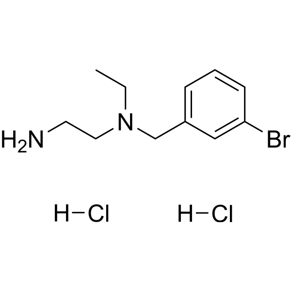
Brophenexin (compound 8) is a potent NMDAR/TRPM4 interaction interface inhibitor. Brophenexin shows neuroprotective activity. Brophenexin prevents NMDA-induced cell death and mitochondrial dysfunction in hippocampal neurons, with an IC50 of 2.1 μM. Brophenexin protects mice from MCAO-induced brain damage and NMDA-induced retinal ganglion cell loss.
For research use only. We do not sell to patients.
- Purity : 98.74%
- CAS No.: 2243506-33-2
- Formula: C11H19BrCl2N2
- Molecular Weight:330.09
-
Storage:
4°C, sealed storage, away from moisture and light
* In solvent : -80°C, 6 months; -20°C, 1 month (sealed storage, away from moisture and light)

To place orders, for customer services and technical support, please contact: MedChemExpress USA
Tel: 609-228-6898 E-mail: [email protected] [email protected]


-
 Biological Activity
-
Chemical Information
-
Solvent & Solubility
- Protocols
- Purity & Documentation
- References
-
 Help & FAQs


-
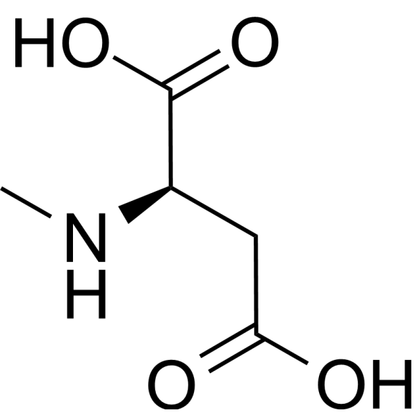
HY-17551
NMDAEndogenous Metabolite -
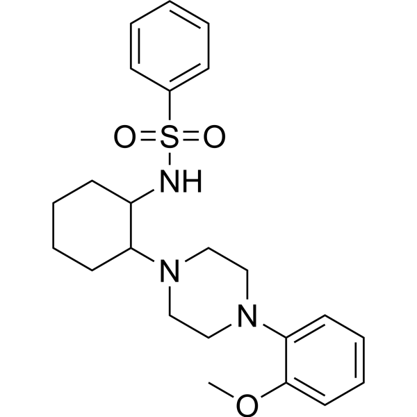
HY-139426
ML-SI3TRPML Modulator -
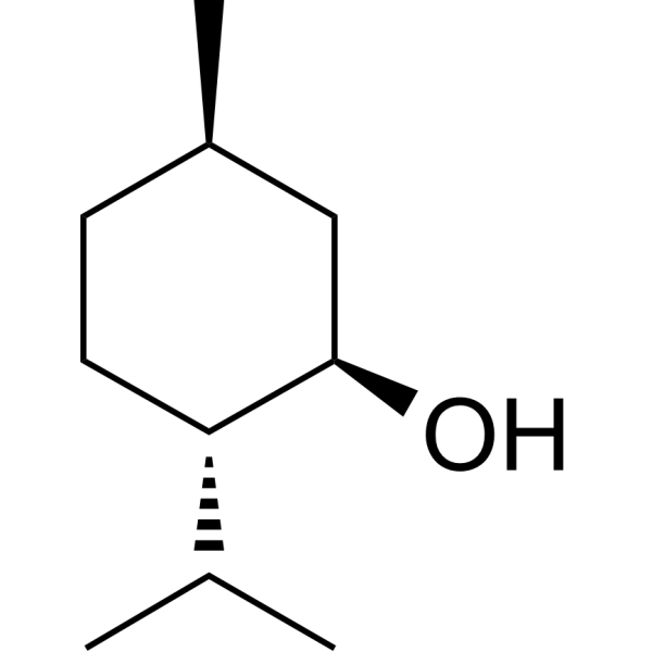
HY-75161
(-)-MentholEndogenous Metabolite -
HY-100973A
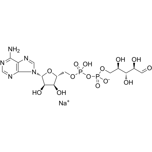
Adenosine 5′-diphosphoribose sodiumTRPM2 Activator -
HY-B0151S2
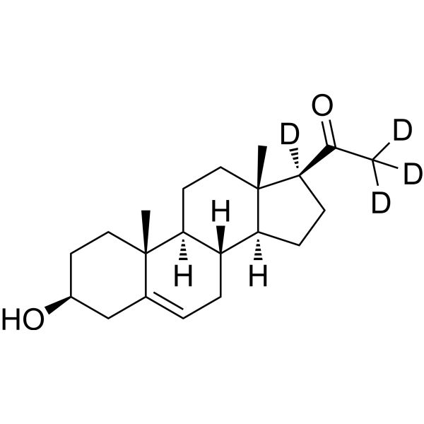
Pregnenolone-d4-1Stable Isotope -
HY-107535
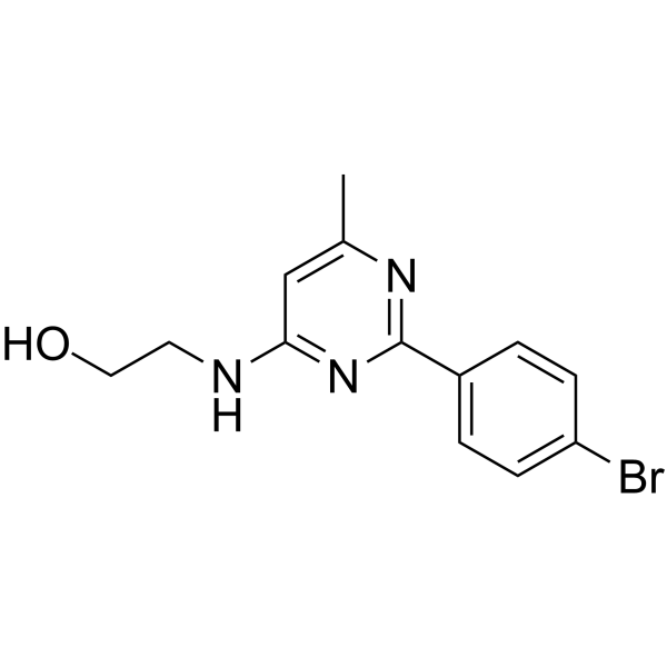
AS1269574GPR119 Agonist -

HY-75161R
(-)-Menthol (Standard)Endogenous Metabolite -
HY-122051
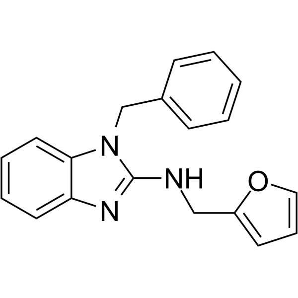
AC1903TRPC5 Inhibitor -
HY-B0285B
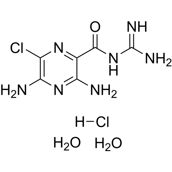
Amiloride hydrochloride dihydrateENaC/uTPA Inhibitor -
HY-108461
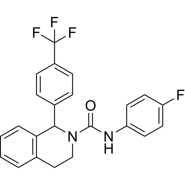
TRPM8-IN-1TRPM8 Inhibitor -
HY-121539
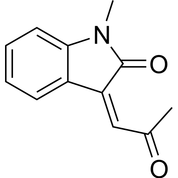
SupercinnamaldehydeTRPA1 Activator -

HY-100973AR
Adenosine 5′-diphosphoribose sodium (Standard)TRPM2 Activator
Publications Citing Use of MedChemExpress (MCE) Brophenexin
MoreAll iGluR Isoforms
More

Biological Activity
Description
In Vitro
NMDAR/TRPM4-IN-2 (compound 8) (0-10 μM) reduces the interactions of GluN2A and GluN2B with TRPM4 in a dose-dependent manner[1].
NMDAR/TRPM4-IN-2 eliminates the CREB shutoff pathway and restores ERK1/2 activation and IEG induction while sparing the synaptic activity-driven, transcription-promoting activities of NMDARs[1].
MedChemExpress (MCE) has not independently confirmed the accuracy of these methods. They are for reference only. Further protocols information, click here.


Chemical Information
-
CAS No. 2243506-33-2
-
Appearance Solid
-
Molecular Weight 330.09
-
Formula C11H19BrCl2N2
-
Color White to yellow
-
SMILES
NCCN(CC1=CC=CC(Br)=C1)CC.[H]Cl.[H]Cl
-
Synonyms
NMDAR/TRPM4-IN-2
-
Shipping
Room temperature in continental US; may vary elsewhere.
-
Storage
4°C, sealed storage, away from moisture and light
* In solvent : -80°C, 6 months; -20°C, 1 month (sealed storage, away from moisture and light)


Publications (3)
-
Journal Impact Factor
-
Most Recent
-
-
Pharmacol Res Perspect
NMDA Receptor-Mediated Ca2+ Flux Attenuated by the NMDA Receptor/TRPM4 Interface Inhibitor Brophenexin. [Abstract]2024 Dec;12(6):e70038. PMID: 39574295 -


Solvent & Solubility
In Vitro:
DMSO : 250 mg/mL (757.37 mM; Need ultrasonic; Hygroscopic DMSO has a significant impact on the solubility of product, please use newly opened DMSO)
H2O : 100 mg/mL (302.95 mM; Need ultrasonic)
Please refer to the solubility information to select the appropriate solvent. Once prepared, please aliquot and store the solution to prevent product inactivation from repeated freeze-thaw cycles.
Storage method and period of stock solution: -80°C, 6 months; -20°C, 1 month (sealed storage, away from moisture and light). When stored at -80°C, please use it within 6 months. When stored at -20°C, please use it within 1 month.
* Note: If you choose water as the stock solution, please dilute it to the working solution, then filter and sterilize it with a 0.22 μm filter before use.
Please refer to the solubility information to select the appropriate solvent. Once prepared, please aliquot and store the solution to prevent product inactivation from repeated freeze-thaw cycles.
Storage method and period of stock solution: -80°C, 6 months; -20°C, 1 month (sealed storage, away from moisture and light). When stored at -80°C, please use it within 6 months. When stored at -20°C, please use it within 1 month.
* Note: If you choose water as the stock solution, please dilute it to the working solution, then filter and sterilize it with a 0.22 μm filter before use.
Concentration (start) × Volume (start) = Concentration (final) × Volume (final)
In Vivo:
Select the appropriate dissolution method based on your experimental animal and administration route.
- For the following dissolution methods, please ensure to first prepare a clear stock solution using an In Vitro approach and then sequentially add co-solvents:
- To ensure reliable experimental results, the clarified stock solution can be appropriately stored based on storage conditions. As for the working solution for In Vivo experiments, it is recommended to prepare freshly and use it on the same day.
- The percentages shown for the solvents indicate their volumetric ratio in the final prepared solution. If precipitation or phase separation occurs during preparation, heat and/or sonication can be used to aid dissolution.
Add each solvent one by one: 10% DMSO 40% PEG300 5% Tween-80 45% Saline
Solubility: ≥ 2.08 mg/mL (6.30 mM); Clear solution
This protocol yields a clear solution of ≥ 2.08 mg/mL (saturation unknown).
Taking 1 mL working solution as an example, add 100 μL DMSO stock solution (20.8 mg/mL) to 400 μL PEG300, and mix evenly; then add 50 μL Tween-80 and mix evenly; then add 450 μL Saline to adjust the volume to 1 mL.
Preparation of Saline: Dissolve 0.9 g sodium chloride in ddH₂O and dilute to 100 mL to obtain a clear Saline solution.
Add each solvent one by one: 10% DMSO 90% (20% SBE-β-CD in Saline)
Solubility: ≥ 2.08 mg/mL (6.30 mM); Clear solution
This protocol yields a clear solution of ≥ 2.08 mg/mL (saturation unknown).
Taking 1 mL working solution as an example, add 100 μL DMSO stock solution (20.8 mg/mL) to 900 μL 20% SBE-β-CD in Saline, and mix evenly.
Preparation of 20% SBE-β-CD in Saline (4°C, storage for one week): 2 g SBE-β-CD powder is dissolved in 10 mL Saline, completely dissolve until clear.
In Vivo Dissolution Calculator
Please enter the basic information of animal experiments:
-
-
-
-
Recommended: Prepare an additional quantity of animals to account for potential losses during experiments.
Working solution concentration: 0.22 mg/mL
This product has good water solubility, please refer to the measured solubility data in water/PBS/Saline for details.

Protocols
-
Cell Cytotoxicity Assay
Cytotoxicity assays are usually based on the assessment of cell membrane damage, which can also be indirectly detected by measuring cell viability. Detection methods include MTT assay, CKK-8 assay, LDH assay and ATP assay, etc.
-
Mammalian live/dead viability and cytotoxicity staining
Live/dead viability and cytotoxicity staining assays are based on the simultaneous detection of intracellular esterase activity in metabolically active (viable) cells and membrane integrity loss in non-viable cells. In commonly used dual-staining approaches, membrane-permeant fluorogenic substrates are converted by intracellular esterases into fluorescent products in live cells, while impermeant DNA-binding dyes selectively enter cells with compromised plasma membranes and label nucleic acids in dead or dying cells, enabling discrimination between viable and non-viable populations by fluorescence microscopy or flow cytometry.
-
Mitochondrial membrane-potential and mitochondrial mass staining
Mitochondrial membrane potential staining measures the electrochemical polarization across the mitochondrial inner membrane in live cells using lipophilic cationic fluorescent probes; early rhodamine-based work showed that selective mitochondrial dye accumulation is lost when the mitochondrial transmembrane potential is dissipated. JC-1 reports mitochondrial polarization by shifting from green monomer fluorescence to red J-aggregate fluorescence as dye concentration increases within energized mitochondria; therefore, the red/green fluorescence ratio is used as a relative readout of mitochondrial membrane potential. TMRE or TMRM staining provides a single-channel relative readout because these cationic rhodamine esters accumulate in polarized mitochondria, and lower fluorescence indicates reduced mitochondrial polarization when acquisition and dye-loading conditions are controlled. Mitochondrial mass staining is commonly performed with MitoTracker Green FM or related MitoTracker dyes as
-
Apoptosis Solutions
Apoptosis is a regulated, generally non-lytic cell-death pathway that removes unwanted, damaged, infected, or abnormal cells through coordinated morphological changes, caspase activation, DNA fragmentation, and membrane remodeling. The intrinsic apoptosis pathway is controlled mainly by mitochondrial outer membrane permeabilization, BCL-2 family proteins, cytochrome c release, apoptosome formation, caspase-9 activation, and downstream executioner caspase-3/7 activation. The extrinsic apoptosis pathway is initiated by death receptors such as Fas, TNFR, and TRAIL receptors, which recruit adaptor proteins and activate caspase-8 before engaging executioner caspases or mitochondrial amplification through BID cleavage. Apoptosis is linked to many phenotypes, including cancer cell killing, tissue homeostasis, immune regulation, neurodegeneration, infection response, and treatment-induced cytotoxicity; unresolved questions include how apoptosis interacts with necroptosis, pyroptosis, ferroptos
-
Primary Embryonic Hippocampal Neuron Culture
Primary embryonic hippocampal neuron culture is an in vitro method in which hippocampi from embryonic rodents are dissected, enzymatically or mechanically dissociated, plated on adhesive substrates, and maintained in defined neuronal medium or in low-density sandwich/co-culture formats to support neuronal attachment, neurite extension, polarity formation, dendritic arborization, and synapse formation. The main readouts are cell survival, neuronal purity, neurite outgrowth, axon-dendrite polarization, synaptic marker development, and functional neuronal activity, assessed by phase-contrast microscopy, immunocytochemistry for neuronal/glial markers, live imaging, or electrophysiology depending on the downstream experiment.


Purity & Documentation
-
Data Sheet (271 KB)
-
SDS (393 KB)
- English - EN (393 KB)
- Français - FR (393 KB)
- Deutsch - DE (393 KB)
- Norwegian - NO (393 KB)
- Español - ES (393 KB)
- Swedish - SV (393 KB)
- Italian - IT (393 KB)
- Korean - KR (393 KB)
- Portuguese - PT (393 KB)
-
Handling Instructions (2659 KB)
References


Complete Stock Solution Preparation Table
Please refer to the solubility information to select the appropriate solvent. Once prepared, please aliquot and store the solution to prevent product inactivation from repeated freeze-thaw cycles.
Storage method and period of stock solution: -80°C, 6 months; -20°C, 1 month (sealed storage, away from moisture and light). When stored at -80°C, please use it within 6 months. When stored at -20°C, please use it within 1 month.
| Optional Solvent | Concentration Solvent Mass | 1 mg | 5 mg | 10 mg | 25 mg |
|---|---|---|---|---|---|
| H2O / DMSO | 1 mM | 3.0295 mL | 15.1474 mL | 30.2948 mL | 75.7369 mL |
| 5 mM | 0.6059 mL | 3.0295 mL | 6.0590 mL | 15.1474 mL | |
| 10 mM | 0.3029 mL | 1.5147 mL | 3.0295 mL | 7.5737 mL | |
| 15 mM | 0.2020 mL | 1.0098 mL | 2.0197 mL | 5.0491 mL | |
| 20 mM | 0.1515 mL | 0.7574 mL | 1.5147 mL | 3.7868 mL | |
| 25 mM | 0.1212 mL | 0.6059 mL | 1.2118 mL | 3.0295 mL | |
| 30 mM | 0.1010 mL | 0.5049 mL | 1.0098 mL | 2.5246 mL | |
| 40 mM | 0.0757 mL | 0.3787 mL | 0.7574 mL | 1.8934 mL | |
| 50 mM | 0.0606 mL | 0.3029 mL | 0.6059 mL | 1.5147 mL | |
| 60 mM | 0.0505 mL | 0.2525 mL | 0.5049 mL | 1.2623 mL | |
| 80 mM | 0.0379 mL | 0.1893 mL | 0.3787 mL | 0.9467 mL | |
| 100 mM | 0.0303 mL | 0.1515 mL | 0.3029 mL | 0.7574 mL |
* Note: If you choose water as the stock solution, please dilute it to the working solution, then filter and sterilize it with a 0.22 μm filter before use.


Brophenexin Related Classifications
HY-139192 Related Classifications
 Powered by Bioz