



Pacritinib
Based on 20 publication(s) in Google Scholar
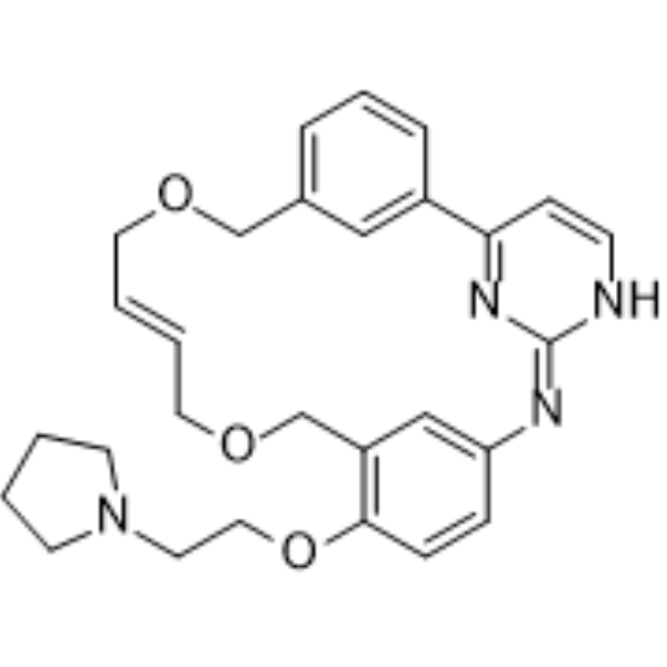
Pacritinib (SB1518) is an orally active, selective JAK2/FLT3 inhibitor with IC50 values of 23 nM and 22 nM, respectively. Pacritinib inhibits downstream STAT5, ERK1/2, and AKT signaling, and induces Apoptosis and G1 phase arrest. Pacritinib normalizes tumor-induced cytokines/chemokines, and reduces metastasis, tumor growth, shrinkage, migration, liver injury, inflammation, and steatosis. Pacritinib can be used in research on acute myeloid leukemia, myeloproliferative neoplasms, leukemia/lymphoma, GVHD, and alcoholic/non-alcoholic liver fibrosis.
For research use only. We do not sell to patients.
- Purity : 99.88%
- CAS No.: 937272-79-2
- Formula: C28H32N4O3
- Molecular Weight:472.58
-
Storage:Powder -20°C, 3 years , 4°C, 2 years ; In solvent -80°C, 2 years , -20°C, 1 year

To place orders, for customer services and technical support, please contact: MedChemExpress USA
Tel: 609-228-6898 E-mail: [email protected] [email protected]


-
 Biological Activity
-
Chemical Information
-
Solvent & Solubility
- Protocols
- Purity & Documentation
- References
-
 Help & FAQs


-
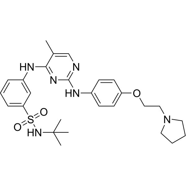
HY-10409
FedratinibJAK2 Inhibitor -
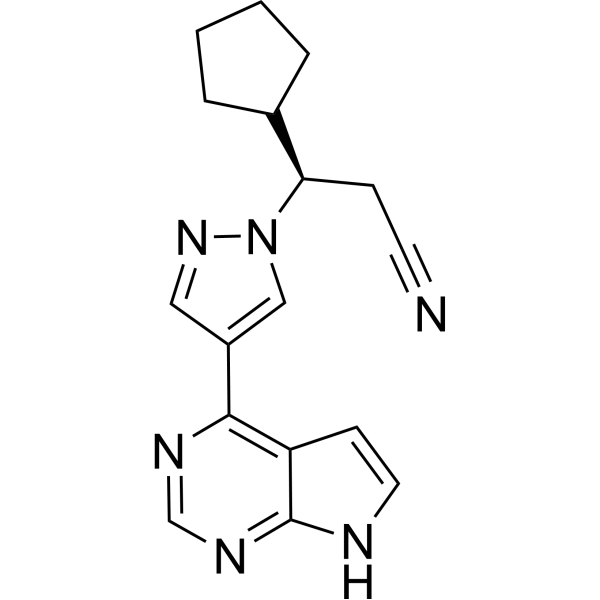
HY-50856
RuxolitinibJAK1/2 Inhibitor -
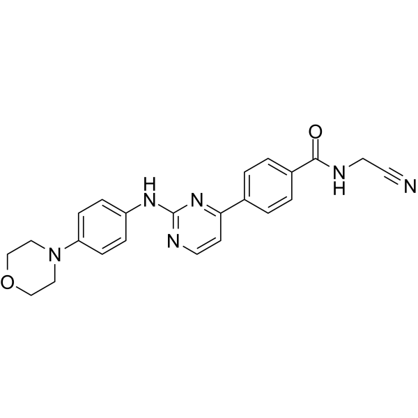
HY-10961
MomelotinibJAK1/2 Inhibitor -
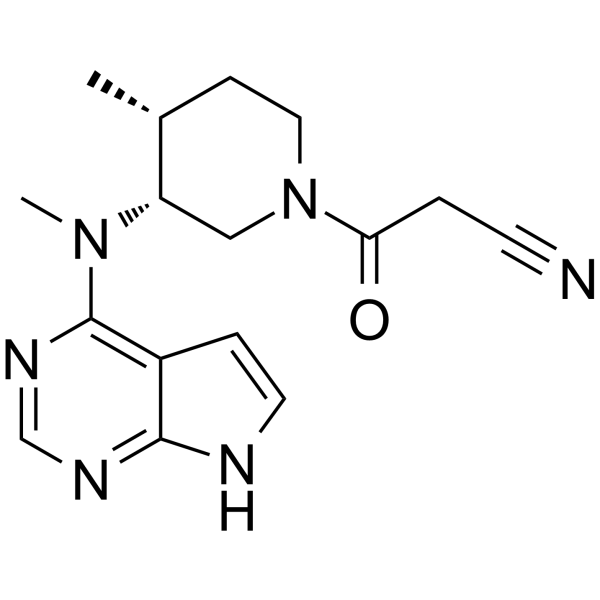
HY-40354
TofacitinibJAK1/JAK3 Inhibitor -
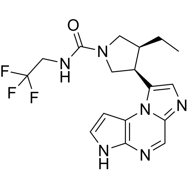
HY-19569
UpadacitinibJAK1 Inhibitor -
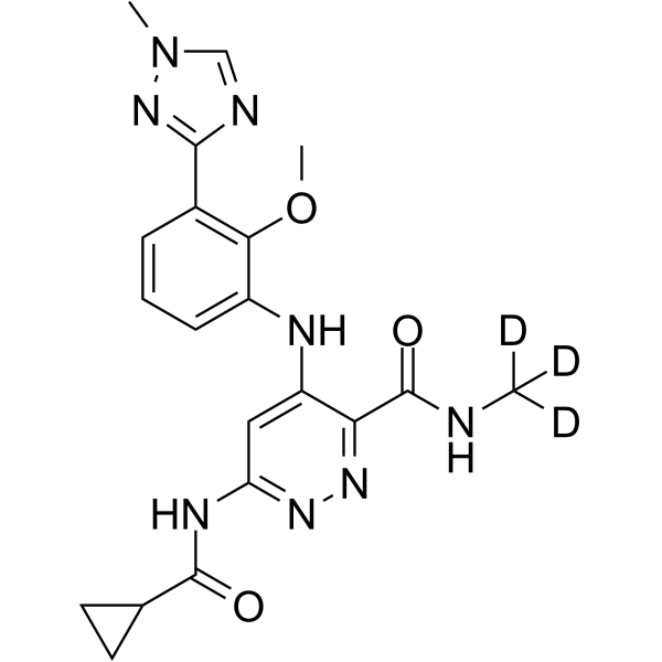
HY-117287
DeucravacitinibTYK2 Inhibitor -
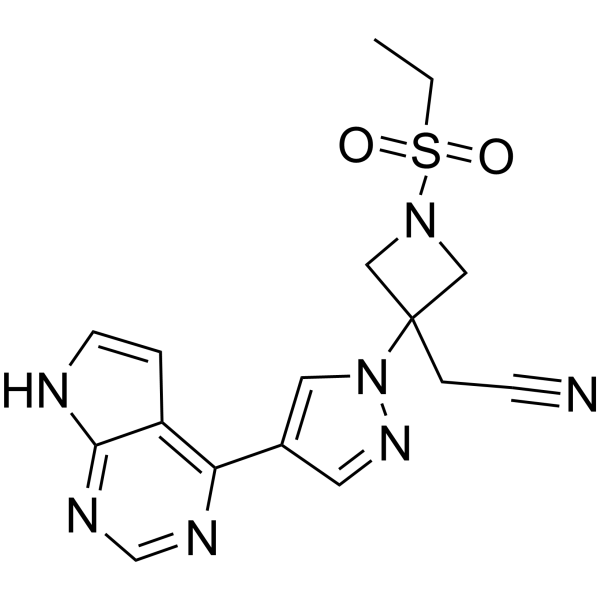
HY-15315
BaricitinibJAK1/2 Inhibitor -
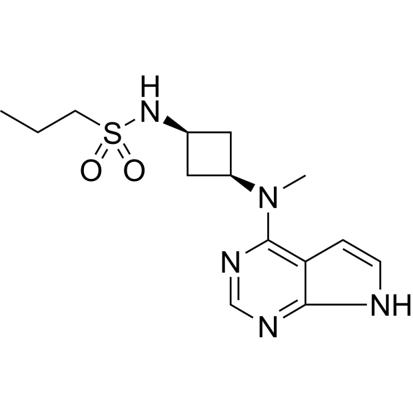
HY-107429
AbrocitinibJAK1 Inhibitor -
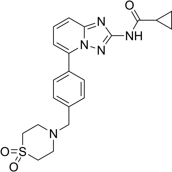
HY-18300
FilgotinibJAK1 Inhibitor -
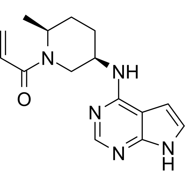
HY-100754
RitlecitinibJAK3 Inhibitor -
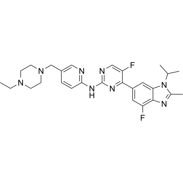
HY-16297A
AbemaciclibCDK4/6 Inhibitor -
HY-13818
StatticSTAT3 Inhibitor
Publications Citing Use of MedChemExpress (MCE) Pacritinib
More- Nat Med. 2017 Nov;23(11):1319-1330. [Abstract]
- Science. 2026 Feb 5;391(6785):eadz4075. [Abstract]
- Science. 2017 Dec 1;358(6367):eaan4368. [Abstract]
- Nat Commun. 2021 Aug 13;12(1):4917. [Abstract]
- Nat Commun. 2021 Jul 21;12(1):4441. [Abstract]
- Sci Transl Med. 2018 Jul 18;10(450):eaaq1093. [Abstract]
- Cell Death Dis. 2021 Dec 20;13(1):16. [Abstract]
- Mol Cancer Ther. 2026 Jul 14.
- Mol Syst Biol. 2024 Jan;20(1):28-55. [Abstract]
- Inflamm Res. 2020 Jan;69(1):51-62. [Abstract]
- Drug Des Devel Ther. 2018 Apr 30:12:1009-1017. [Abstract]
- Eur J Pharmacol. 2025 Sep 15:1003:177942. [Abstract]
- PLoS Pathog. 2025 Jun 23;21(6):e1013281. [Abstract]
- PLoS Genet. 2024 May 28;20(5):e1011293. [Abstract]
- BMJ Open Ophthalmol. 2026 Jan 12;11(1):e002307. [Abstract]
- Fundam Clin Pharmacol. 2021 Oct;35(5):919-929. [Abstract]
- Eur J Drug Metab Pharmacokinet. 2021 Sep;46(5):625-635. [Abstract]
- bioRxiv. 2025 June 03.
- bioRxiv. 2025 February 21.
- Patent. US20220323444A1.
 Customer Validation & Images

-
WB
-
RT-PCR
-
In Vivo Efficacy Study
-
Cell Proliferation/Viability Assay
-
WB


Biological Activity
Description
IC50 & Target
[1]|
JAK2 23 nM (IC50) |
FLT3 22 nM (IC50) |
STAT5 |
ERK1 |
ERK2 |
Cellular Effect
|
Cell Line

|
Type | Value | Description | References |
|---|---|---|---|---|
| A2780 | IC50 |
0.664 μM
Compound: Chemical Probe: SB1518
|
Antiproliferative activity against human A2780 cells assessed as inhibition of cell viability incubated for 48 hrs by CellTiterGlo assay
Antiproliferative activity against human A2780 cells assessed as inhibition of cell viability incubated for 48 hrs by CellTiterGlo assay
|
[PMID: 21691275] |
| A549 | IC50 |
2.547 μM
Compound: Chemical Probe: SB1518
|
Antiproliferative activity against human A549 cells assessed as inhibition of cell viability incubated for 48 hrs by CellTiterGlo assay
Antiproliferative activity against human A549 cells assessed as inhibition of cell viability incubated for 48 hrs by CellTiterGlo assay
|
[PMID: 21691275] |
| BaF3 | GI50 |
<434 nM
Compound: Pacritinib
|
Growth inhibition of mouse BaF3 cells transfected in FLT3-TDK mutant measured after 72 hrs by CCK8 assay
Growth inhibition of mouse BaF3 cells transfected in FLT3-TDK mutant measured after 72 hrs by CCK8 assay
|
[PMID: 35923716] |
| BaF3 | GI50 |
0.16 μM
Compound: 21c, SB1518, 85:15 trans/cis mixture
|
Antiproliferative activity against mouse BA/F3 cells harboring JAK2 V617F mutation assessed as cell viability after 48 hrs by Cell titer Glo assay
Antiproliferative activity against mouse BA/F3 cells harboring JAK2 V617F mutation assessed as cell viability after 48 hrs by Cell titer Glo assay
|
[PMID: 21604762] |
| BaF3 | GI50 |
0.16 μM
Compound: 5; SB1518,VONJO
|
Growth inhibition of mouse BaF3 cells harboring JAK2 V617F mutant assessed as reduction in cell viability incubated for 72 hrs by MTT assay
Growth inhibition of mouse BaF3 cells harboring JAK2 V617F mutant assessed as reduction in cell viability incubated for 72 hrs by MTT assay
|
[PMID: 38389874] |
| BaF3 | GI50 |
133 nM
Compound: Pacritinib
|
Growth inhibition of mouse BaF3 cells transfected in FLT3-ITD mutant measured after 72 hrs by CCK8 assay
Growth inhibition of mouse BaF3 cells transfected in FLT3-ITD mutant measured after 72 hrs by CCK8 assay
|
[PMID: 35923716] |
| BaF3 | IC50 |
0.16 μM
Compound: Chemical Probe: SB1518
|
Antiproliferative activity against mouse BaF3 cells harboring JAK2 V617F mutant assessed as inhibition of cell viability incubated for 48 hrs by CellTiterGlo assay
Antiproliferative activity against mouse BaF3 cells harboring JAK2 V617F mutant assessed as inhibition of cell viability incubated for 48 hrs by CellTiterGlo assay
|
[PMID: 21691275] |
| BaF3 | IC50 |
1061.3 nM
Compound: SB1518
|
Inhibition of JAK2 V617F mutant in BAF3 cells assessed as reduction in cell growth measured after 72 hrs by MTT assay
Inhibition of JAK2 V617F mutant in BAF3 cells assessed as reduction in cell growth measured after 72 hrs by MTT assay
|
[PMID: 31670517] |
| BaF3 | IC50 |
133 nM
Compound: 21; SB1518
|
Cytotoxicity against mouse BaF3 cells expressing FLT3-ITD measured after 72 hrs by cell titre glo assay
Cytotoxicity against mouse BaF3 cells expressing FLT3-ITD measured after 72 hrs by cell titre glo assay
|
[PMID: 33719439] |
| BaF3 | IC50 |
19 nM
Compound: 21c, SB1518, 85:15 trans/cis mixture
|
Inhibition of JAK2 V617F mutant expressed in mouse Ba/f3 cells
Inhibition of JAK2 V617F mutant expressed in mouse Ba/f3 cells
|
[PMID: 21604762] |
| BaF3 | IC50 |
291 nM
Compound: 21; SB1518
|
Cytotoxicity against mouse BaF3 cells expressing FLT3-ITD-F691L measured after 72 hrs by cell titre glo assay
Cytotoxicity against mouse BaF3 cells expressing FLT3-ITD-F691L measured after 72 hrs by cell titre glo assay
|
[PMID: 33719439] |
| BaF3 | IC50 |
306 nM
Compound: 21; SB1518
|
Cytotoxicity against mouse BaF3 cells expressing FLT3-ITD-D835H measured after 72 hrs by cell titre glo assay
Cytotoxicity against mouse BaF3 cells expressing FLT3-ITD-D835H measured after 72 hrs by cell titre glo assay
|
[PMID: 33719439] |
| BaF3 | IC50 |
434 nM
Compound: 21; SB1518
|
Cytotoxicity against mouse BaF3 cells expressing FLT3-ITD-D835Y measured after 72 hrs by cell titre glo assay
Cytotoxicity against mouse BaF3 cells expressing FLT3-ITD-D835Y measured after 72 hrs by cell titre glo assay
|
[PMID: 33719439] |
| CHL-1 | IC50 |
1.399 μM
Compound: Chemical Probe: SB1518
|
Antiproliferative activity against human CHL-1 cells assessed as inhibition of cell viability incubated for 48 hrs by CellTiterGlo assay
Antiproliferative activity against human CHL-1 cells assessed as inhibition of cell viability incubated for 48 hrs by CellTiterGlo assay
|
[PMID: 21691275] |
| COLO 205 | IC50 |
2.615 μM
Compound: Chemical Probe: SB1518
|
Antiproliferative activity against human COLO 205 cells assessed as inhibition of cell viability incubated for 48 hrs by CellTiterGlo assay
Antiproliferative activity against human COLO 205 cells assessed as inhibition of cell viability incubated for 48 hrs by CellTiterGlo assay
|
[PMID: 21691275] |
| HCT-116 | IC50 |
0.88 μM
Compound: 2; SB1518
|
Antiproliferative activity against human HCT116 cells after 72 hrs by MTT assay
Antiproliferative activity against human HCT116 cells after 72 hrs by MTT assay
|
[PMID: 27541357] |
| HCT-116 | IC50 |
1.688 μM
Compound: Chemical Probe: SB1518
|
Antiproliferative activity against human HCT-116 cells assessed as inhibition of cell viability incubated for 48 hrs by CellTiterGlo assay
Antiproliferative activity against human HCT-116 cells assessed as inhibition of cell viability incubated for 48 hrs by CellTiterGlo assay
|
[PMID: 21691275] |
| HCT-116 | IC50 |
1.69 μM
Compound: 2; SB1518
|
Antiproliferative activity against human HCT116 cells after 48 hrs by CellTiter-Glo assay
Antiproliferative activity against human HCT116 cells after 48 hrs by CellTiter-Glo assay
|
[PMID: 27541357] |
| HEL | GI50 |
1.1 μM
Compound: IV; SB1518
|
Antiproliferative activity against human HEL cells assessed as growth inhibition after 72 hrs by CCK-8 assay
Antiproliferative activity against human HEL cells assessed as growth inhibition after 72 hrs by CCK-8 assay
|
[PMID: 31408808] |
| HEL 92.1.7 | IC50 |
1.17 μM
Compound: 2; SB1518
|
Antiproliferative activity against HEL 92.1.7 cells harboring JAK2 V617F mutant after 36 hrs by PrestoBlue dye based assay
Antiproliferative activity against HEL 92.1.7 cells harboring JAK2 V617F mutant after 36 hrs by PrestoBlue dye based assay
|
[PMID: 27541357] |
| HEL 92.1.7 | IC50 |
1.17 μM
Compound: Pacritinib
|
Antiproliferative activity against human HEL 92.1.7 cells after 36 hrs by PrestoBlue dye based assay
Antiproliferative activity against human HEL 92.1.7 cells after 36 hrs by PrestoBlue dye based assay
|
[PMID: 28953386] |
| HEL 92.1.7 | IC50 |
1.726 μM
Compound: 2; SB1518
|
Antiproliferative activity against HEL 92.1.7 cells harboring JAK2 V617F mutant after 48 hrs by CellTiter-Glo assay
Antiproliferative activity against HEL 92.1.7 cells harboring JAK2 V617F mutant after 48 hrs by CellTiter-Glo assay
|
[PMID: 27541357] |
| HEL 92.1.7 | IC50 |
1.726 μM
Compound: Chemical Probe: SB1518
|
Antiproliferative activity against human HEL 92.1.7 cells harboring JAK2 V617F mutant assessed as inhibition of cell viability incubated for 48 hrs by CellTiterGlo assay
Antiproliferative activity against human HEL 92.1.7 cells harboring JAK2 V617F mutant assessed as inhibition of cell viability incubated for 48 hrs by CellTiterGlo assay
|
[PMID: 21691275] |
| HL-60 | GI50 |
1.4 μM
Compound: IV; SB1518
|
Antiproliferative activity against human HL60 cells assessed as growth inhibition after 72 hrs by CCK-8 assay
Antiproliferative activity against human HL60 cells assessed as growth inhibition after 72 hrs by CCK-8 assay
|
[PMID: 31408808] |
| HL-60 | IC50 |
0.52 μM
Compound: 2; SB1518
|
Antiproliferative activity against human HL60 cells after 48 hrs by CellTiter-Glo assay
Antiproliferative activity against human HL60 cells after 48 hrs by CellTiter-Glo assay
|
[PMID: 27541357] |
| HL-60 | IC50 |
0.52 μM
Compound: Chemical Probe: SB1518
|
Antiproliferative activity against human HL-60 cells assessed as inhibition of cell viability incubated for 48 hrs by CellTiterGlo assay
Antiproliferative activity against human HL-60 cells assessed as inhibition of cell viability incubated for 48 hrs by CellTiterGlo assay
|
[PMID: 21691275] |
| HL-60 | IC50 |
1.78 μM
Compound: 2; SB1518
|
Antiproliferative activity against human HL60 cells
Antiproliferative activity against human HL60 cells
|
[PMID: 27541357] |
| HL-60 | IC50 |
1.78 μM
Compound: Pacritinib
|
Antiproliferative activity against human HL60 cells
Antiproliferative activity against human HL60 cells
|
[PMID: 28953386] |
| Jurkat | IC50 |
0.839 μM
Compound: 2; SB1518
|
Antiproliferative activity against human Jurkat cells after 48 hrs by CellTiter-Glo assay
Antiproliferative activity against human Jurkat cells after 48 hrs by CellTiter-Glo assay
|
[PMID: 27541357] |
| Jurkat | IC50 |
1.09 μM
Compound: 2; SB1518
|
Antiproliferative activity against human Jurkat cells
Antiproliferative activity against human Jurkat cells
|
[PMID: 27541357] |
| Jurkat | IC50 |
1.09 μM
Compound: Pacritinib
|
Antiproliferative activity against human Jurkat cells
Antiproliferative activity against human Jurkat cells
|
[PMID: 28953386] |
| Jurkat E6.1 | IC50 |
0.839 μM
Compound: Chemical Probe: SB1518
|
Antiproliferative activity against human Jurkat E6.1 cells assessed as inhibition of cell viability incubated for 48 hrs by CellTiterGlo assay
Antiproliferative activity against human Jurkat E6.1 cells assessed as inhibition of cell viability incubated for 48 hrs by CellTiterGlo assay
|
[PMID: 21691275] |
| Karpas-1106P | EC50 |
1.122 μM
Compound: Chemical Probe: SB1518
|
Induction of apoptosis in human KARPAS-1106P cells assessed as activation of caspase-3/7 incubated for 16 hrs
Induction of apoptosis in human KARPAS-1106P cells assessed as activation of caspase-3/7 incubated for 16 hrs
|
[PMID: 21691275] |
| Karpas-1106P | IC50 |
0.348 μM
Compound: Chemical Probe: SB1518
|
Antiproliferative activity against human KARPAS-1106P cells harboring JAK2 assessed as inhibition of cell viability incubated for 48 hrs by CellTiterGlo assay
Antiproliferative activity against human KARPAS-1106P cells harboring JAK2 assessed as inhibition of cell viability incubated for 48 hrs by CellTiterGlo assay
|
[PMID: 21691275] |
| KG-1 | IC50 |
1.48 μM
Compound: 2; SB1518
|
Antiproliferative activity against human KG1 cells after 48 hrs by CellTiter-Glo assay
Antiproliferative activity against human KG1 cells after 48 hrs by CellTiter-Glo assay
|
[PMID: 27541357] |
| KG-1 | IC50 |
1.48 μM
Compound: Pacritinib
|
Antiproliferative activity against human KG1 cells after 48 hrs by CellTiter-Glo luminescent assay
Antiproliferative activity against human KG1 cells after 48 hrs by CellTiter-Glo luminescent assay
|
[PMID: 28953386] |
| KMS-12-BM | IC50 |
0.75 μM
Compound: 2; SB1518
|
Antiproliferative activity against human KMS-12-BM cells after 48 hrs by CellTiter-Glo assay
Antiproliferative activity against human KMS-12-BM cells after 48 hrs by CellTiter-Glo assay
|
[PMID: 27541357] |
| KMS-12-BM | IC50 |
0.75 μM
Compound: Pacritinib
|
Antiproliferative activity against human KMS-12-BM cells after 48 hrs by CellTiter-Glo luminescent assay
Antiproliferative activity against human KMS-12-BM cells after 48 hrs by CellTiter-Glo luminescent assay
|
[PMID: 28953386] |
| Leukemia cell | IC50 |
0.18 μM
Compound: Chemical Probe: Pacritinib, SB1518
|
Antiproliferative activity against relapsed human derived M2 Fab type Acute myeloid leukemic cells assessed as reduction in cell viability incubated for 48 hr by CellTiter-Glo assay
Antiproliferative activity against relapsed human derived M2 Fab type Acute myeloid leukemic cells assessed as reduction in cell viability incubated for 48 hr by CellTiter-Glo assay
|
[PMID: 22829080] |
| Leukemia cell | IC50 |
0.21 μM
Compound: Chemical Probe: Pacritinib, SB1518
|
Antiproliferative activity against relapsed human derived M3 Fab type Acute myeloid leukemic cells assessed as reduction in cell viability incubated for 48 hr by CellTiter-Glo assay
Antiproliferative activity against relapsed human derived M3 Fab type Acute myeloid leukemic cells assessed as reduction in cell viability incubated for 48 hr by CellTiter-Glo assay
|
[PMID: 22829080] |
| Leukemia cell | IC50 |
0.24 μM
Compound: Chemical Probe: Pacritinib, SB1518
|
Antiproliferative activity against human derived M5b Fab type Acute myeloid leukemic cells assessed as reduction in cell viability incubated for 48 hr by CellTiter-Glo assay
Antiproliferative activity against human derived M5b Fab type Acute myeloid leukemic cells assessed as reduction in cell viability incubated for 48 hr by CellTiter-Glo assay
|
[PMID: 22829080] |
| Leukemia cell | IC50 |
0.25 μM
Compound: Chemical Probe: Pacritinib, SB1518
|
Antiproliferative activity against relapsed human derived M5b Fab type Acute myeloid leukemic cells harboring FLT3-ITD mutant assessed as reduction in cell viability incubated for 48 hr by CellTiter-Glo assay
Antiproliferative activity against relapsed human derived M5b Fab type Acute myeloid leukemic cells harboring FLT3-ITD mutant assessed as reduction in cell viability incubated for 48 hr by CellTiter-Glo assay
|
[PMID: 22829080] |
| Leukemia cell | IC50 |
0.268 μM
Compound: Chemical Probe: Pacritinib, SB1518
|
Induction of apoptosis in human derived Acute myeloid leukemic cells assessed increase in cleaved caspase-3/7 after 16 hrs by Caspase-Glo 3/7 assay
Induction of apoptosis in human derived Acute myeloid leukemic cells assessed increase in cleaved caspase-3/7 after 16 hrs by Caspase-Glo 3/7 assay
|
[PMID: 22829080] |
| Leukemia cell | IC50 |
0.34 μM
Compound: Chemical Probe: Pacritinib, SB1518
|
Antiproliferative activity against relapsed human derived M2 Fab type Acute myeloid leukemic cells harboring FLT3-ITD mutant assessed as reduction in cell viability incubated for 48 hr by CellTiter-Glo assay
Antiproliferative activity against relapsed human derived M2 Fab type Acute myeloid leukemic cells harboring FLT3-ITD mutant assessed as reduction in cell viability incubated for 48 hr by CellTiter-Glo assay
|
[PMID: 22829080] |
| Leukemia cell | IC50 |
0.35 μM
Compound: Chemical Probe: Pacritinib, SB1518
|
Antiproliferative activity against human derived M2 Fab type Acute myeloid leukemic cells assessed as reduction in cell viability incubated for 48 hr by CellTiter-Glo assay
Antiproliferative activity against human derived M2 Fab type Acute myeloid leukemic cells assessed as reduction in cell viability incubated for 48 hr by CellTiter-Glo assay
|
[PMID: 22829080] |
| Leukemia cell | IC50 |
0.46 μM
Compound: Chemical Probe: Pacritinib, SB1518
|
Antiproliferative activity against human derived Acute myeloid leukemic cells assessed as reduction in cell viability incubated for 48 hr by CellTiter-Glo assay
Antiproliferative activity against human derived Acute myeloid leukemic cells assessed as reduction in cell viability incubated for 48 hr by CellTiter-Glo assay
|
[PMID: 22829080] |
| Leukemia cell | IC50 |
0.46 μM
Compound: Chemical Probe: Pacritinib, SB1518
|
Antiproliferative activity against relapsed human derived M5b Fab type Acute myeloid leukemic cells assessed as reduction in cell viability incubated for 48 hr by CellTiter-Glo assay
Antiproliferative activity against relapsed human derived M5b Fab type Acute myeloid leukemic cells assessed as reduction in cell viability incubated for 48 hr by CellTiter-Glo assay
|
[PMID: 22829080] |
| Leukemia cell | IC50 |
0.84 μM
Compound: Chemical Probe: Pacritinib, SB1518
|
Antiproliferative activity against human derived M4 Fab type Acute myeloid leukemic cells assessed as reduction in cell viability incubated for 48 hr by CellTiter-Glo assay
Antiproliferative activity against human derived M4 Fab type Acute myeloid leukemic cells assessed as reduction in cell viability incubated for 48 hr by CellTiter-Glo assay
|
[PMID: 22829080] |
| Leukemia cell | IC50 |
1.33 μM
Compound: Chemical Probe: Pacritinib, SB1518
|
Antiproliferative activity against relapsed human derived Acute myeloid leukemic cells assessed as reduction in cell viability incubated for 48 hr by CellTiter-Glo assay
Antiproliferative activity against relapsed human derived Acute myeloid leukemic cells assessed as reduction in cell viability incubated for 48 hr by CellTiter-Glo assay
|
[PMID: 22829080] |
| LN-18 | CC50 |
>1 μM
Compound: 44
|
Cytotoxicity against human LN-18 cells
Cytotoxicity against human LN-18 cells
|
[PMID: 33539089] |
| LNCaP | IC50 |
0.917 μM
Compound: Chemical Probe: SB1518
|
Antiproliferative activity against human LNCaP cells assessed as inhibition of cell viability incubated for 48 hrs by CellTiterGlo assay
Antiproliferative activity against human LNCaP cells assessed as inhibition of cell viability incubated for 48 hrs by CellTiterGlo assay
|
[PMID: 21691275] |
| MCF7 | IC50 |
0.29 μM
Compound: 2; SB1518
|
Antiproliferative activity against human MCF7 cells after 72 hrs by MTT assay
Antiproliferative activity against human MCF7 cells after 72 hrs by MTT assay
|
[PMID: 27541357] |
| MCF7 | IC50 |
0.85 μM
Compound: 2; SB1518
|
Antiproliferative activity against human MCF7 cells after 48 hrs by CellTiter-Glo assay
Antiproliferative activity against human MCF7 cells after 48 hrs by CellTiter-Glo assay
|
[PMID: 27541357] |
| MCF7 | IC50 |
0.853 μM
Compound: Chemical Probe: SB1518
|
Antiproliferative activity against human MCF7 cells assessed as inhibition of cell viability incubated for 48 hrs by CellTiterGlo assay
Antiproliferative activity against human MCF7 cells assessed as inhibition of cell viability incubated for 48 hrs by CellTiterGlo assay
|
[PMID: 21691275] |
| MDA-MB-231 | IC50 |
2.43 μM
Compound: 2; SB1518
|
Antiproliferative activity against human MDA-MB-231 cells after 72 hrs by MTT assay
Antiproliferative activity against human MDA-MB-231 cells after 72 hrs by MTT assay
|
[PMID: 27541357] |
| MKN-7 | IC50 |
2.042 μM
Compound: Chemical Probe: SB1518
|
Antiproliferative activity against human MKN-7 cells assessed as inhibition of cell viability incubated for 48 hrs by CellTiterGlo assay
Antiproliferative activity against human MKN-7 cells assessed as inhibition of cell viability incubated for 48 hrs by CellTiterGlo assay
|
[PMID: 21691275] |
| MOLM-13 | GI50 |
173 nM
Compound: Pacritinib
|
Growth inhibition of human MOLM-13 cells harboring FLT3 resistant inhibitor assessed as growth inhibition
Growth inhibition of human MOLM-13 cells harboring FLT3 resistant inhibitor assessed as growth inhibition
|
[PMID: 35923716] |
| MOLM-13 | GI50 |
73 nM
Compound: Pacritinib
|
Growth inhibition of human MOLM-13 cells by WST-8 assay
Growth inhibition of human MOLM-13 cells by WST-8 assay
|
[PMID: 35923716] |
| MOLM-13 | IC50 |
0.067 μM
Compound: Chemical Probe: SB1518
|
Antiproliferative activity against human MOLM-13 cells harboring FLT3-ITD mutant assessed as inhibition of cell viability incubated for 48 hrs by CellTiterGlo assay
Antiproliferative activity against human MOLM-13 cells harboring FLT3-ITD mutant assessed as inhibition of cell viability incubated for 48 hrs by CellTiterGlo assay
|
[PMID: 21691275] |
| MOLM-13 | IC50 |
173 nM
Compound: 21; SB1518
|
Cytotoxicity against human MOLM-13 cells expressing FLT3-D835Y measured after 72 hrs by cell titre glo assay
Cytotoxicity against human MOLM-13 cells expressing FLT3-D835Y measured after 72 hrs by cell titre glo assay
|
[PMID: 33719439] |
| MOLM-13 | IC50 |
47 nM
Compound: 3; SB1518
|
Antiproliferative activity against human MOLM13 cells after 48 hrs by CellTiter-Glo assay
Antiproliferative activity against human MOLM13 cells after 48 hrs by CellTiter-Glo assay
|
[PMID: 31207462] |
| MOLM-13 | IC50 |
67 nM
Compound: Chemical Probe: Pacritinib, SB1518
|
Antiproliferative activity against human MOLM-13 cells harboring FLT3-ITD mutant assessed as reduction in cell viability incubated for 48 hr by CellTiter-Glo assay
Antiproliferative activity against human MOLM-13 cells harboring FLT3-ITD mutant assessed as reduction in cell viability incubated for 48 hr by CellTiter-Glo assay
|
[PMID: 22829080] |
| MOLM-13 | IC50 |
73 nM
Compound: 21; SB1518
|
Cytotoxicity against human MOLM-13 cells measured after 72 hrs by cell titre glo assay
Cytotoxicity against human MOLM-13 cells measured after 72 hrs by cell titre glo assay
|
[PMID: 33719439] |
| MOLM-14 | IC50 |
0.079 μM
Compound: 2; SB1518
|
Antiproliferative activity against human MOLM14 cells harboring FLT3-ITD mutant after 48 hrs by CellTiter-Glo assay
Antiproliferative activity against human MOLM14 cells harboring FLT3-ITD mutant after 48 hrs by CellTiter-Glo assay
|
[PMID: 27541357] |
| MOLM-14 | IC50 |
0.079 μM
Compound: Pacritinib
|
Antiproliferative activity against human MOLM14 cells after 48 hrs by CellTiter-Glo luminescent assay
Antiproliferative activity against human MOLM14 cells after 48 hrs by CellTiter-Glo luminescent assay
|
[PMID: 28953386] |
| MOLT-4 | IC50 |
0.75 μM
Compound: Chemical Probe: SB1518
|
Antiproliferative activity against human MOLT-4 cells assessed as inhibition of cell viability incubated for 48 hrs by CellTiterGlo assay
Antiproliferative activity against human MOLT-4 cells assessed as inhibition of cell viability incubated for 48 hrs by CellTiterGlo assay
|
[PMID: 21691275] |
| MV4-11 | EC50 |
0.96 μM
Compound: Chemical Probe: Pacritinib, SB1518
|
Induction of apoptosis in human MV4-11 cells harboring FLT3-ITD mutant assessed increase in cleaved caspase-3/7 after 16 hrs by Caspase-Glo 3/7 assay
Induction of apoptosis in human MV4-11 cells harboring FLT3-ITD mutant assessed increase in cleaved caspase-3/7 after 16 hrs by Caspase-Glo 3/7 assay
|
[PMID: 22829080] |
| MV4-11 | GI50 |
0.047 μM
Compound: 21c, SB1518, 85:15 trans/cis mixture
|
Antiproliferative activity against human MV4-11 cells harboring FLT3 D835Y mutant assessed as cell viability after 48 hrs by Cell titer Glo assay
Antiproliferative activity against human MV4-11 cells harboring FLT3 D835Y mutant assessed as cell viability after 48 hrs by Cell titer Glo assay
|
[PMID: 21604762] |
| MV4-11 | GI50 |
0.047 μM
Compound: 5; SB1518,VONJO
|
Growth inhibition of human MV4-11 cells by WST-8 assay
Growth inhibition of human MV4-11 cells by WST-8 assay
|
[PMID: 38389874] |
| MV4-11 | GI50 |
1.6 μM
Compound: IV; SB1518
|
Antiproliferative activity against human MV4-11 cells assessed as growth inhibition after 72 hrs by CCK-8 assay
Antiproliferative activity against human MV4-11 cells assessed as growth inhibition after 72 hrs by CCK-8 assay
|
[PMID: 31408808] |
| MV4-11 | GI50 |
33 nM
Compound: Pacritinib
|
Growth inhibition of human MV4-11 cells by WST-8 assay
Growth inhibition of human MV4-11 cells by WST-8 assay
|
[PMID: 35923716] |
| MV4-11 | IC50 |
0.047 μM
Compound: Chemical Probe: SB1518
|
Antiproliferative activity against human MV4-11 cells harboring FLT3-ITD mutant assessed as inhibition of cell viability incubated for 48 hrs by CellTiterGlo assay
Antiproliferative activity against human MV4-11 cells harboring FLT3-ITD mutant assessed as inhibition of cell viability incubated for 48 hrs by CellTiterGlo assay
|
[PMID: 21691275] |
| MV4-11 | IC50 |
47 nM
Compound: Chemical Probe: Pacritinib, SB1518
|
Antiproliferative activity against human MV4-11 cells harboring FLT3-ITD mutant assessed as reduction in cell viability incubated for 48 hr by CellTiter-Glo assay
Antiproliferative activity against human MV4-11 cells harboring FLT3-ITD mutant assessed as reduction in cell viability incubated for 48 hr by CellTiter-Glo assay
|
[PMID: 22829080] |
| MV4-11 | IC50 |
6 nM
Compound: 21c, SB1518, 85:15 trans/cis mixture
|
Inhibition of FLT3 D835Y mutant expressed in human MV4-11 cells
Inhibition of FLT3 D835Y mutant expressed in human MV4-11 cells
|
[PMID: 21604762] |
| MV4-11 | IC50 |
67 nM
Compound: 3; SB1518
|
Antiproliferative activity against human MV4-11 cells after 48 hrs by CellTiter-Glo assay
Antiproliferative activity against human MV4-11 cells after 48 hrs by CellTiter-Glo assay
|
[PMID: 31207462] |
| NCI-H322M | IC50 |
2.18 μM
Compound: Chemical Probe: SB1518
|
Antiproliferative activity against human NCI-H322M cells assessed as inhibition of cell viability incubated for 48 hrs by CellTiterGlo assay
Antiproliferative activity against human NCI-H322M cells assessed as inhibition of cell viability incubated for 48 hrs by CellTiterGlo assay
|
[PMID: 21691275] |
| OPM-2 | IC50 |
1.21 μM
Compound: 2; SB1518
|
Antiproliferative activity against human OPM2 cells after 48 hrs by CellTiter-Glo assay
Antiproliferative activity against human OPM2 cells after 48 hrs by CellTiter-Glo assay
|
[PMID: 27541357] |
| OPM-2 | IC50 |
1.21 μM
Compound: Pacritinib
|
Antiproliferative activity against human OPM2 cells after 48 hrs by CellTiter-Glo luminescent assay
Antiproliferative activity against human OPM2 cells after 48 hrs by CellTiter-Glo luminescent assay
|
[PMID: 28953386] |
| PC-3 | IC50 |
0.77 μM
Compound: 2; SB1518
|
Antiproliferative activity against human PC3 cells after 48 hrs by CellTiter-Glo assay
Antiproliferative activity against human PC3 cells after 48 hrs by CellTiter-Glo assay
|
[PMID: 27541357] |
| PC-3 | IC50 |
0.77 μM
Compound: Chemical Probe: SB1518
|
Antiproliferative activity against human PC-3 cells assessed as inhibition of cell viability incubated for 48 hrs by CellTiterGlo assay
Antiproliferative activity against human PC-3 cells assessed as inhibition of cell viability incubated for 48 hrs by CellTiterGlo assay
|
[PMID: 21691275] |
| PC-3 | IC50 |
2.41 μM
Compound: 2; SB1518
|
Antiproliferative activity against human PC3 cells after 72 hrs by MTT assay
Antiproliferative activity against human PC3 cells after 72 hrs by MTT assay
|
[PMID: 27541357] |
| RS4-11 | IC50 |
930 nM
Compound: Chemical Probe: Pacritinib, SB1518
|
Antiproliferative activity against human RS4-11 cells assessed as reduction in cell viability incubated for 48 hr by CellTiter-Glo assay
Antiproliferative activity against human RS4-11 cells assessed as reduction in cell viability incubated for 48 hr by CellTiter-Glo assay
|
[PMID: 22829080] |
| SET-2 | EC50 |
0.571 μM
Compound: Chemical Probe: SB1518
|
Induction of apoptosis in human SET2 cells assessed as activation of caspase-3/7 incubated for 16 hrs
Induction of apoptosis in human SET2 cells assessed as activation of caspase-3/7 incubated for 16 hrs
|
[PMID: 21691275] |
| SET-2 | IC50 |
0.217 μM
Compound: Chemical Probe: SB1518
|
Antiproliferative activity against human SET2 cells harboring JAK2 V617F mutant assessed as inhibition of cell viability incubated for 48 hrs by CellTiterGlo assay
Antiproliferative activity against human SET2 cells harboring JAK2 V617F mutant assessed as inhibition of cell viability incubated for 48 hrs by CellTiterGlo assay
|
[PMID: 21691275] |
| SET-2 | IC50 |
0.22 μM
Compound: Chemical Probe: Pacritinib, SB1518
|
Antiproliferative activity against human SET2 cells harboring JAK2 V617F mutant assessed as reduction in cell viability incubated for 48 hr by CellTiter-Glo assay
Antiproliferative activity against human SET2 cells harboring JAK2 V617F mutant assessed as reduction in cell viability incubated for 48 hr by CellTiter-Glo assay
|
[PMID: 22829080] |
| SET-2 | IC50 |
915.7 nM
Compound: SB1518
|
Antiproliferative activity against human SET2 cells overexpressing JAK2 V716F mutant assessed as reduction in cell growth after 72 hrs by MTT assay
Antiproliferative activity against human SET2 cells overexpressing JAK2 V716F mutant assessed as reduction in cell growth after 72 hrs by MTT assay
|
[PMID: 31670517] |
| SNU-387 | IC50 |
2.03 μM
Compound: Chemical Probe: SB1518
|
Antiproliferative activity against human SNU-387 cells assessed as inhibition of cell viability incubated for 48 hrs by CellTiterGlo assay
Antiproliferative activity against human SNU-387 cells assessed as inhibition of cell viability incubated for 48 hrs by CellTiterGlo assay
|
[PMID: 21691275] |
| SNU-475 | IC50 |
2.44 μM
Compound: Chemical Probe: SB1518
|
Antiproliferative activity against human SNU-475 cells assessed as inhibition of cell viability incubated for 48 hrs by CellTiterGlo assay
Antiproliferative activity against human SNU-475 cells assessed as inhibition of cell viability incubated for 48 hrs by CellTiterGlo assay
|
[PMID: 21691275] |
| T47D | IC50 |
1.275 μM
Compound: Chemical Probe: SB1518
|
Antiproliferative activity against human T47D cells assessed as inhibition of cell viability incubated for 48 hrs by CellTiterGlo assay
Antiproliferative activity against human T47D cells assessed as inhibition of cell viability incubated for 48 hrs by CellTiterGlo assay
|
[PMID: 21691275] |
| TAMH | IC50 |
3.68 μM
Compound: 2; SB1518
|
Antiproliferative activity against mouse TAMH cells after 24 hrs by CellTiter-Glo assay
Antiproliferative activity against mouse TAMH cells after 24 hrs by CellTiter-Glo assay
|
[PMID: 27541357] |
| TAMH | IC50 |
3.68 μM
Compound: Pacritinib
|
Cytotoxicity against TAMH cells assessed as cell viability after 24 hrs by CellTiter-Glo assay
Cytotoxicity against TAMH cells assessed as cell viability after 24 hrs by CellTiter-Glo assay
|
[PMID: 28953386] |
| U-266 | IC50 |
1.35 μM
Compound: Chemical Probe: SB1518
|
Antiproliferative activity against human U-266 cells assessed as inhibition of cell viability incubated for 48 hrs by CellTiterGlo assay
Antiproliferative activity against human U-266 cells assessed as inhibition of cell viability incubated for 48 hrs by CellTiterGlo assay
|
[PMID: 21691275] |
In Vitro
Pacritinib has comparable potency against JAK2 and FLT3 in cell-free kinase assays, with IC50 values of 23 nM and 22 nM, respectively[1].
Pacritinib (1 μM; 24 h) reduces fibrosis marker expression and proliferation in primary mouse wild-type and SM22Cre+-Jak2f/f HSCs as well as LX-2 and TWNT-4 HSCs, with JAK2-dependent effects in mouse HSCs[3].
Pacritinib inhibits JAK2, JAK2 V617F, FLT3, and FLT3 D835Y with IC50 values of 6-22 nM[4].
Pacritinib (up to 10 μM; 48 h) inhibits proliferation of AML cell lines, with IC50 values of 47 nM in MV4-11, 67 nM in MOLM-13, 220 nM in SET-2, and 930 nM in RS4;11[1].
Pacritinib (8-1000 nM; 3 h) inhibits FLT3 autophosphorylation and downstream STAT5, MAPK, and PI3K signaling in FLT3-ITD and FLT3-wt AML cell lines, with pSTAT5 IC50 values of 40 nM in MV4-11, 20 nM in MOLM-13, and 8 nM in RS4;11[1].
Pacritinib inhibits the proliferation of leukemia and lymphoma cell lines dependent on JAK2 or FLT3 activation, with an IC50 of 35-240 nM[4].
Pacritinib inhibits JAK/STAT signaling and proliferation in cancer cell lines, including Karpas 1106P and Karpas1160, with an IC50 of 110 ng/mL for Karpas1160 proliferation[7].
Pacritinib (0.5 nM-10 μM; 16 h) induces caspase-3/7 activation in MV4-11 cells with an EC50 of 0.96 μM[1].
Pacritinib (0.03-0.15 μM; 48-72 h) dose-dependently increases early and late apoptosis in MV4-11 cells without inducing necrosis[1].
Pacritinib combined with Pracinostat (HY-13322) completely eliminates JAK2 autophosphorylation in SET-2 cells and enhances apoptosis markers in SET-2, HEL92.1.7, MV4-11, and MOLM-13 cells[2].
Pacritinib combined with Sirolimus (HY-10219) inhibits the production of IFNγ, IL13, and GMCSF in DC alloantigen-stimulated human T cells[5].
Pacritinib (10 µM; 5-7 days) inhibits TH1, TH17, and Treg differentiation and CFSE-based T cell proliferation in MNC cultures from MPN samples and normal controls[6].
MedChemExpress (MCE) has not independently confirmed the accuracy of these methods. They are for reference only. Further protocols information, click here.
-
Cell Line:AML cell lines, including SET-2, MV4-11, MOLM-13, RS4;11, and other AML-derived lines
-
Concentration:up to 10 μM
-
Incubation Time:48 h
-
Result:Showed the highest potency in FAB M5 subtype lines (THP-1, MV4-11, and MOLM-13).
SET-2 (JAK2V617F) IC50 = 0.22 μM; MV4-11 IC50 = 47 nM; MOLM-13 IC50 = 67 nM; RS4;11 IC50 = 930 nM; MV4-11-P IC50 = 153 nM; MV4-11-R IC50 = 434 nM, a 2.9-fold difference between parental and resistant cells.
-
Cell Line:MV4-11 cells
-
Concentration:0.5 nM-10 μM
-
Incubation Time:16 h
-
Result:Dose-dependently activated caspase-3/7, with an EC50 of 0.96 μM.
-
Cell Line:MV4-11 cells
-
Concentration:0.03 and 0.15 μM
-
Incubation Time:48 h and 72 h
-
Result:At 48 h, 0.03 mM increased early and late apoptotic cells to 24.50% and 5.58%, respectively; 0.15 mM increased them to 39.39% and 20.83%.
At 72 h, 0.03 mM increased early and late apoptotic cells to 22.69% and 3.74%; 0.15 mM increased them to 58.69% and 15.10%.
No necrosis induction was observed.
-
Cell Line:MV4-11, MOLM-13, and RS4;11 cells
-
Concentration:8, 40, 200, 1000 nM
-
Incubation Time:3 h (MOLM-13 and RS4;11 plus 10 ng/mL FLT3 ligand for 3 min)
-
Result:In MV4-11, dose-dependent decreases occurred in pFLT3, pSTAT5, pERK1/2, and pAkt with IC50 values of 80, 40, 33, and 29 nM, respectively.
In MOLM-13, IC50 values were 180 nM for pFLT3 and 20 nM for pSTAT5.
In RS4;11, IC50 values were 600 nM for pFLT3 and 8 nM for pSTAT5.
In Vivo
Pacritinib (Pacritinib hydrochloride; 150 mg/kg; oral; twice daily; for 7 consecutive days) inhibits MOLM-13 tumor growth and reduces lung metastasis in the MOLM-13 AML xenograft model in vivo[1].
Pacritinib (oral) demonstrates target inhibition and antitumor activity in the MV4-11 leukemia model in nude mice[4].
Pacritinib (150 mg/kg; oral; twice daily; 19 days (d0-d18)) inhibits SET-2 AML xenograft tumor growth, reduces pSTAT5 and the incidence of lymph node metastasis, and normalizes plasma levels of IL-6, IP-10, and KC[2].
Pacritinib (65 μg/kg; intravenous injection; twice weekly; 4 weeks) attenuates liver injury and fibrosis in the CCl4/ethanol-induced ASH mouse model[3].
Pacritinib (50 mg/kg; 14 days) reduces pathogenic CD4+ pSTAT3+ and Th1/Th17 cells and preserves Tregs in xenogeneic GVHD NSG mice, and PAC/SIR (50 mg/kg plus sirolimus; 14 days) improves survival and GVHD scores[5].
MedChemExpress (MCE) has not independently confirmed the accuracy of these methods. They are for reference only.
-
Animal Model:BALB/c nude (BALB/cOlaHsd-Foxn1nu; female; athymic; 9-16 weeks of age at tumor implantation)[1]
-
Dosage:25 mg/kg, 50 mg/kg, and 100 mg/kg (efficacy); 150 mg/kg (target engagement)
-
Administration:oral; once daily; 21 consecutive days; single dose (target engagement)
-
Result:Induced dose-dependent tumor growth inhibition: 38% at 25 mg/kg, 92% at 50 mg/kg, and 121% at 100 mg/kg.
Complete regression was observed in 3/10 mice at 50 mg/kg/day and 8/8 mice at 100 mg/kg/day.
A single 150 mg/kg dose blocked FLT3 and downstream signaling in tumor tissues at 2 and 4 h; pSTAT5 (Y694) was decreased in tumor lysates at 2 and 4 h compared with vehicle.
-
Animal Model:C.B-17/IcrHan (Hsd-Prkdcscid; female; severe combined immunodeficiency)[1]
-
Dosage:150 mg/kg (efficacy); 150 mg/kg (target engagement)
-
Administration:oral; b.i.d.; 7 consecutive days (methods state 8 consecutive days); single dose (target engagement)
-
Result:Dosing resulted in 83% tumor growth inhibition after 7 days of dosing.
Metastatic incidence (mostly in the lungs) was reduced from 0.83 to 0.33 in the high-dose pacritinib group.
A single 150 mg/kg dose blocked FLT3 and downstream signaling at 3 h; in tumor lysates, pSTAT5 (Y694) was fully inhibited 3 h after the last dose on day 7.
-
Animal Model:C.B-17.Cg-PrkdcscidLystbg/Crl (female, 9 weeks old)[2]
-
Dosage:150 mg/kg (doses refer to free base)
-
Administration:p.o. gavage; twice daily; for 19 days (d0-d18)
-
Result:Led to 61% tumor growth inhibition based on tumor volume and 47% tumor growth inhibition based on tumor weight.
pSTAT5 levels in tumors were decreased by about 50% after chronic monotherapy.
No lymph node metastases were observed in mice dosed with pacritinib alone.
Plasma MCP-1 levels in tumor-bearing mice were reduced by 10% after pacritinib monotherapy; IL-6, IP-10, and KC were normalized.
-
Animal Model:C57BL/6J (wild-type, male, 10-12 weeks old)[3]
-
Dosage:65 μg/kg body weight
-
Administration:i.v. injection; twice per week; starting 3 weeks after first CCl4 injection for 4 weeks
-
Result:Reduced serum ALT levels.
Reduced serum AST levels.
Improved liver parenchyma by H&E staining.
Decreased Sirius red collagen staining.
α-SMA immunostaining showed a trend toward reduction.
Collagen-1 and p-moesin protein levels were reduced.
Col1a1 mRNA expression showed a trend toward reduction, while Acta2 and Tgfb showed no changes.
No changes were observed in steatosis markers, oil red O staining, hepatic triglyceride content, or liver-to-body weight ratio.
-
Animal Model:NSG (male or female, age 6 to 24 weeks old)[5]
-
Dosage:50 mg/kg (PAC); 50 mg/kg plus sirolimus (PAC/SIR)
-
Administration:for 14 days
-
Result:Decreased the frequency of CD4+, pSTAT3+ human T cells in mouse spleens at day 14.
Increased the amount of Tregs in mouse spleen at day 14.
Improved survival from xenogeneic GVHD compared with either inhibitor alone.
Reduced GVHD scores.
Decreased the frequency of CD4+, pSTAT3+ human T cells at day 14.
Decreased CD4+, pS6+ T cells at day 14.
Decreased both CD4+, pSTAT3+ and pS6+ T cells.
Decreased the frequency of pathogenic Th1 cells in the spleen at day 14.
Decreased human Th17 cells at day 14.
Had similar Th2 cell frequency.
Did not increase Treg frequency in mice transplanted with standard human PBMCs.
In mice transplanted with Treg-depleted PBMCs, PAC/SIR produced more human Tregs at day 14 in mouse spleens compared with sirolimus.
PAC/SIR-treated mice retained high frequencies of CD4+, pH3ser10+ T cells compared with controls.


Chemical Information
-
CAS No. 937272-79-2
-
Appearance Solid
-
Molecular Weight 472.58
-
Formula C28H32N4O3
-
Color Light yellow to yellow
-
SMILES
C1(COC/C=C/COCC2=C(OCCN3CCCC3)C=CC4=C2)=CC=CC(C5=N/C(NC=C5)=N\4)=C1
-
Synonyms
SB1518
-
Shipping
Room temperature in continental US; may vary elsewhere.
-
Storage
Powder -20°C 3 years 4°C 2 years In solvent -80°C 2 years -20°C 1 year


Publications (20)
-
Journal Impact Factor
-
Most Recent
-
Nat Med
Chromosome 1q21.3 amplification is a trackable biomarker and actionable target for breast cancer recurrence. [Abstract]2017 Nov;23(11):1319-1330. PMID: 28967919
Pacritinib purchased from MedChemExpress. Usage Cited in: Nat Med. 2017 Nov;23(11):1319-1330. [Abstract]
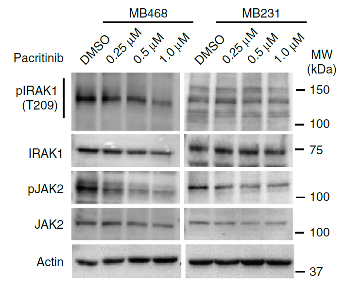
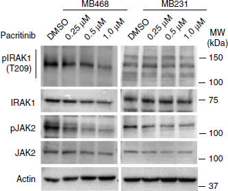
Pacritinib effectively disrupts the S100A7/8/9–IRAK1 feedback loop to inhibit tumorsphere growth. Representative western blot (n=2) showing inhibition of phosphorylated IRAK1 and phosphorylated JAK2 (pJAK2) within 6 h of Pacritinib treatment in MB468 and MB231 cells. Actin is used as a loading control.
-
Science
2026 Feb 5;391(6785):eadz4075. PMID: 41643022 -
Science
2017 Dec 1;358(6367):eaan4368. PMID: 29191878 -
Nat Commun
Genotoxic stress and viral infection induce transient expression of APOBEC3A and pro-inflammatory genes through two distinct pathways. [Abstract]2021 Aug 13;12(1):4917. PMID: 34389714
Pacritinib purchased from MedChemExpress. Usage Cited in: Nat Commun. 2021 Aug 13;12(1):4917. [Abstract]
Pacritinib (0.25-1 µM; 6 h) treatment caused inhibition of phosphorylated IRAK1 and phosphorylated JAK2 (pJAK2) in MB468 and MB231 cells.
Pacritinib purchased from MedChemExpress. Usage Cited in: Nat Commun. 2021 Aug 13;12(1):4917. [Abstract]
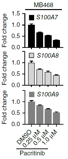
Real-time PCR analysis of S100A7, S100A8, and S100A9 gene expression after Pacritinib (0.25-1 µM; 24 h) treatment in MB468 cells.
Pacritinib purchased from MedChemExpress. Usage Cited in: Nat Commun. 2021 Aug 13;12(1):4917. [Abstract]
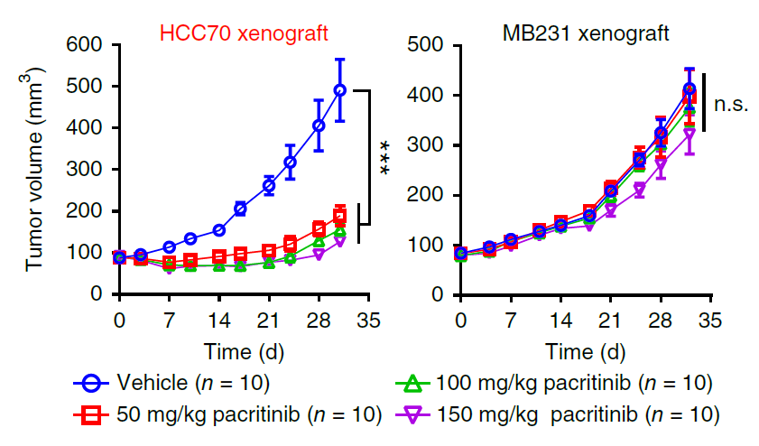
NOD-SCID mice bearing MB231 and HCC70 tumors were treated with pacritinib (50, 100, or 150 mg/kg; p.o.; once daily for 21 d). Pacritinib treatment in different doses resulted in substantial inhibition of tumor growth for HCC70 xenograft tumors but had no obvious effect on the growth of MB231 xenografts.
Pacritinib purchased from MedChemExpress. Usage Cited in: Nat Commun. 2021 Aug 13;12(1):4917. [Abstract]
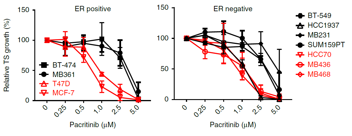
Growth curve of tumorspheres from ER-positive and ER-negative breast cancer cell lines treated with increasing doses of Pacritinib (0.25-5 µM). Pacritinib treatment was more effective in impairment of tumorsphere growth in cancer cell lines positive for 1q21.3 amplification than in cell lines without amplification.
-
Nat Commun
Stromal induction of BRD4 phosphorylation Results in Chromatin Remodeling and BET inhibitor Resistance in Colorectal Cancer. [Abstract]2021 Jul 21;12(1):4441. PMID: 34290255 -
Sci Transl Med
PP2A inhibition is a druggable MEK inhibitor resistance mechanism in KRAS-mutant lung cancer cells. [Abstract]2018 Jul 18;10(450):eaaq1093. PMID: 30021885 -
Cell Death Dis
Colorectal cancer-associated fibroblasts promote metastasis by up-regulating LRG1 through stromal IL-6/STAT3 signaling. [Abstract]2021 Dec 20;13(1):16. PMID: 34930899 -
-
Mol Syst Biol
Illuminating phenotypic drug responses of sarcoma cells to kinase inhibitors by phosphoproteomics. [Abstract]2024 Jan;20(1):28-55. PMID: 38177929 -
Inflamm Res
Potent repression of C-reactive protein (CRP) expression by the JAK1/2 inhibitor ruxolitinib in inflammatory human hepatocytes. [Abstract]2020 Jan;69(1):51-62. PMID: 31654094 -
Drug Des Devel Ther
Advances in the drug therapies of acute myeloid leukemia (except acute wpromyelocytic leukemia). [Abstract]2018 Apr 30:12:1009-1017. PMID: 29750014 -
Eur J Pharmacol
VOPP1, a determinant of the sensitivity of non-small cell lung cancer cells to NAE inhibitors. [Abstract]2025 Sep 15:1003:177942. PMID: 40651787 -
PLoS Pathog
Kaposi sarcoma-associated herpesvirus cooperates with Epstein-Barr virus to co-transform a small set of human B cells oncogenically. [Abstract]2025 Jun 23;21(6):e1013281. PMID: 40549805 -
PLoS Genet
Her2 amplification, Rel-A, and Bach1 can influence APOBEC3A expression in breast cancer cells. [Abstract]2024 May 28;20(5):e1011293. PMID: 38805570 -
BMJ Open Ophthalmol
Impact of anticancer drugs on human Tenon's fibroblast proliferation: implications for glaucoma surgery. [Abstract]2026 Jan 12;11(1):e002307. PMID: 41526033 -
Fundam Clin Pharmacol
2021 Oct;35(5):919-929. PMID: 33523504 -
Eur J Drug Metab Pharmacokinet
Differential Inhibition of Equilibrative Nucleoside Transporter 1 (ENT1) Activity by Tyrosine Kinase Inhibitors. [Abstract]2021 Sep;46(5):625-635. PMID: 34275128 -
-
-


Solvent & Solubility
In Vitro:
1 M HCl : ≥ 100 mg/mL (211.60 mM)
DMSO : 10 mg/mL (21.16 mM; ultrasonic and warming and heat to 60°C; Hygroscopic DMSO has a significant impact on the solubility of product, please use newly opened DMSO)
0.1 M HCl : 2 mg/mL (4.23 mM; Need ultrasonic)
* "≥" means soluble, but saturation unknown.
Please refer to the solubility information to select the appropriate solvent. Once prepared, please aliquot and store the solution to prevent product inactivation from repeated freeze-thaw cycles.
Storage method and period of stock solution: -80°C, 2 years; -20°C, 1 year. When stored at -80°C, please use it within 2 years. When stored at -20°C, please use it within 1 year.
Please refer to the solubility information to select the appropriate solvent. Once prepared, please aliquot and store the solution to prevent product inactivation from repeated freeze-thaw cycles.
Storage method and period of stock solution: -80°C, 2 years; -20°C, 1 year. When stored at -80°C, please use it within 2 years. When stored at -20°C, please use it within 1 year.
Concentration (start) × Volume (start) = Concentration (final) × Volume (final)
In Vivo:
Select the appropriate dissolution method based on your experimental animal and administration route.
- For the following dissolution methods, please ensure to first prepare a clear stock solution using an In Vitro approach and then sequentially add co-solvents:
- To ensure reliable experimental results, the clarified stock solution can be appropriately stored based on storage conditions. As for the working solution for In Vivo experiments, it is recommended to prepare freshly and use it on the same day.
- The percentages shown for the solvents indicate their volumetric ratio in the final prepared solution. If precipitation or phase separation occurs during preparation, heat and/or sonication can be used to aid dissolution.
Add each solvent one by one: 10% DMSO 40% PEG300 5% Tween-80 45% Saline
Solubility: ≥ 1 mg/mL (2.12 mM); Clear solution
This protocol yields a clear solution of ≥ 1 mg/mL (saturation unknown).
Taking 1 mL working solution as an example, add 100 μL DMSO stock solution (10.0 mg/mL) to 400 μL PEG300, and mix evenly; then add 50 μL Tween-80 and mix evenly; then add 450 μL Saline to adjust the volume to 1 mL.
Preparation of Saline: Dissolve 0.9 g sodium chloride in ddH₂O and dilute to 100 mL to obtain a clear Saline solution.
Add each solvent one by one: 10% DMSO 90% (20% SBE-β-CD in Saline)
Solubility: 1 mg/mL (2.12 mM); Suspended solution; Need ultrasonic
This protocol yields a suspended solution of 1 mg/mL. Suspended solution can be used for oral and intraperitoneal injection.
Taking 1 mL working solution as an example, add 100 μL DMSO stock solution (10.0 mg/mL) to 900 μL 20% SBE-β-CD in Saline, and mix evenly.
Preparation of 20% SBE-β-CD in Saline (4°C, storage for one week): 2 g SBE-β-CD powder is dissolved in 10 mL Saline, completely dissolve until clear.
In Vivo Dissolution Calculator
Please enter the basic information of animal experiments:
-
-
-
-
Recommended: Prepare an additional quantity of animals to account for potential losses during experiments.
Please enter your animal formula composition:
-
%DMSO +
Recommended: Keep the proportion of DMSO in working solution below 2% if your animal is weak.
-
%+
-
+%Tween-80 + +
-
%Saline +
The co-solvents required include: DMSO, . All of co-solvents are available by MedChemExpress (MCE). , Tween 80. All of co-solvents are available by MedChemExpress (MCE).
Working solution concentration: 0.22 mg/mL
Method for preparing stock solution: mg drug dissolved in μL DMSO. Stock solution concentration: mg/mL.
1. Take μL DMSO stock solution;
2. Add μL .
μL , mix evenly;
3. Then add μL Tween 80, mix evenly;
4. Then add μL
Please ensure that the stock solution in the first step is dissolved to a clear state, and add co-solvents in sequence. You can use ultrasonic heating (ultrasonic cleaner, recommended frequency 20-40 kHz), vortexing, etc. to assist dissolution.

Protocols
-
Collagen: Sirius Red Staining
Sirius Red or picrosirius red staining is a histochemical method for visualizing collagen-rich extracellular matrix in tissue sections, and collagen fibers are detected as red-stained structures under bright-field microscopy with enhanced birefringence under polarized light. Picrosirius red is useful for assessing total collagen organization, distribution, and fibrosis burden, but polarized color should not be interpreted as a definitive collagen type I versus type III readout because color is affected by fiber orientation, thickness, and packing.
-
Annexin V plus membrane-impermeant dye apoptosis staining
Annexin V-based apoptosis assays rely on the detection of phosphatidylserine (PS) externalization from the inner leaflet of the plasma membrane to the outer leaflet, an early biochemical hallmark of apoptosis. Fluorescently labeled Annexin V binds PS in a calcium-dependent manner, enabling identification of early apoptotic cells by flow cytometry or fluorescence microscopy. When combined with a membrane-impermeant DNA-binding dye (e. g. , propidium iodide), this approach allows discrimination between viable (Annexin V−/dye−), early apoptotic (Annexin V+/dye−), and late apoptotic or necrotic (Annexin V+/dye+) cell populations by assessing membrane integrity and PS exposure.
-
Cell migration
Cell migration is a method that plays an important role in wound healing, cell differentiation, embryonic development, etc.
-
Connective Tissue: Masson's Trichrome/Collagen Trichrome Staining
Masson’s Trichrome (collagen/trichrome staining) is a histological technique that differentially stains tissue compartments using sequential acidic dyes to distinguish collagen from muscle and cytoplasmic components based on dye affinity and tissue permeability differences, enabling visualization of fibrosis and connective tissue architecture in histological sections. The classical formulation typically uses Weigert's iron hematoxylin for nuclear staining, Biebrich scarlet-acid fuchsin for cytoplasm and muscle, and aniline blue (or light green variants) for collagen, producing a characteristic blue/green collagen signal contrasted against red cytoplasm and dark nuclei. The staining principle relies on selective displacement of smaller dye molecules by larger anionic dyes in collagen-rich regions under controlled acidified conditions, which enhances collagen-specific dye retention. This property makes the method widely used for fibrosis assessment in organs such as heart, liver, lung, a
-
Patient-Derived Orthotopic Xenograft (PDOX)
Patient-derived orthotopic xenograft (PDOX) modeling implants fresh patient tumor tissue or patient-derived tumor cells into the anatomically corresponding organ or tissue site of immunodeficient mice, usually by surgical orthotopic implantation, to preserve patient tumor histology, local microenvironmental context, invasion, metastatic behavior, and treatment-response features better than subcutaneous implantation. PDOX readouts include tumor engraftment, orthotopic tumor growth, local invasion, metastasis, recurrence after resection, histologic similarity to the donor tumor, biomarker retention, molecular concordance, survival, and response or resistance to therapy. PDOX models are used for preclinical drug testing and individualized therapy evaluation, but engraftment success varies by tumor type and specimen quality.
-
Research Protocol for Inflammation-related Diseases
The NLRP3 inflammasome is a cytosolic innate immune signaling platform that integrates priming signals and danger-signal activation to promote caspase-1 activation, maturation of IL-1β and IL-18, and gasdermin D-mediated pyroptotic cell death. The core experimental logic is to determine whether inflammatory disease phenotypes are driven by increased NLRP3 expression, ASC-containing inflammasome assembly, caspase-1 cleavage, GSDMD cleavage, and extracellular release of IL-1β/IL-18 rather than by nonspecific cell injury alone. The pathway is strongly linked to inflammation-related disease phenotypes because monosodium urate crystals activate NALP3/NLRP3 inflammasome signaling in gout-like crystal inflammation, cholesterol crystals activate NLRP3 inflammasomes in atherogenesis models, and DSS-induced intestinal inflammation has been reported to involve NLRP3 inflammasome activity. However, experimental colitis studies also show context-dependent protective effects of NLRP3 inflammasome co
-
Liver Histomorphometry
Liver histomorphometry is a quantitative histological approach used to measure structural alterations in hepatic tissue, including parenchymal loss, steatosis, fibrosis, and vascular remodeling, by combining stained tissue section analysis with stereological or computerized image-based measurements. Classical morphometric frameworks quantify volume fractions of liver compartments and fibrotic regions using systematic sampling and image analysis, enabling objective comparison of pathological changes across experimental groups. These approaches are widely applied in liver cirrhosis and fibrosis studies to reduce subjectivity in histological scoring and improve reproducibility of tissue evaluation. Recent methodological advances integrate automated image analysis and radiomics-based extraction of histological features from standard liver stains (e. g. , H&E and fibrotic stains), enabling quantitative correlation between morphometric features and fibrosis stages in non-alcoholic fatty live
-
Fibrosis/Collagen Morphometry
Fibrosis and collagen morphometry is based on the quantitative visualization of fibrillar collagen deposition in tissue sections using histochemical stains such as Sirius Red (Picrosirius Red) or Masson's trichrome, followed by image-based or polarization-enhanced analysis to estimate collagen proportional area as a surrogate of extracellular matrix accumulation during fibrotic remodeling. Sirius Red combined with polarized light microscopy enhances detection of collagen fibers due to birefringence properties, enabling more specific visualization of collagen type I and III fibrils compared to conventional bright-field histology, while whole-section or region-restricted digital morphometry reduces field-selection bias in fibrosis assessment. Alternative quantitative approaches include second harmonic generation (SHG) and two-photon excited fluorescence microscopy, which enable label-free detection of fibrillar collagen and have been validated against histological staining and biochemica
-
Detection of Apoptosis
Apoptosis, also called programmed cell death, is generally characterized by distinct morphological characteristics.
-
TUNEL staining for apoptotic DNA fragmentation
TUNEL staining detects DNA strand breaks by using terminal deoxynucleotidyl transferase to add labeled nucleotides to exposed 3′-OH DNA termini, generating either microscopic staining in fixed cells or tissue sections, or fluorescence/cytometric signal in cell suspensions. TUNEL positivity reflects DNA fragmentation but should not be interpreted alone as definitive apoptosis, because TUNEL can also label necrotic, autolytic, mechanically damaged, or DNA-repair-associated DNA breaks.
-
Patient-Derived Xenograft (PDX)
Patient-derived xenograft (PDX) models are generated by engrafting primary human tumor tissue directly into immunodeficient mice, allowing in vivo propagation of patient tumor biology without initial in vitro adaptation. These models are used to preserve key histopathological and molecular characteristics of the original tumor and enable assessment of tumor growth dynamics and therapeutic response in a living organism. The biological readout is tumor engraftment and subsequent growth in the murine host, which reflects the ability of human tumor cells to survive, vascularize, and expand in an immunocompromised microenvironment.
-
Apoptosis Solutions
Apoptosis is a regulated, generally non-lytic cell-death pathway that removes unwanted, damaged, infected, or abnormal cells through coordinated morphological changes, caspase activation, DNA fragmentation, and membrane remodeling. The intrinsic apoptosis pathway is controlled mainly by mitochondrial outer membrane permeabilization, BCL-2 family proteins, cytochrome c release, apoptosome formation, caspase-9 activation, and downstream executioner caspase-3/7 activation. The extrinsic apoptosis pathway is initiated by death receptors such as Fas, TNFR, and TRAIL receptors, which recruit adaptor proteins and activate caspase-8 before engaging executioner caspases or mitochondrial amplification through BID cleavage. Apoptosis is linked to many phenotypes, including cancer cell killing, tissue homeostasis, immune regulation, neurodegeneration, infection response, and treatment-induced cytotoxicity; unresolved questions include how apoptosis interacts with necroptosis, pyroptosis, ferroptos
-
Subcutaneous Cell-Line-Derived Xenograft
Subcutaneous cell-line-derived xenograft (CDX) models are established by implanting cultured human cancer cell lines into immunodeficient mice, where the injected cells form localized tumors that can be monitored in vivo as a measure of tumorigenic potential, growth kinetics, and treatment response. These models are widely used in oncology research because they allow reproducible tumor formation and enable comparative assessment of tumor growth between different cell lines or genetic manipulations in a controlled in vivo microenvironment. Subcutaneous implantation of cancer cells in immunodeficient mice is a standard approach for evaluating tumor growth behavior and therapeutic response across multiple cancer types, including prostate, esophageal, pancreatic, and colon cancer models.
-
Orthotopic Cell-Line Xenograft
Orthotopic cell-line xenograft models involve implantation of human cancer cell lines into the anatomically corresponding organ of immunodeficient mice to reproduce tumor growth within a native microenvironment, enabling more clinically relevant tumor behavior compared with subcutaneous models. These models are widely used because orthotopic placement better recapitulates tumor progression, including invasion and metastatic spread, which are often underrepresented in heterotopic implantation systems. Compared with conventional xenografts, orthotopic implantation is described as more technically complex but provides improved simulation of tumor-microenvironment interactions and metastatic behavior, making it particularly valuable for translational oncology research. Surgical orthotopic implantation approaches have been emphasized as enabling faithful reproduction of clinical cancer features, including metastasis and disease progression patterns that align with the tumor’s organ of origi


Purity & Documentation
-
Data Sheet (297 KB)
-
SDS (396 KB)
- English - EN (396 KB)
- Français - FR (396 KB)
- Deutsch - DE (396 KB)
- Norwegian - NO (396 KB)
- Español - ES (396 KB)
- Swedish - SV (396 KB)
- Italian - IT (396 KB)
- Korean - KR (396 KB)
- Portuguese - PT (396 KB)
-
Handling Instructions (2659 KB)
References
[1]. Hart S, et al. Pacritinib (SB1518), a JAK2/FLT3 inhibitor for the treatment of acute myeloid leukemia. Blood cancer journal. 2011 Nov;1(11):e44. [Content Brief]
[2]. Novotny-Diermayr V, et al. The oral HDAC inhibitor pracinostat (SB939) is efficacious and synergistic with the JAK2 inhibitor pacritinib (SB1518) in preclinical models of AML. Blood cancer journal. 2012 May;2(5):e69. [Content Brief]


Complete Stock Solution Preparation Table
Please refer to the solubility information to select the appropriate solvent. Once prepared, please aliquot and store the solution to prevent product inactivation from repeated freeze-thaw cycles.
Storage method and period of stock solution: -80°C, 2 years; -20°C, 1 year. When stored at -80°C, please use it within 2 years. When stored at -20°C, please use it within 1 year.
| Optional Solvent | Concentration Solvent Mass | 1 mg | 5 mg | 10 mg | 25 mg |
|---|---|---|---|---|---|
| 0.1 M HCl / DMSO / 1 M HCl | 1 mM | 2.1160 mL | 10.5802 mL | 21.1604 mL | 52.9011 mL |
| DMSO / 1 M HCl | 5 mM | 0.4232 mL | 2.1160 mL | 4.2321 mL | 10.5802 mL |
| 10 mM | 0.2116 mL | 1.0580 mL | 2.1160 mL | 5.2901 mL | |
| 15 mM | 0.1411 mL | 0.7053 mL | 1.4107 mL | 3.5267 mL | |
| 20 mM | 0.1058 mL | 0.5290 mL | 1.0580 mL | 2.6451 mL | |
| 1 M HCl | 25 mM | 0.0846 mL | 0.4232 mL | 0.8464 mL | 2.1160 mL |
| 30 mM | 0.0705 mL | 0.3527 mL | 0.7053 mL | 1.7634 mL | |
| 40 mM | 0.0529 mL | 0.2645 mL | 0.5290 mL | 1.3225 mL | |
| 50 mM | 0.0423 mL | 0.2116 mL | 0.4232 mL | 1.0580 mL | |
| 60 mM | 0.0353 mL | 0.1763 mL | 0.3527 mL | 0.8817 mL | |
| 80 mM | 0.0265 mL | 0.1323 mL | 0.2645 mL | 0.6613 mL | |
| 100 mM | 0.0212 mL | 0.1058 mL | 0.2116 mL | 0.5290 mL |


Pacritinib Related Classifications
HY-16379 Related Classifications
 Powered by Bioz