



Vacquinol-1
Based on 1 publication(s) in Google Scholar
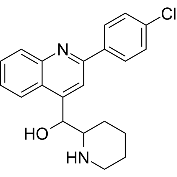
Vacquinol-1 (NSC13316) is a MKK4-specific activator that activates the MAPK pathway. Vacquinol-1 inhibits the growth, migration and colony formation, and induces apoptosis of cancer cells. Vacquinol-1 is applicable to research related to cancers such as hepatocellular carcinoma.
For research use only. We do not sell to patients.
- Purity : 99.33%
- CAS No.: 5428-80-8
- Formula: C21H21ClN2O
- Molecular Weight:352.86
-
Storage:Powder -20°C, 3 years , 4°C, 2 years ; In solvent -80°C, 6 months , -20°C, 1 month

To place orders, for customer services and technical support, please contact: MedChemExpress USA
Tel: 609-228-6898 E-mail: [email protected] [email protected]


-
 Biological Activity
-
Chemical Information
-
Solvent & Solubility
- Protocols
- Purity & Documentation
- References
-
 Help & FAQs


-
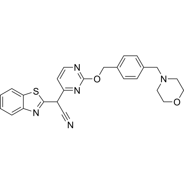
HY-14761
BentamapimodJNK Inhibitor -
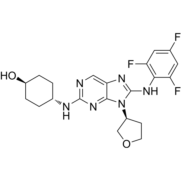
HY-15495
Tanzisertib -
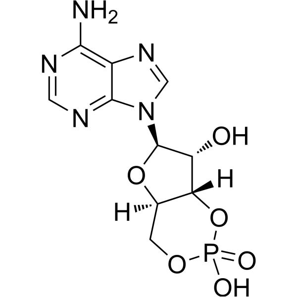
HY-B1511
Cyclic AMPSecond Messenger -
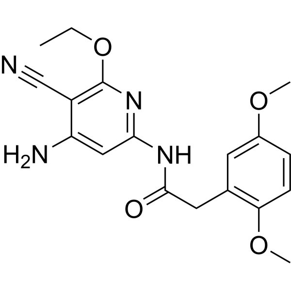
HY-107598
JNK Inhibitor VIIIJNK Inhibitor -
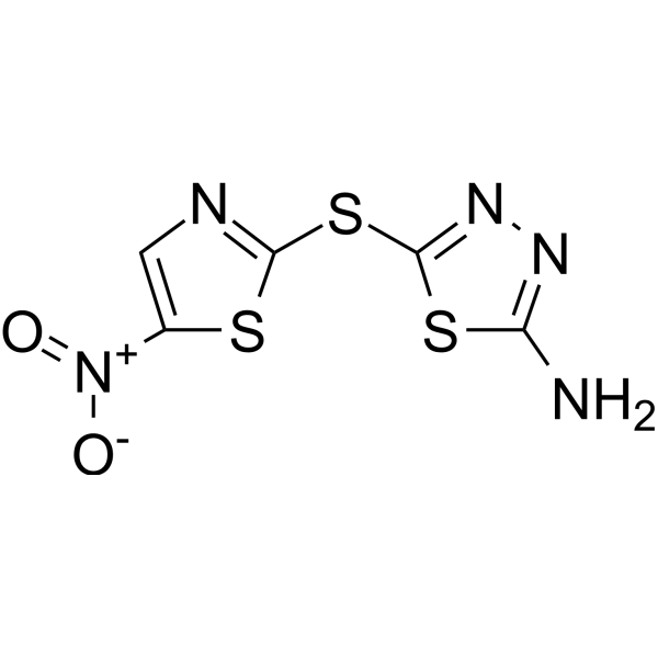
HY-107597
HalicinJNK Inhibitor -
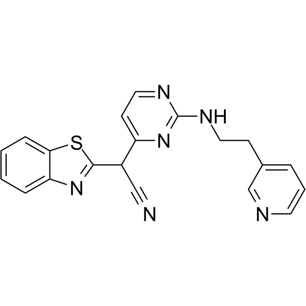
HY-11010
AS601245JNK Inhibitor -
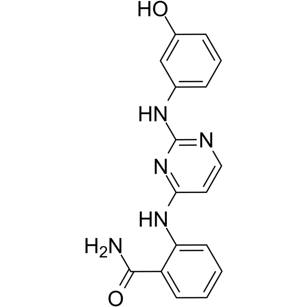
HY-15737
DB07268JNK Inhibitor -
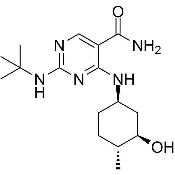
HY-138304
CC-90001JNK Inhibitor -
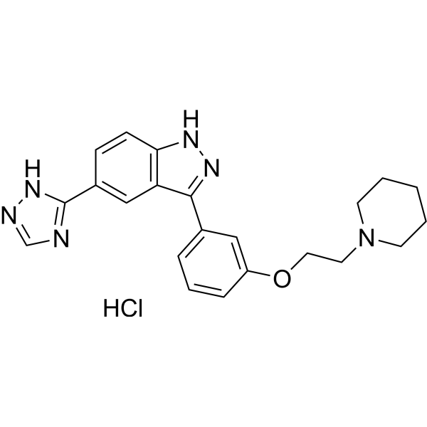
HY-13022
CC-401 hydrochlorideJNK Inhibitor -
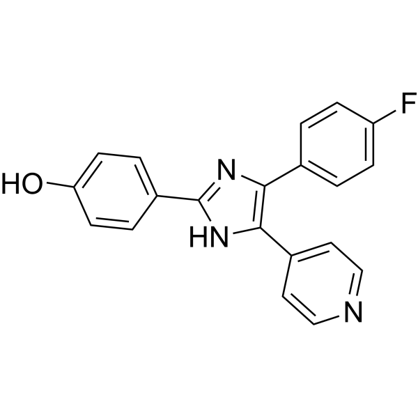
HY-10295
SB 202190p38 MAPK Inhibitor -
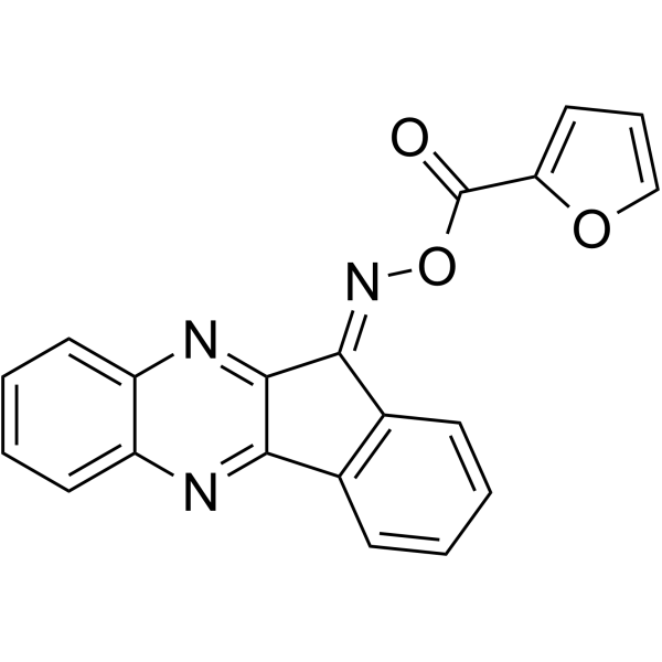
HY-107600
IQ-3c-JNK Inhibitor -
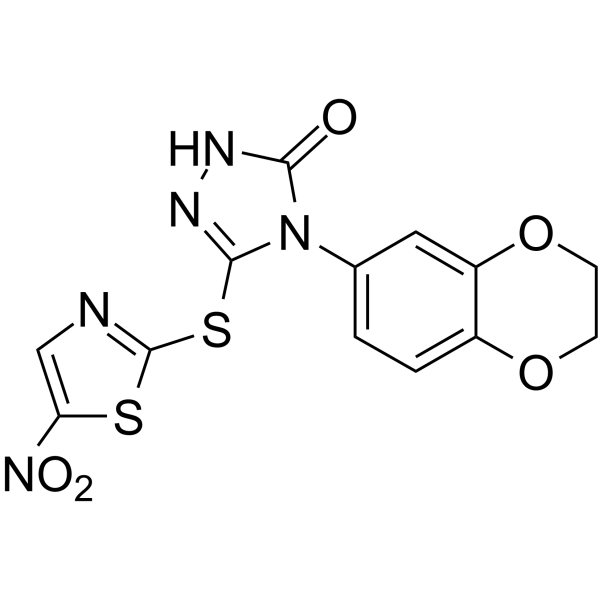
HY-10366
BI-78D3JNK Inhibitor
Publications Citing Use of MedChemExpress (MCE) Vacquinol-1
MoreAll Caspase Isoforms
More

Biological Activity
Description
Cellular Effect
|
Cell Line

|
Type | Value | Description | References |
|---|---|---|---|---|
| U3013MG | IC50 |
3.14 μM
Compound: 1
|
Cytotoxicity against patient-derived U3013MG cells assessed as reduction in cell viability by Celltiter-Glo reagent based assay
Cytotoxicity against patient-derived U3013MG cells assessed as reduction in cell viability by Celltiter-Glo reagent based assay
|
[PMID: 27607569] |
In Vitro
Vacquinol-1 (1.56-25 μM; 24-72 h) inhibits the viability of BEL7402/Huh7 hepatocellular carcinoma cells in vitro[1].
Vacquinol-1 (0-24 μM; 24 h) inhibits the colony-forming ability of BEL7402/Huh7 hepatocellular carcinoma cells and induces cell apoptosis[1].
Vacquinol-1 (10 μM; 24 h) activates the endogenous apoptotic pathway in BEL7402 hepatocellular carcinoma cells, induces the cleavage of caspase-3, caspase-9 and PARP-1, upregulates the expression of Bax and Bim, and downregulates the expression of Bcl-2[1].
Vacquinol-1 (10 μM; 1-12 h) induces the translocation of the pro-apoptotic protein Bax from the cytoplasm to mitochondria in BEL7402 hepatocellular carcinoma cells[1].
Vacquinol-1 (15 μM; 5 h) activates the JNK and p38 MAPK pathways, upregulates α-SMA, an activation marker of the hepatic stellate cell line HSC-T6, promotes collagen production, and induces phosphorylation of the linker region of Smad2/3[2].
MedChemExpress (MCE) has not independently confirmed the accuracy of these methods. They are for reference only. Further protocols information, click here.
-
Cell Line:BEL7402 (hepatocellular carcinoma, HCC)
-
Concentration:1.56 μM, 3.13 μM, 6.26 μM, 12.5 μM, 25 μM
-
Incubation Time:24, 48, 72 h
-
Result:Inhibited BEL7402 cell proliferation in a dose-dependent manner.
Reached half maximal inhibitory concentration (IC50) values of 12.35 μM (24 h), 11.48 μM (48 h), and 10.21 μM (72 h).
-
Cell Line:Huh7 (hepatocellular carcinoma, HCC)
-
Concentration:1.56 μM, 3.13 μM, 6.26 μM, 12.5 μM, 25 μM
-
Incubation Time:24, 48, 72 h
-
Result:Inhibited Huh7 cell proliferation in a dose-dependent manner.
Reached half maximal inhibitory concentration (IC50) values of 6.21 μM (24 h), 5.58 μM (48 h), and 4.89 μM (72 h).
-
Cell Line:BEL7402 (hepatocellular carcinoma, HCC)
-
Concentration:10 μM
-
Incubation Time:24 h
-
Result:Increased the percentage of apoptotic cells.
Resulted in nuclear fragmentation and apoptotic body formation in BEL7402 cells, whereas control cells displayed round, normal nuclei.
-
Cell Line:rat hepatic stellate HSC-T6 cells
-
Concentration:15 μM
-
Incubation Time:5 h
-
Result:Upregulated the protein levels of phosphorylated (P)-JNK1/2 and P-p38 (but not P-ERK1/2) in HSC-T6 cells.
Increased the protein levels of α-SMA, Collagen I, P-Smad2L, and P-Smad3L in HSC-T6 cells.


Chemical Information
-
CAS No. 5428-80-8
-
Appearance Solid
-
Molecular Weight 352.86
-
Formula C21H21ClN2O
-
Color White to off-white
-
SMILES
OC(C1NCCCC1)C2=CC(C3=CC=C(Cl)C=C3)=NC4=CC=CC=C24
-
Synonyms
NSC13316
-
Shipping
Room temperature in continental US; may vary elsewhere.
-
Storage
Powder -20°C 3 years 4°C 2 years In solvent -80°C 6 months -20°C 1 month


Publications (1)
-
Journal Impact Factor
-
Most Recent


Solvent & Solubility
In Vitro:
DMSO : 12.5 mg/mL (35.42 mM; Need ultrasonic; Hygroscopic DMSO has a significant impact on the solubility of product, please use newly opened DMSO)
Please refer to the solubility information to select the appropriate solvent. Once prepared, please aliquot and store the solution to prevent product inactivation from repeated freeze-thaw cycles.
Storage method and period of stock solution: -80°C, 6 months; -20°C, 1 month. When stored at -80°C, please use it within 6 months. When stored at -20°C, please use it within 1 month.
Please refer to the solubility information to select the appropriate solvent. Once prepared, please aliquot and store the solution to prevent product inactivation from repeated freeze-thaw cycles.
Storage method and period of stock solution: -80°C, 6 months; -20°C, 1 month. When stored at -80°C, please use it within 6 months. When stored at -20°C, please use it within 1 month.
Concentration (start) × Volume (start) = Concentration (final) × Volume (final)

Protocols
-
Apoptosis
Apoptosis, also called programmed cell death, is generally characterized by distinct morphological characteristics.
-
TUNEL staining for apoptotic DNA fragmentation
TUNEL staining detects DNA strand breaks by using terminal deoxynucleotidyl transferase to add labeled nucleotides to exposed 3′-OH DNA termini, generating either microscopic staining in fixed cells or tissue sections, or fluorescence/cytometric signal in cell suspensions. TUNEL positivity reflects DNA fragmentation but should not be interpreted alone as definitive apoptosis, because TUNEL can also label necrotic, autolytic, mechanically damaged, or DNA-repair-associated DNA breaks.
-
Annexin V plus membrane-impermeant dye apoptosis staining
Annexin V-based apoptosis assays rely on the detection of phosphatidylserine (PS) externalization from the inner leaflet of the plasma membrane to the outer leaflet, an early biochemical hallmark of apoptosis. Fluorescently labeled Annexin V binds PS in a calcium-dependent manner, enabling identification of early apoptotic cells by flow cytometry or fluorescence microscopy. When combined with a membrane-impermeant DNA-binding dye (e. g. , propidium iodide), this approach allows discrimination between viable (Annexin V−/dye−), early apoptotic (Annexin V+/dye−), and late apoptotic or necrotic (Annexin V+/dye+) cell populations by assessing membrane integrity and PS exposure.
-
Cell migration
Cell migration is a method that plays an important role in wound healing, cell differentiation, embryonic development, etc.
-
Apoptosis Solutions
Apoptosis is a regulated, generally non-lytic cell-death pathway that removes unwanted, damaged, infected, or abnormal cells through coordinated morphological changes, caspase activation, DNA fragmentation, and membrane remodeling. The intrinsic apoptosis pathway is controlled mainly by mitochondrial outer membrane permeabilization, BCL-2 family proteins, cytochrome c release, apoptosome formation, caspase-9 activation, and downstream executioner caspase-3/7 activation. The extrinsic apoptosis pathway is initiated by death receptors such as Fas, TNFR, and TRAIL receptors, which recruit adaptor proteins and activate caspase-8 before engaging executioner caspases or mitochondrial amplification through BID cleavage. Apoptosis is linked to many phenotypes, including cancer cell killing, tissue homeostasis, immune regulation, neurodegeneration, infection response, and treatment-induced cytotoxicity; unresolved questions include how apoptosis interacts with necroptosis, pyroptosis, ferroptos
-
Soft Agar Colony Formation Assay
Soft agar colony formation assay measures anchorage-independent growth, in which transformed or tumorigenic cells proliferate as colonies in a semisolid agar matrix while many non-transformed adherent cells fail to proliferate without attachment; classic studies showed that growth in semisolid medium correlates with tumorigenicity in nude mice, and later protocol papers describe the method as a stringent in vitro assay for malignant transformation. The readout is the number, size, morphology, or signal intensity of colonies formed within agar after incubation; published formats include manual colony counting after staining, 96-well or 384-well quantitative formats, DNA-binding dye detection, MTT/tetrazolium-based detection, digital image analysis, and PCR-based marker detection from soft agar cultures.
-
Liver Cancer Modeling
Liver cancer can be classified into primary liver cancer and secondary liver cancer. Secondary liver cancer is the metastatic liver cancer. Primary liver cancer includes hepatocellular carcinoma (HCC), intrahepatic cholangiocarcinoma (ICC) and fibrolamellar HCC, of which HCC is the most common form, accounting for approximately 90% of primary liver cancers[1]. HCC mouse models include chemical agent-induced models, transplanted tumor models, and genetic engineered models.
-
Colony Formation (Clonogenic) Assay
The clonogenic (colony formation) assay measures the ability of a single cell to retain reproductive viability and form a macroscopic colony, typically defined as a cluster derived from one progenitor cell after a defined growth period. This assay is widely used to evaluate cell survival after exposure to ionizing radiation or cytotoxic treatments and is considered a standard method in radiation biology for generating dose-response relationships of reproductive cell death. Colony formation reflects long-term proliferative capacity rather than short-term metabolic activity, and survival is quantified by comparing treated versus untreated conditions based on colony number and derived survival fractions.


Purity & Documentation
-
Data Sheet (280 KB)
-
SDS (394 KB)
- English - EN (394 KB)
- Français - FR (394 KB)
- Deutsch - DE (394 KB)
- Norwegian - NO (394 KB)
- Español - ES (394 KB)
- Swedish - SV (394 KB)
- Italian - IT (394 KB)
- Korean - KR (394 KB)
- Portuguese - PT (394 KB)
-
Handling Instructions (2659 KB)
References
[1]. Sun SL, et al. Vacquinol‑1 induces apoptosis in hepatocellular carcinoma cell. Mol Med Rep. 2018;18(1):557-563. [Content Brief]
[2]. Wu C, et al. Salvianolic acid B exerts anti-liver fibrosis effects via inhibition of MAPK-mediated phospho-Smad2/3 at linker regions in vivo and in vitro. Life Sci. 2019;239:116881. [Content Brief]


Complete Stock Solution Preparation Table
Please refer to the solubility information to select the appropriate solvent. Once prepared, please aliquot and store the solution to prevent product inactivation from repeated freeze-thaw cycles.
Storage method and period of stock solution: -80°C, 6 months; -20°C, 1 month. When stored at -80°C, please use it within 6 months. When stored at -20°C, please use it within 1 month.
| Optional Solvent | Concentration Solvent Mass | 1 mg | 5 mg | 10 mg | 25 mg |
|---|---|---|---|---|---|
| DMSO | 1 mM | 2.8340 mL | 14.1699 mL | 28.3399 mL | 70.8496 mL |
| 5 mM | 0.5668 mL | 2.8340 mL | 5.6680 mL | 14.1699 mL | |
| 10 mM | 0.2834 mL | 1.4170 mL | 2.8340 mL | 7.0850 mL | |
| 15 mM | 0.1889 mL | 0.9447 mL | 1.8893 mL | 4.7233 mL | |
| 20 mM | 0.1417 mL | 0.7085 mL | 1.4170 mL | 3.5425 mL | |
| 25 mM | 0.1134 mL | 0.5668 mL | 1.1336 mL | 2.8340 mL | |
| 30 mM | 0.0945 mL | 0.4723 mL | 0.9447 mL | 2.3617 mL |


Vacquinol-1 Related Classifications
HY-19345 Related Classifications
 Powered by Bioz