



WS-384

Based on 1 Customer Validation
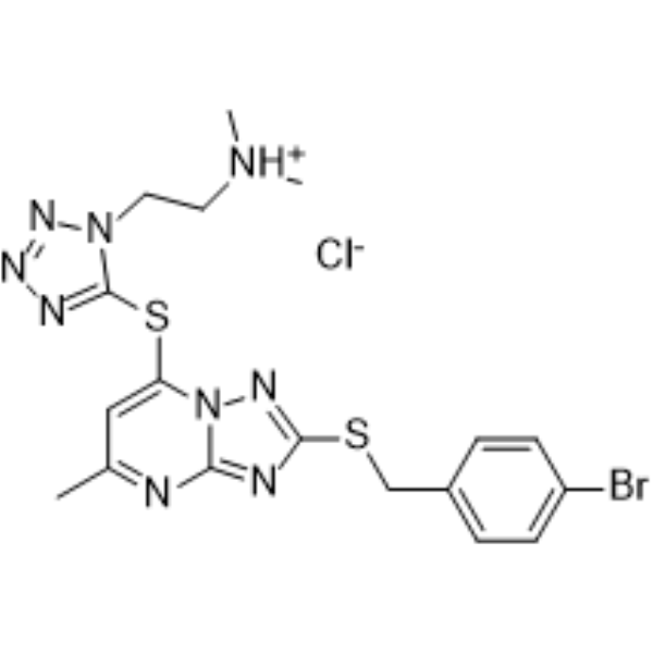
WS-384 is a dual LSD1 and DCN1-UBC12 protein-protein interaction inhibitor with oral activity, with IC50 values of 338.79 nM and 14.81 nM, respectively. WS-384 possesses anticancer activity and can cause cell cycle arrest, DNA damage, and induce apoptosis. WS-384 can be used in the research of non-small cell lung cancer (NSCLC).
Nur für Forschungszwecke. Wir verkaufen nicht an Patienten.
- Reinheit : 98.23%
- CAS. Nr.: 2247544-03-0
- Formel: C18H21BrClN9S2
- Molecular Weight:542.91
-
Speicherung:
4°C, sealed storage, away from moisture
* In solvent : -80°C, 6 months; -20°C, 1 month (sealed storage, away from moisture)

To place orders, for customer services and technical support, please contact: MedChemExpress USA
Tel: 609-228-6898 E-mail: [email protected] [email protected]


-
 Biologische Aktivität
-
Chemical Information
-
Lösungsmittel & Löslichkeit
- Protokoll
- Reinheit & Dokumentation
- Verweise
-
 Help & FAQs


Alle DNA/RNA Synthesis Isoform-spezifische Produkte anzeigen
MoreAlle Caspase Isoform-spezifische Produkte anzeigen
More

Biologische Aktivität
Beschreibung
IC50 & Target
IC50: 338.79 nM (LSD1)[1].
IC50: 14.81 nM (DCN1-UBC12 Protein-protein interaction)[1].
In Vitro
WS-384 (1-32 μM; 24-72 h) inhibits the growth of A549 and H1975 cells in a time- and dose-dependent manner, with an IC50 ranging from 2.15 to 6.67 μM, demonstrating anticancer activity[1].
WS-384 (1-8 μM; 48 h) enhances the expression levels of p21 gene and protein in A549 and H1975 cells by inhibiting the neddylation of cullin 1 and reducing the H3K4 demethylation at the CDKN1A promoter, thus inducing cell cycle arrest at the G2/M phase and apoptosis. Additionally, WS-384 (1-8 μM; 48 h) can also cause DNA damage[1].
MedChemExpress (MCE) has not independently confirmed the accuracy of these methods. They are for reference only.
-
Cell Line:A549, H1975
-
Concentration:1 μM, 2 μM, 4 μM, 8 μM (A549);
0.5 μM, 1 μM, 2 μM, 4 μM (H1975) -
Incubation Time:48 h
-
Result:Increased the apoptosis rate in a concentration-dependent manner, with the apoptosis rate of A549 cells reaching 44.9% at a concentration of 8 μM and the apoptosis rate of H1975 cells reaching 36.42% at a concentration of 4 μM.
-
Cell Line:A549, H1975
-
Concentration:2 μM, 4 μM, 8 μM (A549);
1 μM, 2 μM, 4 μM (H1975) -
Incubation Time:48 h
-
Result:Blocked the neddylation of cullin1 and cullin3 but had no effect on the neddylation of other cullin members (cullin2, cullin4A, and cullin5).
Increased the proteins expression of cleaved caspase 3, cleaved caspase 7, cleaved caspase 9, and cleaved PARP.
Decreased the protein expression of p-CDC2, cyclin B1. CDK4 and CDK6.
Increased the protein expression γ-H2AX (a marker protein of DNA damage).
In Vivo
MedChemExpress (MCE) has not independently confirmed the accuracy of these methods. They are for reference only.
-
Animal Model:BALB/c nude mice xenograft model[1].
-
Dosage:25 mg/kg; 50 mg/kg;
-
Administration:Oral gavage (p.o.); Once daily for 36 consecutive days
-
Result:Significantly reduced tumor weight and volume in mice in the 50 mg/kg group.
Had no significant toxic effects on the heart, liver, spleen, lungs, and kidneys.
- Annexin V plus membrane-impermeant dye apoptosis staining
- Immunoprecipitation
- BrdU Incorporation Assay
- Protocol for Bimolecular Fluorescence Complementation (BiFC) Assay
- Protocol for Cell Cycle
- Detection of Apoptosis
- TUNEL staining for apoptotic DNA fragmentation
- Flow cytometric DNA-content cell-cycle staining
- Pull-down
- Apoptosis Solutions
- Genotoxicity/Mutagenicity Study
- Co-Immunoprecipitation
- Protocol for Yeast Two-Hybrid (Y2H) Assay


Chemical Information
-
CAS. Nr. 2247544-03-0
-
Appearance Solid
-
Molecular Weight 542.91
-
Formel C18H21BrClN9S2
-
Color Off-white to light yellow
-
SMILES
BrC1=CC=C(CSC2=NN3C(SC4=NN=NN4CCN(C)C)=CC(C)=NC3=N2)C=C1.Cl
-
Versand
Room temperature in continental US; may vary elsewhere.
-
Speicherung
4°C, sealed storage, away from moisture
* In solvent : -80°C, 6 months; -20°C, 1 month (sealed storage, away from moisture)


Lösungsmittel & Löslichkeit
In Vitro:
DMSO : 17.86 mg/mL (32.90 mM; ultrasonic and warming and heat to 60°C; Hygroscopic DMSO has a significant impact on the solubility of product, please use newly opened DMSO)
Please refer to the solubility information to select the appropriate solvent. Once prepared, please aliquot and store the solution to prevent product inactivation from repeated freeze-thaw cycles.
Storage method and period of stock solution: -80°C, 6 months; -20°C, 1 month (sealed storage, away from moisture). When stored at -80°C, please use it within 6 months. When stored at -20°C, please use it within 1 month.
Please refer to the solubility information to select the appropriate solvent. Once prepared, please aliquot and store the solution to prevent product inactivation from repeated freeze-thaw cycles.
Storage method and period of stock solution: -80°C, 6 months; -20°C, 1 month (sealed storage, away from moisture). When stored at -80°C, please use it within 6 months. When stored at -20°C, please use it within 1 month.
Konzentration (Stammlösung) × Volumen (Stammlösung) = Konzentration (Ziellösung) × Volumen (Ziellösung)
In Vivo:
Select the appropriate dissolution method based on your experimental animal and administration route.
- For the following dissolution methods, please ensure to first prepare a clear stock solution using an In Vitro approach and then sequentially add co-solvents:
- To ensure reliable experimental results, the clarified stock solution can be appropriately stored based on storage conditions. As for the working solution for In Vivo experiments, it is recommended to prepare freshly and use it on the same day.
- The percentages shown for the solvents indicate their volumetric ratio in the final prepared solution. If precipitation or phase separation occurs during preparation, heat and/or sonication can be used to aid dissolution.
Add each solvent one by one: 10% DMSO 40% PEG300 5% Tween-80 45% Saline
Solubility: ≥ 1.79 mg/mL (3.30 mM); Clear solution
This protocol yields a clear solution of ≥ 1.79 mg/mL (saturation unknown).
Taking 1 mL working solution as an example, add 100 μL DMSO stock solution (17.9 mg/mL) to 400 μL PEG300, and mix evenly; then add 50 μL Tween-80 and mix evenly; then add 450 μL Saline to adjust the volume to 1 mL.
Preparation of Saline: Dissolve 0.9 g sodium chloride in ddH₂O and dilute to 100 mL to obtain a clear Saline solution.
Add each solvent one by one: 10% DMSO 90% (20% SBE-β-CD in Saline)
Solubility: ≥ 1.79 mg/mL (3.30 mM); Clear solution
This protocol yields a clear solution of ≥ 1.79 mg/mL (saturation unknown).
Taking 1 mL working solution as an example, add 100 μL DMSO stock solution (17.9 mg/mL) to 900 μL 20% SBE-β-CD in Saline, and mix evenly.
Preparation of 20% SBE-β-CD in Saline (4°C, storage for one week): 2 g SBE-β-CD powder is dissolved in 10 mL Saline, completely dissolve until clear.
In Vivo Dissolution Calculator
Please enter the basic information of animal experiments:
-
-
-
-
Recommended: Prepare an additional quantity of animals to account for potential losses during experiments.
Please enter your animal formula composition:
-
%DMSO +
Recommended: Keep the proportion of DMSO in working solution below 2% if your animal is weak.
-
%+
-
+%Tween-80 + +
-
%Saline +
The co-solvents required include: DMSO, . All of co-solvents are available by MedChemExpress (MCE). , Tween 80. All of co-solvents are available by MedChemExpress (MCE).
Working solution concentration: 0.22 mg/mL
Method for preparing stock solution: mg drug dissolved in μL DMSO. Stock solution concentration: mg/mL. * In solvent : -80°C, 6 months; -20°C, 1 month (sealed storage, away from moisture)
1. Take μL DMSO stock solution;
2. Add μL .
μL , mix evenly;
3. Then add μL Tween 80, mix evenly;
4. Then add μL
Please ensure that the stock solution in the first step is dissolved to a clear state, and add co-solvents in sequence. You can use ultrasonic heating (ultrasonic cleaner, recommended frequency 20-40 kHz), vortexing, etc. to assist dissolution.


Reinheit & Dokumentation
-
Data Sheet (278 KB)
-
SDS (251 KB)
- English - EN (251 KB)
- Français - FR (251 KB)
- Deutsch - DE (251 KB)
- Norwegian - NO (251 KB)
- Español - ES (251 KB)
- Swedish - SV (251 KB)
- Italian - IT (251 KB)
- Korean - KR (251 KB)
- Portuguese - PT (251 KB)
-
Handling Instructions (2659 KB)
Verweise


Complete Stock Solution Preparation Table
Please refer to the solubility information to select the appropriate solvent. Once prepared, please aliquot and store the solution to prevent product inactivation from repeated freeze-thaw cycles.
Storage method and period of stock solution: -80°C, 6 months; -20°C, 1 month (sealed storage, away from moisture). When stored at -80°C, please use it within 6 months. When stored at -20°C, please use it within 1 month.
| Optional Solvent | Concentration Solvent Mass | 1 mg | 5 mg | 10 mg | 25 mg |
|---|---|---|---|---|---|
| DMSO | 1 mM | 1.8419 mL | 9.2096 mL | 18.4193 mL | 46.0482 mL |
| 5 mM | 0.3684 mL | 1.8419 mL | 3.6839 mL | 9.2096 mL | |
| 10 mM | 0.1842 mL | 0.9210 mL | 1.8419 mL | 4.6048 mL | |
| 15 mM | 0.1228 mL | 0.6140 mL | 1.2280 mL | 3.0699 mL | |
| 20 mM | 0.0921 mL | 0.4605 mL | 0.9210 mL | 2.3024 mL | |
| 25 mM | 0.0737 mL | 0.3684 mL | 0.7368 mL | 1.8419 mL | |
| 30 mM | 0.0614 mL | 0.3070 mL | 0.6140 mL | 1.5349 mL |


WS-384 Related Classifications
HY-161470 Related Classifications
 Powered by Bioz