



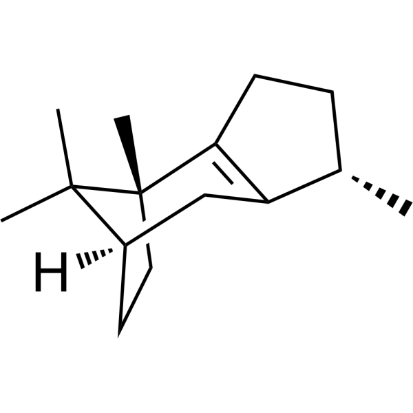
β-Patchoulene
β-Patchoulene is an orally active anti-inflammatory, antioxidant, and anti-apoptotic agent. β-Patchoulene inhibits the NF-κB, TLR4, and cAMP/PKA/CREB signaling pathways; activates the Sirt1/Nrf2 and AMPK signaling pathways; and targets Fas/FasL, Caspase-3, ERK1/2, ROCK1/MLC2 for inhibition. β-Patchoulene regulates cytokine secretion, inflammatory cell infiltration, lipid peroxidation, cell polarization, gut microbiota, and lipid metabolism, restores barrier function, mitochondrial function, and cell viability, and exhibits repellent activity against Spodoptera exigua larvae. β-Patchoulene can be used in research related to various inflammatory, ischemic, fibrosis-associated diseases, as well as hepatocellular carcinoma.
For research use only. We do not sell to patients.
- CAS No.: 514-51-2
- Formula: C15H24
- Molecular Weight:204.35
-
Storage:
-20°C, protect from light
* In solvent : -80°C, 6 months; -20°C, 1 month (protect from light)

To place orders, for customer services and technical support, please contact: MedChemExpress USA
Tel: 609-228-6898 E-mail: [email protected] [email protected]


-
 Biological Activity
-
Chemical Information
- Protocols
- Purity & Documentation
- References
-
 Help & FAQs




Biological Activity
Description
|
NF-κB |
TLR4 |
PKA |
SIRT1 |
AMPK |
Caspase-3 |
ERK1 |
ERK2 |
ROCK-1 |
In Vitro
β-Patchoulene (10-100 μmol/L) exhibits no significant cytotoxicity to human GES-1 gastric epithelial cells at concentrations up to 80 μmol/L[2].
β-Patchoulene (10-40 μmol/L; 4 h pre-incubation, 24 h co-cultivation with ethanol) pre-treatment significantly improves cell viability of ethanol-injured human GES-1 gastric epithelial cells[2].
β-patchoulene (5-50 μM; 1 h pre-incubation) dose-dependently suppresses pro-inflammatory cytokine production, oxidative stress, and apoptosis in H/R-injured cultured macrophages by enhancing Nrf2 nuclear translocation and HO-1 expression while inhibiting NF-κB p-P65 expression[5].
β-Patchoulene (0.1-160 μM; 24 or 48 h) exhibits no cytotoxicity to rat intestinal epithelial IEC-6 cells[8].
β-Patchoulene (20-80 μM; 24 h) significantly recovers the viability of 5-Fluorouracil (HY-90006)-injured rat intestinal epithelial IEC-6 cells[8].
β-Patchoulene (20 μM; 24 h) suppresses AQP3 expression and inactivates the cAMP/PKA/CREB signaling pathway in 5-FU-treated rat intestinal epithelial IEC-6 cells[8].
β-Patchoulene (10-100 μM; 24 h) is non-cytotoxic to human hepatocyte L02 cells with significant cytotoxicity observed only at 100 μM[9].
β-Patchoulene (40 μM; 24 h) significantly attenuates FFA-induced lipid accumulation in human hepatocyte L02 cells via activating the AMPK signaling pathway[9].
β-Patchoulene (0.3125-20 μM; 48 h) suppresses the viability and proliferation of human HCC Huh-7 and MHCC97 cells in a dose-dependent manner[10].
β-Patchoulene (2.5 μM; 48 h) restrains proliferation, facilitates apoptosis, suppresses invasion and EMT, and inactivates the NF-κB/HIF-1α signaling pathway in hypoxia-stimulated human HCC Huh-7 and MHCC97 cells via regulating related protein expressions[10].
β-Patchoulene was the main product of the reaction where recombinant DoPAES protein[12].
MedChemExpress (MCE) has not independently confirmed the accuracy of these methods. They are for reference only.
-
Cell Line:human GES-1 gastric epithelial cells (ethanol-induced injury)
-
Concentration:10, 20, 40 μmol/L
-
Incubation Time:4 h (pre-incubation); 24 h (co-cultivation with ethanol)
-
Result:Increased cell viability in ethanol-exposed GES-1 cells to 66.96% at 10 μmol/L.
Increased cell viability in ethanol-exposed GES-1 cells to 69.81% at 20 μmol/L.
Increased cell viability in ethanol-exposed GES-1 cells to 73.31% at 40 μmol/L.
-
Cell Line:Cultured Macrophages
-
Concentration:5 μM, 50 μM
-
Incubation Time:1 h (pre-incubation)
-
Result:Promoted nuclear Nrf2 translocation in H/R-injured macrophages in a dose-dependent manner.
Upregulated HO-1 protein expression in H/R-injured macrophages in a dose-dependent manner.
Inhibited NF-κB p-P65 protein expression in H/R-injured macrophages in a dose-dependent manner.
-
Cell Line:rat intestinal epithelial IEC-6 cells
-
Concentration:0.1, 1, 10, 20, 40, 60, 80, 160 μM
-
Incubation Time:24 h; 48 h
-
Result:Showed no cytotoxicity to IEC-6 cells at all tested concentrations over 24 and 48 h, with cell viability remaining near 100% relative to the control group.
-
Cell Line:5-fluorouracil (5-FU)-injured rat intestinal epithelial IEC-6 cells
-
Concentration:20, 40, 80 μM
-
Incubation Time:24 h
-
Result:Significantly restored the viability of 5-FU-injured IEC-6 cells.
-
Cell Line:5-FU-treated rat intestinal epithelial IEC-6 cells
-
Concentration:20 μM
-
Incubation Time:24 h (post 8-Br-cAMP induction)
-
Result:Suppressed overexpression of AQP3 and PKA, and inhibited phosphorylation of MEK1/2, MSK1, and CREB compared to the 5-FU-only group.
Had effects counteracted by the PKA agonist 8-Br-cAMP via upregulation of related protein expression or phosphorylation.
-
Cell Line:human hepatocyte L02 cells
-
Concentration:10 μM, 20 μM, 40 μM, 80 μM, 100 μM
-
Incubation Time:24 h
-
Result:Shows no significant reduction in cell viability at concentrations up to 80 μM relative to the normal control group.
Causes a significant decrease in cell viability at 100 μM concentration.
-
Cell Line:human hepatocellular carcinoma Huh-7, MHCC97 cells
-
Concentration:0.3125, 0.625, 1.25, 2.5, 5, 10 μM
-
Incubation Time:48 h
-
Result:Reduced the viability of Huh-7 and MHCC97 cells in a concentration-dependent manner.
-
Cell Line:human hepatocellular carcinoma Huh-7, MHCC97 cells
-
Concentration:0.3125, 0.625, 1.25, 2.5, 5, 10 μM
-
Incubation Time:48 h (treatment), followed by 2 weeks of culture
-
Result:Decreased the colony-forming ability of Huh-7 and MHCC97 cells in a concentration-dependent manner.
-
Cell Line:hypoxia-induced human hepatocellular carcinoma Huh-7, MHCC97 cells
-
Concentration:2.5 μM
-
Incubation Time:48 h
-
Result:Significantly increased the apoptotic rate of hypoxia-induced Huh-7 and MHCC97 cells compared to the hypoxia-only group.
Downregulated the expression of the anti-apoptotic protein Survivin, which was upregulated by hypoxia.
In Vivo
β-Patchoulene (10-40 mg/kg; i.g.; daily; 7 days) dose-dependently protects against ethanol-induced gastric ulcer in male Sprague Dawley rats, anti-inflammatory, anti-apoptotic, and signalling pathway-modulating mechanisms[2].
β-Patchoulene (0.2-1 mg/kg; i.c.v.; single dose immediately post-CLP) significantly improves cognitive function, reduces neuroinflammation and oxidative stress, inhibits microglia activation and M1 polarization, enhances Sirt1/Nrf2/HO-1 pathway activity, reduces splenic lymphocyte apoptosis[3].
β-Patchoulene (10-40 mg/kg; i.g.; daily; 7 days) dose-dependently inhibits xylene-induced ear edema in KM mice[4].
β-Patchoulene (10-40 mg/kg; i.g.; daily; 7 days) dose-dependently reduces acetic acid-induced vascular permeability in KM mice[4].
β-Patchoulene (10-40 mg/kg; i.g.; daily; 7 days) exerts potent, dose-dependent anti-inflammatory effects in carrageenan-induced paw edema in KM mice, mediated via suppression of pro-inflammatory mediators, oxidative stress, and NF-κB signaling[4].
β-Patchoulene (10 mg/kg; i.v.; single dose 2 hours before surgery) preconditioning protects male C57BL/6 mice against hepatic ischemia-reperfusion injury by reducing liver enzyme release, inflammation, oxidative stress, and apoptosis, with concurrent activation of the Nrf2/HO-1 pathway and inhibition of NF-ƘB signaling[5].
β-Patchoulene (10 mg/kg; i.v.; single pretreatment dose 1 h before surgery) exerts neuroprotective effects against cerebral ischemia-reperfusion injury in Sprague-Dawley rats, as evidenced by reduced infarct volume, brain edema, neurological deficits, apoptosis, inflammation, and oxidative stress, via inactivation of the TLR4/NF-κB signaling pathway[6].
β-Patchoulene (5-20 mg/kg; p.o.; daily; 7 days) dose-dependently ameliorates DSS-induced ulcerative colitis and secondary liver injury in male BALB/c mice by suppressing colonic leakage, inhibiting inflammatory signaling pathways, and restoring gut microbiota homeostasis, with the 20 mg/kg dose producing the most robust effects across all measured endpoints[7].
β-Patchoulene (10-40 mg/kg; p.o.; daily; 7 days) dose-dependently ameliorates 5-Fluorouracil (HY-90006)-induced intestinal mucositis in male Sprague Dawley rats, with 40 mg/kg oral gavage achieving the strongest effects on improving body characteristics, intestinal histopathology, suppressing inflammation, restoring the mucus barrier, and inhibiting the cAMP/PKA/CREB signaling pathway to reduce AQP3 expression[8].
β-Patchoulene (40 mg/kg; p.o.; daily; 7 days) ameliorates 5-Fluorouracil (HY-90006)-induced intestinal mucositis in male Sprague Dawley rats[8].
β-Patchoulene (10-40 mg/kg; i.g.; daily; 4 weeks) activates AMPK signaling to inhibit hepatic lipid synthesis and promote mitochondrial lipid oxidation, significantly alleviating HFD-induced NAFLD in rats[9].
β-Patchoulene (5-20 μmol/kg) dose-dependently reduces hepatocellular carcinoma tumor growth, induces tumor cell apoptosis, inhibits epithelial-mesenchymal transition, and inactivates the NF-κB/HIF-1α pathway in hypoxic nude mouse models[10].
MedChemExpress (MCE) has not independently confirmed the accuracy of these methods. They are for reference only.
-
Animal Model:Kunming (KM) (male, 18-22 g, LPS-induced ALI)[1]
-
Dosage:2.5 mg/kg; 5 mg/kg; 10 mg/kg
-
Administration:p.o.; once daily; 7 days
-
Result:Decreased LPS-induced mouse mortality effectively.
Lessened pulmonary edema and improved multiple pathological lesions in lung tissues.
Downregulated pulmonary oxidative stress levels and inflammatory cytokine secretion.
Suppressed the activation of NF-κB inflammatory signaling pathway.
Promoted the activation of Nrf2 antioxidant signaling pathway.
Upregulated the expression level of miR-146a in lung tissues.
-
Animal Model:Sprague Dawley (male, 180-220 g, ethanol-induced gastric ulcer)[2]
-
Dosage:10 mg/kg; 20 mg/kg; 40 mg/kg
-
Administration:i.g.; daily; 7 days
-
Result:Reduced gastric ulcer area in a dose-dependent manner with obvious inhibition effect.
Lowered histological total microscopic score in a dose-dependent manner, alleviating epithelial damage and submucosal oedema.
Improved SOD activity, enhanced CAT activity, increased GSH level, decreased MDA level, and reduced serum levels of TNF-α, IL-1β and IL-6 at all doses.
Downregulated Fas, FasL, and caspase-3 IODs in a dose-dependent manner at all doses.
Inhibited increased p-p65/p65 and p-IκB/IκB ratios at the highest dose.
Enhanced p-ERK1/2/ERK1/2 ratio at the highest dose.
Upregulated c-fos mRNA, c-jun mRNA, and miR-21 expression at the highest dose.
-
Animal Model:C57BL/6 (male, 6-10 weeks old, sepsis associated encephalopathy induced by cecal ligation and puncture surgery)[3]
-
Dosage:0.2 mg/kg; 1 mg/kg
-
Administration:i.c.v.; single dose immediately post-CLP
-
Result:Elevated behavioral and cognitive related indicators at multiple time points after CLP operation at 1 mg/kg, and effectively raised the survival rate of model mice.
Reduced cerebral inflammatory factors and oxidative stress levels without affecting anti-inflammatory factor expression, and inhibited hippocampal microglial M1 polarization.
Regulated the expression of Sirt1/Nrf2 pathway-related proteins and relieved hippocampal neuronal apoptosis.
Alleviated systemic inflammatory response and suppressed splenic lymphocyte apoptosis.
Low-dose treatment could partially ameliorate cognitive dysfunction.
-
Animal Model:Kun Ming (KM) (male and female, 18-22 g)[4]
-
Dosage:10 mg/kg; 20 mg/kg; 40 mg/kg
-
Administration:i.g.; daily; 7 days
-
Result:Inhibited xylene-induced ear edema and acetic acid-triggered vascular permeability in a dose-dependent manner, and relieved carrageenan-caused paw edema with obvious long-term inhibitory effects.
Alleviated pathological swelling, tissue damage and inflammatory cell infiltration, and improved tissue fibrous structure.
Lowered paw tissue MDA and MPO contents effectively.
Reduced the release of multiple pro-inflammatory cytokines and inflammatory mediators.
Downregulated the expression levels of iNOS and COX-2 proteins.
Stabilized IκBα to block NF-κB signaling pathway activation and restrain nuclear translocation of p65.
-
Animal Model:C57BL/6 (male, 8-10 weeks old)[5]
-
Dosage:10 mg/kg
-
Administration:i.v.; single dose 2 hours before surgery
-
Result:Lowered serum ALT and AST levels and alleviated liver pathological injury in hepatic ischemia-reperfusion mice.
Inhibited hepatic inflammatory cytokine expression and reduced inflammatory factor-positive macrophages.
Mitigated hepatic oxidative stress and restrained hepatocyte apoptosis.
Suppressed M1 polarization of Kupffer cells.
Activated Nrf2/HO-1 antioxidant pathway and inhibited NF-κB p65 phosphorylation.
-
Animal Model:Sprague-Dawley (male, 8 weeks old, 80-120 g, middle cerebral artery occlusion for 2 h followed by 24 h reperfusion)[6]
-
Dosage:10 mg/kg
-
Administration:i.v.; single pretreatment dose 1 h before surgery
-
Result:Reduced cerebral infarct volume, brain edema and neurological deficits, and recovered mitochondrial membrane potential in cerebral ischemia-reperfusion injured rats.
Declined neuronal apoptosis rate, regulated BAX/Bcl-2 balance and lowered caspase-3 activity.
Downregulated inflammatory factor expression, relieved oxidative stress damage and improved antioxidant enzyme activity.
Blocked the TLR4/NF-κB pathway via modulating related protein expression and restricting p65 nuclear translocation.
-
Animal Model:BALB/c (male, 21-25 g, induced with 3% dextran sulfate sodium in drinking water ad libitum for 7 days)[7]
-
Dosage:5 mg/kg; 10 mg/kg; 20 mg/kg
-
Administration:p.o.; daily; 7 days
-
Result:Improved general disease conditions and relieved colon tissue damage in a dose-dependent manner, and alleviated colon shortening at medium and high doses.
Inhibited intestinal cell apoptosis and upregulated intestinal barrier-related gene expression at medium and high doses.
Lowered colonic inflammatory mediators and adhesion molecule contents, and restrained multiple inflammatory signaling pathways.
Declined local and systemic lipopolysaccharide levels, and regulated various serum inflammatory and liver function related indicators.
Ameliorated liver inflammatory lesions and reduced hepatic inflammatory factor release in a dose-related way.
Restored intestinal microbial diversity and optimized gut flora structure at high dose.
-
Animal Model:Sprague Dawley (male, 180-220 g, 5-fluorouracil-induced intestinal mucositis)[8]
-
Dosage:10 mg/kg; 20 mg/kg; 40 mg/kg
-
Administration:p.o.; daily; 7 days
-
Result:Promoted body weight gain and improved food intake at medium and high doses, and relieved diarrhea symptoms at all doses in 5-FU induced model rats.
Optimized intestinal tissue morphological structure and alleviated intestinal pathological damage at all doses.
Regulated intestinal inflammatory factor levels effectively at medium and high doses.
Elevated intestinal mucin secretion and increased goblet cell quantity to enhance intestinal mucosal barrier function.
Downregulated the expression of related functional proteins and restrained the activation of multiple downstream signaling pathways in a dose-dependent manner.
-
Animal Model:Sprague Dawley (male, 180-220 g, 5-fluorouracil-induced intestinal mucositis)[8]
-
Dosage:40 mg/kg
-
Administration:p.o.; daily; 7 days
-
Result:Promoted body weight gain and improved food intake at medium and high doses, and relieved diarrhea symptoms at all doses in 5-FU induced model rats.
Optimized intestinal tissue morphological structure and alleviated intestinal pathological damage at all doses.
Reduced intestinal TNF-α, IL-1β, IL-6 and IL-10 contents obviously at medium and high doses.
Elevated intestinal MUC2 mucin level and goblet cell number to strengthen intestinal mucosal barrier function.
Downregulated the expression of related functional proteins and inhibited the activation of MEK/ERK/p38 signaling cascades at all doses with dose-dependent effects.
-
Animal Model:Sprague Dawley (male, 180−220 g, NAFLD induced by 60% fat high-fat diet for 4 weeks)[9]
-
Dosage:10 mg/kg; 20 mg/kg; 40 mg/kg
-
Administration:i.g.; daily; 4 weeks
-
Result:Reduced liver index without affecting body weight gain at low dose, while middle and high doses decreased both body weight gain and liver index in HFD-fed models.
Improved serum levels of ALT, AST, HDL-C, LDL-C, TG and TC, and lowered hepatic TG, TC and NEFA contents at all tested doses.
Alleviated hepatic pathological lesions and decreased lipid deposition area in liver tissues.
Suppressed the expression of hepatic lipid synthesis related proteins and genes including SREBP-1c, HMG-CR, FASN and SCD1, and regulated ACC1 phosphorylation level.
Elevated hepatic antioxidant capacity by decreasing MDA content and raising GSH-Px and SOD activities, and upregulated lipid oxidation markers SIRT1, PPARα and CPT-1a with no obvious influence on ACOX1 expression.
Activated AMPK signaling pathway by elevating p-AMPKα/AMPKα ratio and AMPKα mRNA expression in liver tissues in a dose-dependent manner.
- Collagen: Sirius Red Staining
- Mitochondrial membrane-potential fluorescent assay
- Mammalian live/dead viability and cytotoxicity staining
- Connective Tissue: Masson's Trichrome/Collagen Trichrome Staining
- Research Protocol for Microbiome Analysis
- CCK-8/WST-8 Cell Proliferation Assay
- Research Protocol for Inflammation-related Diseases
- Liver Cancer Modeling
- Cell Viability Determination by MTT Colorimetric Assay
- Fibrosis/Collagen Morphometry
- Detection of Apoptosis
- Cell Cytotoxicity Assay
- Mitochondrial membrane-potential and mitochondrial mass staining
- Apoptosis Solutions
- Ferroptosis Solutions
- MTT Cell Proliferation Assay


Chemical Information
-
CAS No. 514-51-2
-
Appearance Liquid (Density: 0.951±0.10 g/cm3)
-
Molecular Weight 204.35
-
Formula C15H24
-
Color Colorless to light yellow
-
SMILES
C[C@]1(CC2)C(CC[C@@H]3C)=C3C[C@]2([H])C(C)1C
-
Structure Classification
-
Initial Source
-
Shipping
Room temperature in continental US; may vary elsewhere.
-
Storage
-20°C, protect from light
* In solvent : -80°C, 6 months; -20°C, 1 month (protect from light)


Purity & Documentation
-
Data Sheet (310 KB)
-
SDS (393 KB)
- English - EN (393 KB)
- Français - FR (393 KB)
- Deutsch - DE (393 KB)
- Norwegian - NO (393 KB)
- Español - ES (393 KB)
- Swedish - SV (393 KB)
- Italian - IT (393 KB)
- Korean - KR (393 KB)
- Portuguese - PT (393 KB)
-
Handling Instructions (2659 KB)
References
[1]. Chen XY, et al. β-Patchoulene from patchouli oil protects against LPS-induced acute lung injury via suppressing NF-κB and activating Nrf2 pathways. Int Immunopharmacol. 2017;50:270-278. [Content Brief]
[2]. Liu Y, et al. Transformation of patchouli alcohol to β-patchoulene by gastric juice: β-patchoulene is more effective in preventing ethanol-induced gastric injury. Sci Rep. 2017;7(1):5591. [Content Brief]
[3]. Tian Y, et al. β-patchoulene alleviates cognitive dysfunction in a mouse model of sepsis associated encephalopathy by inhibition of microglia activation through Sirt1/Nrf2 signaling pathway. PLoS One. 2023;18(1):e0279964. [Content Brief]
[4]. Zhang Z, et al. Anti-inflammatory activity of β-patchoulene isolated from patchouli oil in mice. Eur J Pharmacol. 2016;781:229-238. [Content Brief]
[5]. Tao T, et al. β-Patchoulene Preconditioning Protects Mice Against Hepatic Ischemia-Reperfusion Injury by Regulating Nrf2/HO-1 Signaling Pathway. J Surg Res. 2022;275:161-171. [Content Brief]
[6]. Zhang FB, et al. Effect of β-patchoulene on cerebral ischemia-reperfusion injury and the TLR4/NF-κB signaling pathway. Exp Ther Med. 2019;17(5):3335-3342. [Content Brief]
[8]. Wu J, et al. β-Patchoulene Ameliorates Water Transport and the Mucus Barrier in 5-Fluorouracil-Induced Intestinal Mucositis Rats via the cAMP/PKA/CREB Signaling Pathway. Front Pharmacol. 2021;12:689491. [Content Brief]
[9]. Xu N, et al. β-patchoulene improves lipid metabolism to alleviate non-alcoholic fatty liver disease via activating AMPK signaling pathway. Biomed Pharmacother. 2021;134:111104. [Content Brief]
[10]. Tu H, et al. β-Patchoulene represses hypoxia-induced proliferation and epithelial-mesenchymal transition of liver cancer cells. Bioengineered. 2022;13(5):11907-11922. [Content Brief]
[11]. Pu Q, et al. A Wheat β-Patchoulene Synthase Confers Resistance Against Herbivory in Transgenic Arabidopsis. Genes (Basel). 2019;10(6):441. [Content Brief]

Calculators
Concentration (start) × Volume (start) = Concentration (final) × Volume (final)


β-Patchoulene Related Classifications
HY-N12378 Related Classifications
 Powered by Bioz
Keywords
- β-Patchoulene
- 514-51-2
- NF-κB
- Toll-like Receptor (TLR)
- PKA
- Epigenetic Reader Domain
- Keap1-Nrf2
- Sirtuin
- AMPK
- Caspase
- FASTK
- ERK
- ROCK
- Apoptosis
- human GES-1 gastric epithelial cells
- gastric ulcer
- rat intestinal epithelial IEC-6 cells
- human HCC MHCC97 cells
- human hepatocyte L02 cells
- sepsis associated encephalopathy
- Nrf2
- acute lung injury
- human HCC Huh-7 cells
- Inhibitor
- inhibitor
- inhibit