



γ-Glutamylvaline

Based on 1 Customer Validation
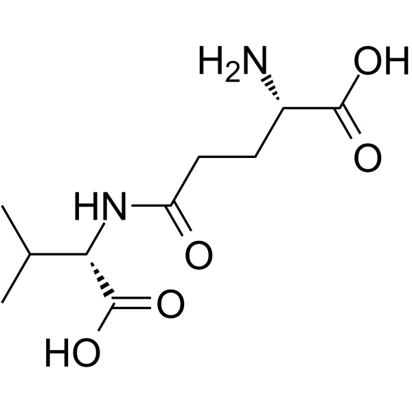
γ‑Glutamylvaline (γ-Glu-Val) is a calcium‑sensing receptor (CaSR) agonist. γ‑Glutamylvaline activates CaSR and facilitates its binding to β‑arrestin 2 to modulate inflammatory and metabolic homeostasis signaling. γ‑Glutamylvaline inhibits TNF‑α‑induced IL‑6/MCP‑1 and enhances adiponectin/PPARγ in adipocytes. γ‑Glutamylvaline upregulates Wnt5a, restores β‑catenin phosphorylation, and reduces serine‑phosphorylated IRS‑1 in adipocytes. γ-Glutamylvaline can be used for the research of low-grade chronic inflammation.
For research use only. We do not sell to patients.
- Purity : 99.94%
- CAS No.: 2746-34-1
- Formula: C10H18N2O5
- Molecular Weight:246.26
-
Storage:
Sealed storage, away from moisture.
Powder -80°C, 2 years , -20°C, 1 year* In solvent : -80°C, 6 months; -20°C, 1 month (sealed storage, away from moisture)

To place orders, for customer services and technical support, please contact: MedChemExpress USA
Tel: 609-228-6898 E-mail: [email protected] [email protected]


-
 Biological Activity
-
Chemical Information
-
Solvent & Solubility
- Protocols
- Purity & Documentation
- References
-
 Help & FAQs


-
HY-P4632
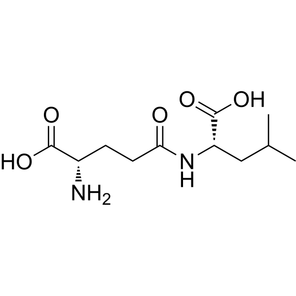
H-γ-Glu-Leu-OHDipeptide -
HY-113402
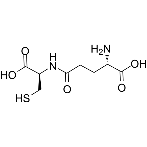
Gamma-glutamylcysteineDipeptide -
HY-N0391
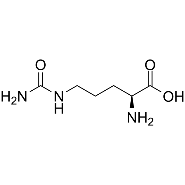
L-CitrullineEndogenous Metabolite -
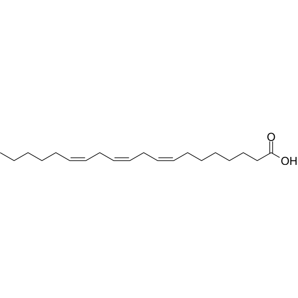
HY-A0143
Dihomo-γ-linolenic acidEndogenous Metabolite -
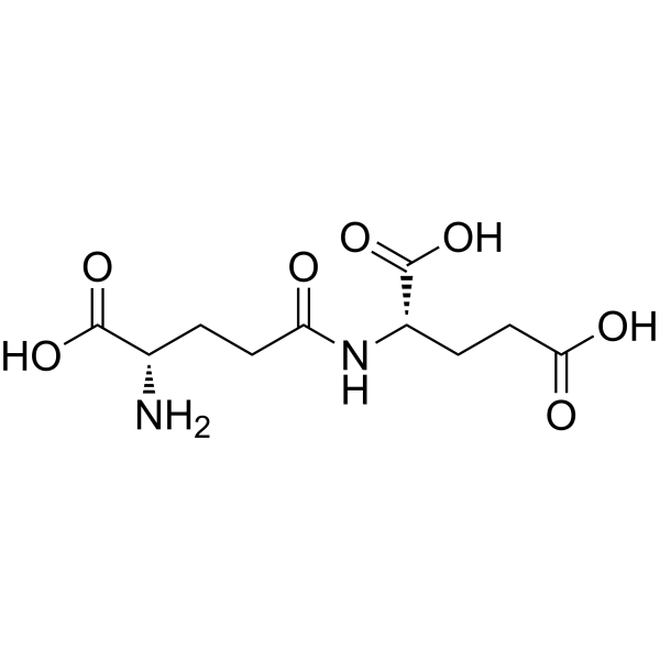
HY-118090
L-γ-Glutamyl-L-glutamic acidPoly-γ-glutamic Acid Hydrolysis Product -
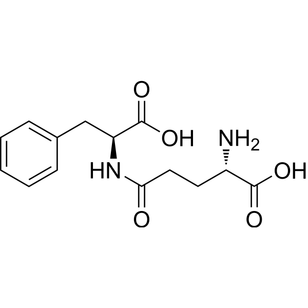
HY-101399
γ-Glu-PheEndogenous Metabolite -
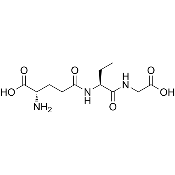
HY-126752
Ophthalmic acidGlutathione S-transferase Inhibitor -
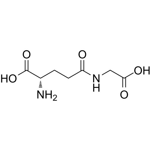
HY-P3280
γ-Glu-GlyHuman Lipid Metabolite -

HY-W724905
gamma-GlutamylisoleucineDipeptide -
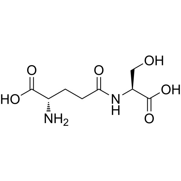
HY-138119
γ-GlutamylserineCalcium Receptor Activator -
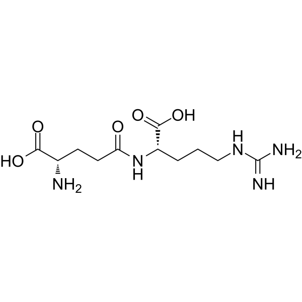
HY-N10477
gamma-Glutamylarginineγ-glutamic Acid
All Endogenous Metabolite Isoforms
More

Biological Activity
Description
IC50 & Target
[1]|
PPAR-γ |
IL-6 |
Wnt5A |
In Vitro
γ-Glutamylvaline (0.01-10 μM; 6 h) activates CaSR in differentiated 3T3-L1 mouse adipocytes, as measured by increased intracellular calcium fluorescence after 6 h of incubation[1].
γ-Glutamylvaline (1-100 μM; 24 h pretreatment, 24 h TNF-α exposure) significantly suppresses TNF-α-induced IL-6 and MCP-1 production and restores adiponectin levels in differentiated 3T3-L1 mouse adipocytes[1].
γ-Glutamylvaline (10 μM; 2 h) promotes the binding interaction between CaSR and β-arrestin 2 in differentiated 3T3-L1 mouse adipocytes[1].
γ-Glutamylvaline (10 μM; 2 h) upregulates Wnt5a gene expression and restores TNF-α-suppressed β-catenin phosphorylation in differentiated 3T3-L1 mouse adipocytes[1].
γ-Glutamylvaline (10 μM; 2 h pretreatment, 30 min TNF-α stimulation) counteracts TNF-α-induced downregulation of PPARγ1+2 in differentiated 3T3-L1 mouse adipocytes[1].
γ-Glutamylvaline (10 μM; 45 min insulin exposure, 2 h treatment, 30 min TNF-α stimulation) inhibits TNF-α-induced serine phosphorylation of IRS-1 in differentiated 3T3-L1 mouse adipocytes[1].
MedChemExpress (MCE) has not independently confirmed the accuracy of these methods. They are for reference only. Further protocols information, click here.
-
Cell Line:differentiated 3T3-L1 mouse adipocytes (TNF-α-induced inflammation model)
-
Concentration:1, 10, 100 μM; 10 μM (IL-6 suppression); 10, 100 μM (MCP-1 suppression; adiponectin restoration)
-
Incubation Time:24 h (pretreatment); 24 h (TNF-α exposure)
-
Result:Suppressed TNF-α-induced IL-6 production to 23.92 ng/mL at 10 μM.
Suppressed TNF-α-induced MCP-1 production to 101.17 ng/mL and 120.36 ng/mL at 10 μM and 100 μM, respectively.
Restored adiponectin production to 7.37 ng/mL and 7.40 ng/mL at 10 μM and 100 μM, respectively.
-
Cell Line:differentiated 3T3-L1 mouse adipocytes (TNF-α-induced model)
-
Concentration:10 μM
-
Incubation Time:2 h (pretreatment); 30 min (TNF-α stimulation)
-
Result:Counteracted TNF-α-induced downregulation of PPARγ1/PPARγ2, increasing expression to 1.249 relative to β-actin at 10 μM.
This effect was reversed by pretreatment with the CaSR antagonist NPS-2143.
-
Cell Line:differentiated 3T3-L1 mouse adipocytes
-
Concentration:10 μM
-
Incubation Time:45 min (insulin exposure); 2 h (γ-glutamylvaline treatment); 30 min (TNF-α stimulation)
-
Result:Inhibited TNF-α-upregulated p-Ser-IRS1 expression to 1.89 relative to total IRS-1 at 10 μM.


Chemical Information
-
CAS No. 2746-34-1
-
Appearance Solid
-
Molecular Weight 246.26
-
Formula C10H18N2O5
-
Color White to off-white
-
Synonyms
γ-Glu-Val
-
Shipping
Room temperature in continental US; may vary elsewhere.
-
Storage
Sealed storage, away from moisture
Powder -80°C 2 years -20°C 1 year * In solvent : -80°C, 6 months; -20°C, 1 month (sealed storage, away from moisture)


Solvent & Solubility
In Vitro:
H2O : ≥ 100 mg/mL (406.07 mM)
* "≥" means soluble, but saturation unknown.
Please refer to the solubility information to select the appropriate solvent. Once prepared, please aliquot and store the solution to prevent product inactivation from repeated freeze-thaw cycles.
Storage method and period of stock solution: -80°C, 6 months; -20°C, 1 month (sealed storage, away from moisture). When stored at -80°C, please use it within 6 months. When stored at -20°C, please use it within 1 month.
* Note: If you choose water as the stock solution, please dilute it to the working solution, then filter and sterilize it with a 0.22 μm filter before use.
Please refer to the solubility information to select the appropriate solvent. Once prepared, please aliquot and store the solution to prevent product inactivation from repeated freeze-thaw cycles.
Storage method and period of stock solution: -80°C, 6 months; -20°C, 1 month (sealed storage, away from moisture). When stored at -80°C, please use it within 6 months. When stored at -20°C, please use it within 1 month.
* Note: If you choose water as the stock solution, please dilute it to the working solution, then filter and sterilize it with a 0.22 μm filter before use.
Concentration (start) × Volume (start) = Concentration (final) × Volume (final)

Protocols
-
Kinase activity and phosphorylation assays
Kinase activity assays measure the ability of kinases to transfer phosphate groups from ATP to specific substrates, while phosphorylation assays detect the presence and levels of phosphorylated proteins. Common methods include radiolabeled ATP incorporation (e. g. ,), ADP release detection via bioluminescence (e. g. ,[3]), enzyme-linked immunosorbent assays (ELISA) for phospho-specific epitopes (e. g. ,[6]), and microtiter-based formats for high-throughput screening (e. g. ,[8]). The ADP-Glo assay quantifies kinase activity by measuring ADP produced during phosphorylation using a luciferase-based system. Radiometric assays involve autoradiography or scintillation counting after incorporation of 32P-labeled ATP into substrate proteins. ELISA-based approaches rely on phospho-specific antibodies to detect activated kinases in cell lysates or purified samples.
-
Western Blot
Western blotting (WB) is a commonly used experimental method in molecular biology, biochemistry, and immunogenetics for identifying and quantifying target proteins. It combines gel electrophoresis with immunoassay, enabling researchers to analyze protein expression, post-translational modifications, and molecular weight.
-
Cotton Pellet Granuloma
Cotton pellet granuloma is a classical in vivo chronic inflammation model used to evaluate the anti-inflammatory potential of test substances by measuring their ability to inhibit granuloma tissue formation around an implanted foreign body (cotton pellet) in rodents. The method is based on the biological response to a sterile implanted material, which induces proliferative phase inflammation characterized by fibroblast proliferation and collagen-rich granuloma formation, and the final readout reflects the extent of chronic inflammatory tissue growth surrounding the pellet. In multiple preclinical pharmacological evaluations, inhibition of cotton pellet-induced granuloma formation has been used as an indicator of anti-inflammatory activity in both synthetic and natural product screening contexts.
-
Large-size fat particle sorting
Large-size fat particle sorting is widely used to isolate cells up to 200 μm in diameter. Single-cell flow sorting will allow greater insight into adipocyte heterogeneity by identifying gene expression, protein composition, and metabolic signatures at the single-cell level.
-
Protocol for Kinase activity and phosphorylation assays
Kinase activity assays measure transfer of phosphate from ATP to a protein or peptide substrate, generating phosphorylated substrate, ADP, or incorporated radiolabeled phosphate as the readout; phosphorylation assays measure site-specific phosphorylation in cells or tissues as a proxy for kinase-pathway activation, inhibition, or substrate regulation. Phosphorylation can be detected by phospho-specific Western blot, immunoprecipitation kinase assay, phospho-immunofluorescence, phospho-flow cytometry, luminescent ADP detection, radiolabeled ATP incorporation, or reporter-based pathway assays, and these readouts can be applied to cancer cells, primary neurons, mouse tumors, organoids, inflammatory macrophages, ferroptosis studies, and mitophagy studies when the kinase target is biologically relevant.
-
Research Protocol for Inflammation-related Diseases
The NLRP3 inflammasome is a cytosolic innate immune signaling platform that integrates priming signals and danger-signal activation to promote caspase-1 activation, maturation of IL-1β and IL-18, and gasdermin D-mediated pyroptotic cell death. The core experimental logic is to determine whether inflammatory disease phenotypes are driven by increased NLRP3 expression, ASC-containing inflammasome assembly, caspase-1 cleavage, GSDMD cleavage, and extracellular release of IL-1β/IL-18 rather than by nonspecific cell injury alone. The pathway is strongly linked to inflammation-related disease phenotypes because monosodium urate crystals activate NALP3/NLRP3 inflammasome signaling in gout-like crystal inflammation, cholesterol crystals activate NLRP3 inflammasomes in atherogenesis models, and DSS-induced intestinal inflammation has been reported to involve NLRP3 inflammasome activity. However, experimental colitis studies also show context-dependent protective effects of NLRP3 inflammasome co


Purity & Documentation
-
Data Sheet (280 KB)
-
SDS (252 KB)
- English - EN (252 KB)
- Français - FR (252 KB)
- Deutsch - DE (252 KB)
- Norwegian - NO (252 KB)
- Español - ES (252 KB)
- Swedish - SV (252 KB)
- Italian - IT (252 KB)
- Korean - KR (252 KB)
- Portuguese - PT (252 KB)
-
Handling Instructions (2659 KB)
References


Complete Stock Solution Preparation Table
Please refer to the solubility information to select the appropriate solvent. Once prepared, please aliquot and store the solution to prevent product inactivation from repeated freeze-thaw cycles.
Storage method and period of stock solution: -80°C, 6 months; -20°C, 1 month (sealed storage, away from moisture). When stored at -80°C, please use it within 6 months. When stored at -20°C, please use it within 1 month.
| Optional Solvent | Concentration Solvent Mass | 1 mg | 5 mg | 10 mg | 25 mg |
|---|---|---|---|---|---|
| H2O | 1 mM | 4.0607 mL | 20.3037 mL | 40.6075 mL | 101.5187 mL |
| 5 mM | 0.8121 mL | 4.0607 mL | 8.1215 mL | 20.3037 mL | |
| 10 mM | 0.4061 mL | 2.0304 mL | 4.0607 mL | 10.1519 mL | |
| 15 mM | 0.2707 mL | 1.3536 mL | 2.7072 mL | 6.7679 mL | |
| 20 mM | 0.2030 mL | 1.0152 mL | 2.0304 mL | 5.0759 mL | |
| 25 mM | 0.1624 mL | 0.8121 mL | 1.6243 mL | 4.0607 mL | |
| 30 mM | 0.1354 mL | 0.6768 mL | 1.3536 mL | 3.3840 mL | |
| 40 mM | 0.1015 mL | 0.5076 mL | 1.0152 mL | 2.5380 mL | |
| 50 mM | 0.0812 mL | 0.4061 mL | 0.8121 mL | 2.0304 mL | |
| 60 mM | 0.0677 mL | 0.3384 mL | 0.6768 mL | 1.6920 mL | |
| 80 mM | 0.0508 mL | 0.2538 mL | 0.5076 mL | 1.2690 mL | |
| 100 mM | 0.0406 mL | 0.2030 mL | 0.4061 mL | 1.0152 mL |
* Note: If you choose water as the stock solution, please dilute it to the working solution, then filter and sterilize it with a 0.22 μm filter before use.


γ-Glutamylvaline Related Classifications
HY-12557 Related Classifications
 Powered by Bioz