



Gynostemma Extract
Based on 1 publication(s) in Google Scholar
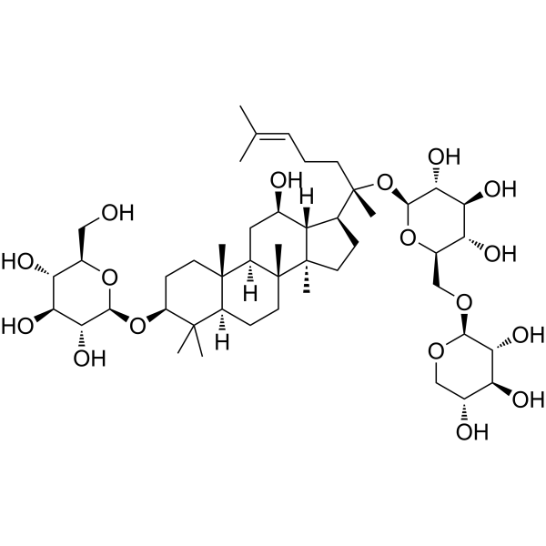
Gynostemma Extract (Gypenoside IX) is a triterpenoid saponin. Gynostemma Extract can be isolated from Panax notoginseng. Gynostemma Extract reduces NFκB nuclear translocation and transcriptional activity by inhibiting the p38 MAPK/Akt/NFκB signaling pathway. Gynostemma Extract reduces the expression of cytokines (COX-2, IL-6) and the production of NO. Gynostemma Extract has anti-inflammatory activity. Gynostemma Extract can be used in the study of liver injury and neuroinflammation.
Para uso exclusivo en investigación. No vendemos a pacientes.
- Pureza : 99.92%
- No. CAS: 80321-63-7
- Fòrmula: C47H80O17
- Peso molecular:917.13
-
Almacenamiento:Powder -20°C, 3 years , 4°C, 2 years ; In solvent -80°C, 6 months , -20°C, 1 month

To place orders, for customer services and technical support, please contact: MedChemExpress USA
Tel: 609-228-6898 E-mail: [email protected] [email protected]


-
 Actividad biológica
-
Chemical Information
-
Solvente y solubilidad
- Protocolo
- Pureza y Documentación
- Referencias
-
 Help & FAQs


Publications Citing Use of MedChemExpress (MCE) Gynostemma Extract
More

Actividad biológica
Descripciòn
|
IL-6 |
COX-2 |
In Vitro
Gynostemma Extract (3.125-50 μM; 24 h) shows no cytotoxicity on C6 cells[2].
Gynostemma Extract (3.125-50 μM; 2 h) dose-dependently reduces NO production in C6 cells stimulated by LPS and TNF-α, with an IC50 of around 25 μM [2].
MedChemExpress (MCE) has not independently confirmed the accuracy of these methods. They are for reference only.
In Vivo
MedChemExpress (MCE) has not independently confirmed the accuracy of these methods. They are for reference only.
-
Animal Model:Five-week-old male C57BL/6 mice with LPS-induced neuroinflammation [2]
-
Dosage:3 mg/kg, 10 mg/kg, 30 mg/kg
-
Administration:Intraperitoneal injection
-
Result:Prevented the increase of mRNA expression of inflammatory genes such as iNOS, TNF-α, IL-6, and COX-2 in brain cortex.
Reduced the production of TNF-α, IL-6, and iNOS at protein levels, alleviated astrogliosis, and impeded the elevation of GFAP.
Attenuated the phosphorylation of NFκB, IκB, p38 MAPK, and Akt in brain cortex.
Ensayo clínico
| NCT Number | Sponsor | Condition | Start Date |
Phase

|
|---|---|---|---|---|
| NCT01329991 | 2011-05 | PHASE1 |


Chemical Information
-
No. CAS 80321-63-7
-
Appearance Solid
-
Peso molecular 917.13
-
Fòrmula C47H80O17
-
Color White to off-white
-
SMILES
O[C@H]1[C@H](O)[C@@H](CO)O[C@@H](O[C@@H]2C(C)(C)[C@@](CC[C@]3(C)[C@]4([H])C[C@@H](O)[C@@]5([H])[C@@]3(C)CC[C@@H]5[C@](O[C@@H]6O[C@H](CO[C@@H]7OC[C@@H](O)[C@H](O)[C@H]7O)[C@@H](O)[C@H](O)[C@H]6O)(C)CC/C=C(C)\C)([H])[C@]4(C)CC2)[C@@H]1O
-
Synonyms
Ginsenoside C-Mx1; Gypenoside IX
-
Structure Classification
-
Envío
Room temperature in continental US; may vary elsewhere.
-
Almacenamiento
Powder -20°C 3 years 4°C 2 years In solvent -80°C 6 months -20°C 1 month


Publications (1)
-
Journal Impact Factor
-
Most Recent


Solvente y solubilidad
In Vitro:
DMSO : 100 mg/mL (109.04 mM; ultrasonic and warming and heat to 60°C; Hygroscopic DMSO has a significant impact on the solubility of product, please use newly opened DMSO)
Please refer to the solubility information to select the appropriate solvent. Once prepared, please aliquot and store the solution to prevent product inactivation from repeated freeze-thaw cycles.
Storage method and period of stock solution: -80°C, 6 months; -20°C, 1 month. When stored at -80°C, please use it within 6 months. When stored at -20°C, please use it within 1 month.
Please refer to the solubility information to select the appropriate solvent. Once prepared, please aliquot and store the solution to prevent product inactivation from repeated freeze-thaw cycles.
Storage method and period of stock solution: -80°C, 6 months; -20°C, 1 month. When stored at -80°C, please use it within 6 months. When stored at -20°C, please use it within 1 month.
Concentration (start) × Volume (start) = Concentration (final) × Volume (final)
In Vivo:
Select the appropriate dissolution method based on your experimental animal and administration route.
- For the following dissolution methods, please ensure to first prepare a clear stock solution using an In Vitro approach and then sequentially add co-solvents:
- To ensure reliable experimental results, the clarified stock solution can be appropriately stored based on storage conditions. As for the working solution for In Vivo experiments, it is recommended to prepare freshly and use it on the same day.
- The percentages shown for the solvents indicate their volumetric ratio in the final prepared solution. If precipitation or phase separation occurs during preparation, heat and/or sonication can be used to aid dissolution.
Add each solvent one by one: 10% DMSO 40% PEG300 5% Tween-80 45% Saline
Solubility: 2.5 mg/mL (2.73 mM); Suspended solution; Need ultrasonic
This protocol yields a suspended solution of 2.5 mg/mL. Suspended solution can be used for oral and intraperitoneal injection.
Taking 1 mL working solution as an example, add 100 μL DMSO stock solution (25.0 mg/mL) to 400 μL PEG300, and mix evenly; then add 50 μL Tween-80 and mix evenly; then add 450 μL Saline to adjust the volume to 1 mL.
Preparation of Saline: Dissolve 0.9 g sodium chloride in ddH₂O and dilute to 100 mL to obtain a clear Saline solution.
Add each solvent one by one: 10% DMSO 90% (20% SBE-β-CD in Saline)
Solubility: ≥ 2.5 mg/mL (2.73 mM); Clear solution
This protocol yields a clear solution of ≥ 2.5 mg/mL (saturation unknown).
Taking 1 mL working solution as an example, add 100 μL DMSO stock solution (25.0 mg/mL) to 900 μL 20% SBE-β-CD in Saline, and mix evenly.
Preparation of 20% SBE-β-CD in Saline (4°C, storage for one week): 2 g SBE-β-CD powder is dissolved in 10 mL Saline, completely dissolve until clear.
In Vivo Dissolution Calculator
Please enter the basic information of animal experiments:
-
-
-
-
Recommended: Prepare an additional quantity of animals to account for potential losses during experiments.
Please enter your animal formula composition:
-
%DMSO +
Recommended: Keep the proportion of DMSO in working solution below 2% if your animal is weak.
-
%+
-
+%Tween-80 + +
-
%Saline +
The co-solvents required include: DMSO, . All of co-solvents are available by MedChemExpress (MCE). , Tween 80. All of co-solvents are available by MedChemExpress (MCE).
Working solution concentration: 0.22 mg/mL
Method for preparing stock solution: mg drug dissolved in μL DMSO. Stock solution concentration: mg/mL.
1. Take μL DMSO stock solution;
2. Add μL .
μL , mix evenly;
3. Then add μL Tween 80, mix evenly;
4. Then add μL
Please ensure that the stock solution in the first step is dissolved to a clear state, and add co-solvents in sequence. You can use ultrasonic heating (ultrasonic cleaner, recommended frequency 20-40 kHz), vortexing, etc. to assist dissolution.


Pureza y Documentación
-
Ficha de datos (280 KB)
-
SDS (393 KB)
- English - EN (393 KB)
- Français - FR (393 KB)
- Deutsch - DE (393 KB)
- Norwegian - NO (393 KB)
- Español - ES (393 KB)
- Swedish - SV (393 KB)
- Italian - IT (393 KB)
- Korean - KR (393 KB)
- Portuguese - PT (393 KB)
-
Instrucciones de manejo (2659 KB)
Referencias
[1]. Yoshikawa M, et al. Structures of new dammarane-type Triterpene Saponins from the flower buds of Panax notoginseng and hepatoprotective effects of principal Ginseng Saponins. J Nat Prod. 2003 Jul;66(7):922-7. [Content Brief]
[2]. Wang X, et al. Gypenoside IX Suppresses p38 MAPK/Akt/NFκB Signaling Pathway Activation and Inflammatory Responses in Astrocytes Stimulated by Proinflammatory Mediators. Inflammation. 2017 Dec;40(6):2137-2150. [Content Brief]


Complete Stock Solution Preparation Table
Please refer to the solubility information to select the appropriate solvent. Once prepared, please aliquot and store the solution to prevent product inactivation from repeated freeze-thaw cycles.
Storage method and period of stock solution: -80°C, 6 months; -20°C, 1 month. When stored at -80°C, please use it within 6 months. When stored at -20°C, please use it within 1 month.
| Optional Solvent | Concentration Solvent Mass | 1 mg | 5 mg | 10 mg | 25 mg |
|---|---|---|---|---|---|
| DMSO | 1 mM | 1.0904 mL | 5.4518 mL | 10.9036 mL | 27.2589 mL |
| 5 mM | 0.2181 mL | 1.0904 mL | 2.1807 mL | 5.4518 mL | |
| 10 mM | 0.1090 mL | 0.5452 mL | 1.0904 mL | 2.7259 mL | |
| 15 mM | 0.0727 mL | 0.3635 mL | 0.7269 mL | 1.8173 mL | |
| 20 mM | 0.0545 mL | 0.2726 mL | 0.5452 mL | 1.3629 mL | |
| 25 mM | 0.0436 mL | 0.2181 mL | 0.4361 mL | 1.0904 mL | |
| 30 mM | 0.0363 mL | 0.1817 mL | 0.3635 mL | 0.9086 mL | |
| 40 mM | 0.0273 mL | 0.1363 mL | 0.2726 mL | 0.6815 mL | |
| 50 mM | 0.0218 mL | 0.1090 mL | 0.2181 mL | 0.5452 mL | |
| 60 mM | 0.0182 mL | 0.0909 mL | 0.1817 mL | 0.4543 mL | |
| 80 mM | 0.0136 mL | 0.0681 mL | 0.1363 mL | 0.3407 mL | |
| 100 mM | 0.0109 mL | 0.0545 mL | 0.1090 mL | 0.2726 mL |


Gynostemma Extract Related Classifications
HY-N0167 Related Classifications
 Powered by Bioz