



A-582941 dihydrochloride

Based on 1 Customer Validation
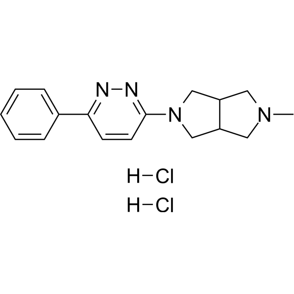
A-582941 dihydrochloride is a selective, orally active, blood-brain barrier-permeable α7 nAChR agonist, with Ki values of 10.8 nM and 17 nM in rat brain and human frontal cortex, respectively. A-582941 dihydrochloride exhibits agonistic activity at 5-HT3 receptors, with a Ki of 150 nM. A-582941 dihydrochloride triggers phosphorylation of ERK1/2 and CREB, inhibits GSK-3β via Ser-9 phosphorylation, increases acetylcholine release, induces the expression of Arc and c-Fos, activates brain regions associated with working memory and attention, and reduces cell death caused by nerve growth factor (NGF) deprivation. A-582941 dihydrochloride is applicable for the research of Alzheimer's disease and schizophrenia.
For research use only. We do not sell to patients.
- Purity : 99.78%
- CAS No.: 848591-90-2
- Formula: C17H22Cl2N4
- Molecular Weight:353.29
-
Storage:
4°C, sealed storage, away from moisture
* In solvent : -80°C, 6 months; -20°C, 1 month (sealed storage, away from moisture)

To place orders, for customer services and technical support, please contact: MedChemExpress USA
Tel: 609-228-6898 E-mail: [email protected] [email protected]


-
 Biological Activity
-
Chemical Information
-
Solvent & Solubility
- Protocols
- Purity & Documentation
- References
-
 Help & FAQs


-
HY-14564A
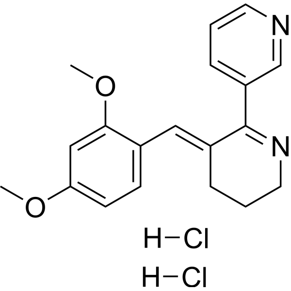
GTS-21 dihydrochlorideα7-nAChR Agonist -
HY-12152
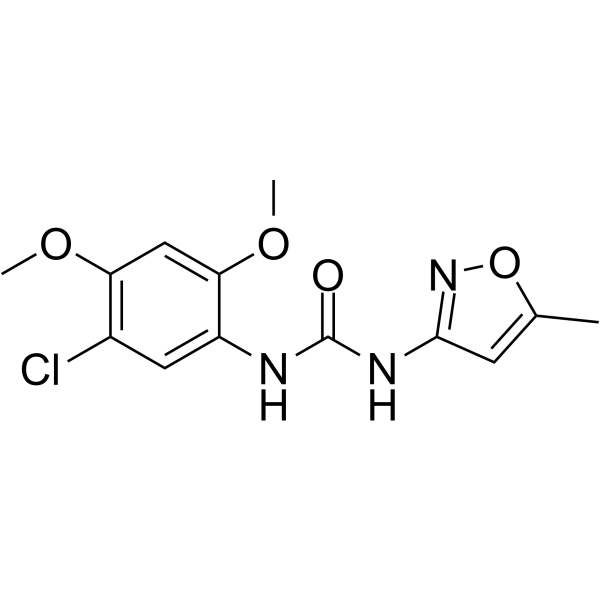
PNU-120596α7 nAChR PMA -
HY-10063
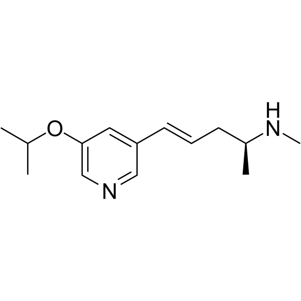
Isproniclineα4β2 nAChR Agonist -
HY-135483A
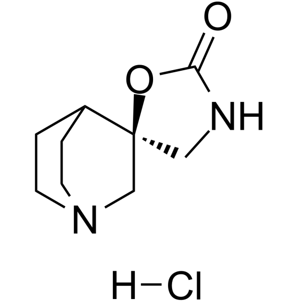
AR-R17779 hydrochloridenAChR Agonist -
HY-11053
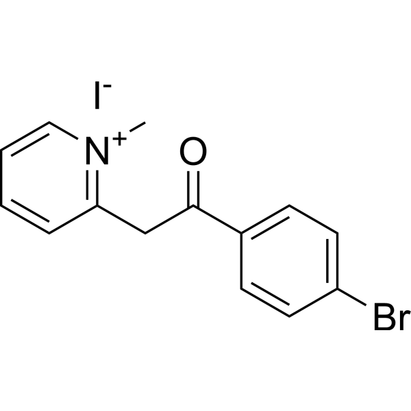
S 24795α7 nAChR partial agonist -
HY-12560A
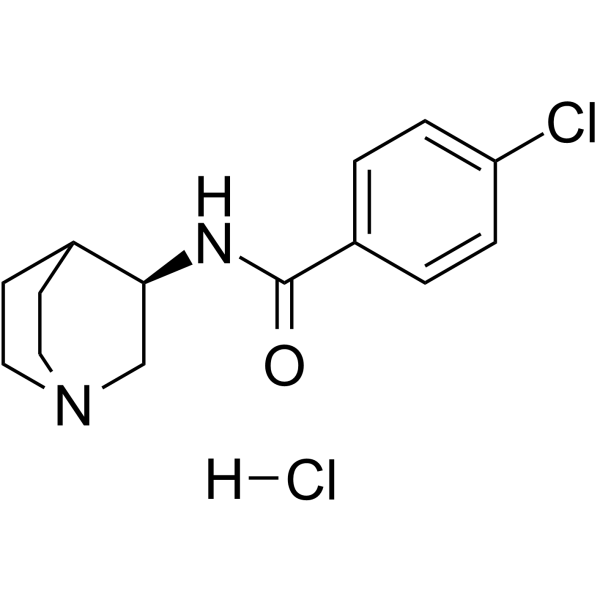
PNU-282987α7 nAChR Agonist/5-HT3 antagonist -
HY-10019
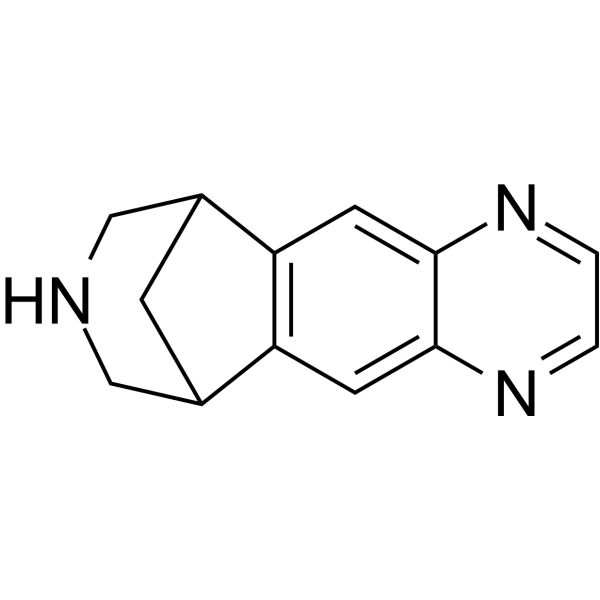
Vareniclineα4β2 nAChR Agonist -
HY-129674
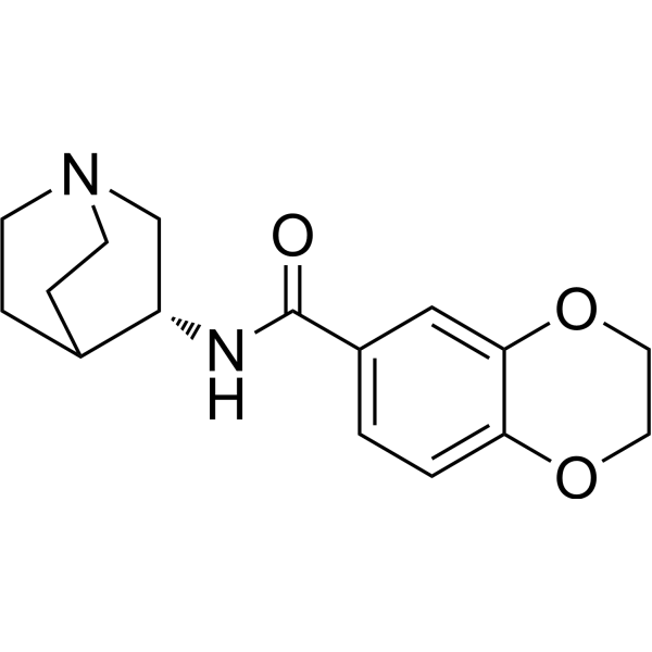
PHA 568487 free baseα-7 nAchR Agonist -
HY-107670
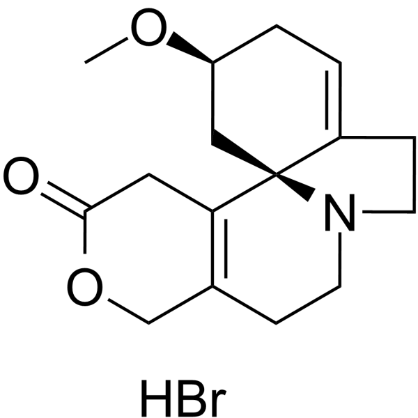
Dihydro-β-erythroidine hydrobromidenAChRs Antagonist -
HY-P1264
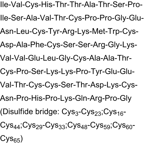
α-BungarotoxinnAChRs Antagonist -
HY-15430A
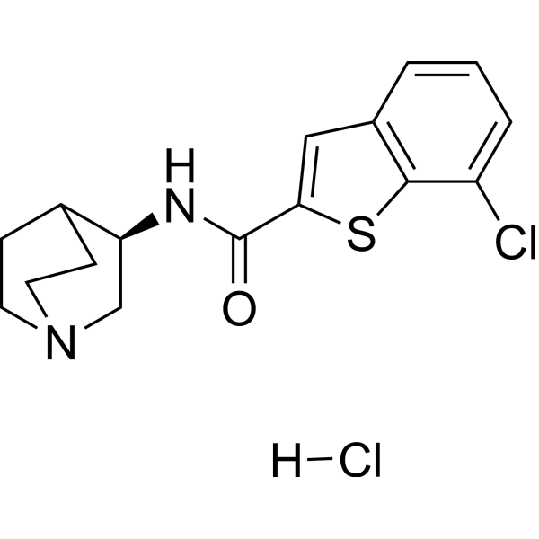
Encenicline hydrochloridenAChR Agonist -
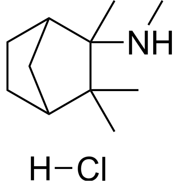
HY-B1395
Mecamylamine hydrochloridenAChR Antagonist
All 5-HT Receptor Isoforms
More

Biological Activity
Description
IC50 & Target
[1]|
α7 nAChR 10.8 nM (Ki, for rat α7 receptors) |
α7 nAChR 16.7 nM (Ki, for human α7 receptors) |
5-HT3 Receptor 150 nM (Ki) |
Cellular Effect
|
Cell Line

|
Type | Value | Description | References |
|---|---|---|---|---|
| HEK293 | EC50 |
>100 μM
Compound: 32
|
Agonist activity at human recombinant alpha4beta2 nicotinic acetylcholine receptor expressed in HEK293 cells assessed as changes in intracellular calcium level by FLIPR
Agonist activity at human recombinant alpha4beta2 nicotinic acetylcholine receptor expressed in HEK293 cells assessed as changes in intracellular calcium level by FLIPR
|
[PMID: 19552432] |
| Oocyte | EC50 |
4.26 μM
Compound: 32
|
Agonist activity at human recombinant alpha7 nicotinic acetylcholine receptor expressed in xenopus laevis oocytes by parallel oocyte electrophysiological assay
Agonist activity at human recombinant alpha7 nicotinic acetylcholine receptor expressed in xenopus laevis oocytes by parallel oocyte electrophysiological assay
|
[PMID: 19552432] |


Chemical Information
-
CAS No. 848591-90-2
-
Appearance Solid
-
Molecular Weight 353.29
-
Formula C17H22Cl2N4
-
Color Light yellow to yellow
-
SMILES
CN1CC(CN(C2=NN=C(C3=CC=CC=C3)C=C2)C4)C4C1.[H]Cl.[H]Cl
-
Shipping
Room temperature in continental US; may vary elsewhere.
-
Storage
4°C, sealed storage, away from moisture
* In solvent : -80°C, 6 months; -20°C, 1 month (sealed storage, away from moisture)


Solvent & Solubility
In Vitro:
DMSO : 100 mg/mL (283.05 mM; Need ultrasonic; Hygroscopic DMSO has a significant impact on the solubility of product, please use newly opened DMSO)
Please refer to the solubility information to select the appropriate solvent. Once prepared, please aliquot and store the solution to prevent product inactivation from repeated freeze-thaw cycles.
Storage method and period of stock solution: -80°C, 6 months; -20°C, 1 month (sealed storage, away from moisture). When stored at -80°C, please use it within 6 months. When stored at -20°C, please use it within 1 month.
Please refer to the solubility information to select the appropriate solvent. Once prepared, please aliquot and store the solution to prevent product inactivation from repeated freeze-thaw cycles.
Storage method and period of stock solution: -80°C, 6 months; -20°C, 1 month (sealed storage, away from moisture). When stored at -80°C, please use it within 6 months. When stored at -20°C, please use it within 1 month.
Concentration (start) × Volume (start) = Concentration (final) × Volume (final)

Protocols
-
Kinase activity and phosphorylation assays
Kinase activity assays measure the ability of kinases to transfer phosphate groups from ATP to specific substrates, while phosphorylation assays detect the presence and levels of phosphorylated proteins. Common methods include radiolabeled ATP incorporation (e. g. ,), ADP release detection via bioluminescence (e. g. ,[3]), enzyme-linked immunosorbent assays (ELISA) for phospho-specific epitopes (e. g. ,[6]), and microtiter-based formats for high-throughput screening (e. g. ,[8]). The ADP-Glo assay quantifies kinase activity by measuring ADP produced during phosphorylation using a luciferase-based system. Radiometric assays involve autoradiography or scintillation counting after incorporation of 32P-labeled ATP into substrate proteins. ELISA-based approaches rely on phospho-specific antibodies to detect activated kinases in cell lysates or purified samples.
-
PC12 NGF-Induced Neuronal Differentiation Culture
PC12 cells are a rat adrenal pheochromocytoma clonal line that responds to NGF by stopping proliferation and extending branching neurite-like processes; after longer NGF exposure, cells develop long processes and neuronal-like ultrastructural and functional features. NGF-induced differentiation is read out mainly by neurite outgrowth, reduced proliferation, microtubule assembly, and neuronal differentiation-associated proteins such as MAPs, tau, GAP-43, and synapsin-1.
-
Western Blot
Western blotting (WB) is a commonly used experimental method in molecular biology, biochemistry, and immunogenetics for identifying and quantifying target proteins. It combines gel electrophoresis with immunoassay, enabling researchers to analyze protein expression, post-translational modifications, and molecular weight.
-
RNA extraction experimental
By lysing cells, releasing RNA, and removing impurities such as proteins and DNA, high-purity RNA products are finally obtained. The commonly used traditional method is the guanidine isothiocyanate/phenol/chloroform method (Trizol), which is suitable for a variety of animal materials including animal tissues, microorganisms, cultured cells, etc., and most plant materials.
-
Cell Cytotoxicity Assay
Cytotoxicity assays are usually based on the assessment of cell membrane damage, which can also be indirectly detected by measuring cell viability. Detection methods include MTT assay, CKK-8 assay, LDH assay and ATP assay, etc.
-
Mammalian live/dead viability and cytotoxicity staining
Live/dead viability and cytotoxicity staining assays are based on the simultaneous detection of intracellular esterase activity in metabolically active (viable) cells and membrane integrity loss in non-viable cells. In commonly used dual-staining approaches, membrane-permeant fluorogenic substrates are converted by intracellular esterases into fluorescent products in live cells, while impermeant DNA-binding dyes selectively enter cells with compromised plasma membranes and label nucleic acids in dead or dying cells, enabling discrimination between viable and non-viable populations by fluorescence microscopy or flow cytometry.
-
Apoptosis Solutions
Apoptosis is a regulated, generally non-lytic cell-death pathway that removes unwanted, damaged, infected, or abnormal cells through coordinated morphological changes, caspase activation, DNA fragmentation, and membrane remodeling. The intrinsic apoptosis pathway is controlled mainly by mitochondrial outer membrane permeabilization, BCL-2 family proteins, cytochrome c release, apoptosome formation, caspase-9 activation, and downstream executioner caspase-3/7 activation. The extrinsic apoptosis pathway is initiated by death receptors such as Fas, TNFR, and TRAIL receptors, which recruit adaptor proteins and activate caspase-8 before engaging executioner caspases or mitochondrial amplification through BID cleavage. Apoptosis is linked to many phenotypes, including cancer cell killing, tissue homeostasis, immune regulation, neurodegeneration, infection response, and treatment-induced cytotoxicity; unresolved questions include how apoptosis interacts with necroptosis, pyroptosis, ferroptos
-
Protocol for Kinase activity and phosphorylation assays
Kinase activity assays measure transfer of phosphate from ATP to a protein or peptide substrate, generating phosphorylated substrate, ADP, or incorporated radiolabeled phosphate as the readout; phosphorylation assays measure site-specific phosphorylation in cells or tissues as a proxy for kinase-pathway activation, inhibition, or substrate regulation. Phosphorylation can be detected by phospho-specific Western blot, immunoprecipitation kinase assay, phospho-immunofluorescence, phospho-flow cytometry, luminescent ADP detection, radiolabeled ATP incorporation, or reporter-based pathway assays, and these readouts can be applied to cancer cells, primary neurons, mouse tumors, organoids, inflammatory macrophages, ferroptosis studies, and mitophagy studies when the kinase target is biologically relevant.
-
PC12 NGF-induced neuronal-like differentiation
PC12 cells are a rat adrenal pheochromocytoma-derived clonal cell line that responds to nerve growth factor by stopping proliferation and extending neurites, producing a sympathetic neuron-like phenotype used to study neuronal differentiation and neurite outgrowth. NGF acts through TrkA-dependent signaling, and neurite outgrowth is associated with ERK/Akt signaling, microtubule organization, neuronal-marker expression, and increased electrophysiological neuronal features such as sodium-channel density. The main assay readout is morphological differentiation, usually measured as the percentage of neurite-bearing cells, neurite length, neurite number, or total neurite length per cell. Additional readouts include GAP-43, tyrosine hydroxylase, βIII-tubulin, neurofilament, synapsin I, synaptophysin, ERK phosphorylation, Akt phosphorylation, and sodium-channel current density.
-
Alzheimer’s Disease Modeling
Alzheimer’s Disease (AD) is a neurodegenerative disorder characterized by a progressive decline in cognitive functions and loss of specific types of neurons and synapses. Alzheimer's symptoms can be simulated in mice by injecting drugs (such as Aβ) or genetically modified.


Purity & Documentation
-
Data Sheet (272 KB)
-
SDS (251 KB)
- English - EN (251 KB)
- Français - FR (251 KB)
- Deutsch - DE (251 KB)
- Norwegian - NO (251 KB)
- Español - ES (251 KB)
- Swedish - SV (251 KB)
- Italian - IT (251 KB)
- Korean - KR (251 KB)
- Portuguese - PT (251 KB)
-
Handling Instructions (2659 KB)
References
[1]. Tietje KR, et al. Preclinical characterization of A-582941: a novel alpha7 neuronal nicotinic receptor agonist with broad spectrum cognition-enhancing properties. CNS Neurosci Ther. 2008;14(1):65-82. [Content Brief]
[2]. Thomsen MS, et al. The selective alpha7 nicotinic acetylcholine receptor agonist A-582941 activates immediate early genes in limbic regions of the forebrain: Differential effects in the juvenile and adult rat. Neuroscience. 2008;154(2):741-753. [Content Brief]
[3]. Buccafusco JJ, et al. Profile of nicotinic acetylcholine receptor agonists ABT-594 and A-582941, with differential subtype selectivity, on delayed matching accuracy by young monkeys. Biochem Pharmacol. 2007;74(8):1202-1211. [Content Brief]


Complete Stock Solution Preparation Table
Please refer to the solubility information to select the appropriate solvent. Once prepared, please aliquot and store the solution to prevent product inactivation from repeated freeze-thaw cycles.
Storage method and period of stock solution: -80°C, 6 months; -20°C, 1 month (sealed storage, away from moisture). When stored at -80°C, please use it within 6 months. When stored at -20°C, please use it within 1 month.
| Optional Solvent | Concentration Solvent Mass | 1 mg | 5 mg | 10 mg | 25 mg |
|---|---|---|---|---|---|
| DMSO | 1 mM | 2.8305 mL | 14.1527 mL | 28.3054 mL | 70.7634 mL |
| 5 mM | 0.5661 mL | 2.8305 mL | 5.6611 mL | 14.1527 mL | |
| 10 mM | 0.2831 mL | 1.4153 mL | 2.8305 mL | 7.0763 mL | |
| 15 mM | 0.1887 mL | 0.9435 mL | 1.8870 mL | 4.7176 mL | |
| 20 mM | 0.1415 mL | 0.7076 mL | 1.4153 mL | 3.5382 mL | |
| 25 mM | 0.1132 mL | 0.5661 mL | 1.1322 mL | 2.8305 mL | |
| 30 mM | 0.0944 mL | 0.4718 mL | 0.9435 mL | 2.3588 mL | |
| 40 mM | 0.0708 mL | 0.3538 mL | 0.7076 mL | 1.7691 mL | |
| 50 mM | 0.0566 mL | 0.2831 mL | 0.5661 mL | 1.4153 mL | |
| 60 mM | 0.0472 mL | 0.2359 mL | 0.4718 mL | 1.1794 mL | |
| 80 mM | 0.0354 mL | 0.1769 mL | 0.3538 mL | 0.8845 mL | |
| 100 mM | 0.0283 mL | 0.1415 mL | 0.2831 mL | 0.7076 mL |


A-582941 dihydrochloride Related Classifications
HY-59201A Related Classifications
 Powered by Bioz