



Agrimonolide
Based on 1 publication(s) in Google Scholar
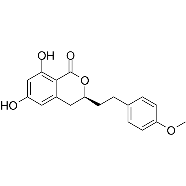
Agrimonolide is a phenethyl isocoumarin compound discovered from Agrimonia pilosa, exhibiting anti-inflammatory, antioxidant, and anticancer activities. Agrimonolide inhibits NF-κB, MAPK, and TLR4-mediated neuroinflammatory signaling, suppresses Notch and JAK2/STAT3 signaling to modulate Th17/Treg balance and maintain the intestinal barrier. Agrimonolide decreases HIF1A expression, inhibits glycolysis, and induces ferroptosis and cell cycle arrest via the mTOR pathway in tumor models, thereby suppressing cancer cell viability, proliferation, and metastasis. Agrimonolide exerts hepatoprotective and anti-fibrotic effects by regulating bile acid transporter expression and reducing hepatic bile acid accumulation. Agrimonolide is used for research on ulcerative colitis, ovarian cancer, non-small cell lung cancer, cholestatic liver injury, and neuroinflammation.
Nos produits utilisent uniquement pour la recherche. Nous ne vendons pas aux patients.
- Pureté : 98%
- CAS No.: 21499-24-1
- Formule: C18H18O5
- Masse moléculaire:314.33
-
Stockage:Powder -20°C, 3 years ; In solvent -80°C, 6 months , -20°C, 1 month

To place orders, for customer services and technical support, please contact: MedChemExpress USA
Tel: 609-228-6898 E-mail: [email protected] [email protected]


-
 Activité biologique
-
Chemical Information
-
Solvant et solubilité
- Protocole
- Pureté et documentation
- Références
-
 Help & FAQs


Publications Citing Use of MedChemExpress (MCE) Agrimonolide
More

Activité biologique
Description
In Vitro
Agrimonolide (10-20 μM; 4 days) promotes Treg cell differentiation from naïve CD4+ T cells isolated from C57BL/6J mice, as shown by increased CD4+CD25+Foxp3+ cell frequency and higher Foxp3 mRNA expression[1].
Agrimonolide (10-40 μM; 0-3 days) inhibits viability of SKOV3 and A2780 ovarian cancer cells[2].
Agrimonolide (10-40 μM; 14 days) suppresses colony formation of SKOV3 and A2780 ovarian cancer cells[2].
Agrimonolide (10-100 μM; 24 h) exhibits no significant cytotoxicity in BV2 microglial and Neuro-2a neuronal cells at concentrations below 100 μM[6].
Agrimonolide (20-40 μM) decreases extracellular acidification rate, indicating reduced glycolytic activity, in SKOV3 and A2780 ovarian cancer cells[2].
Agrimonolide (10-40 μM) decreases protein expression of HIF1A, HK2, and LDHA in SKOV3 and A2780 ovarian cancer cells[2].
Agrimonolide (40 μM) has inhibitory effects on glycolysis and HK2 expression in A2780 ovarian cancer cells that are reversed by overexpression of HIF1A, confirming that Agrimonolide attenuates glycolysis through modulation of HIF1A[2].
Agrimonolide (10-50 μM; 2 h pre-incubation before LPS stimulation) dose-dependently inhibits LPS-induced P65 phosphorylation in BV2 cells with an IC50 of 20.3 μM[6].
Agrimonolide (10-50 μM; 2 h pre-incubation before LPS stimulation) dose-dependently inhibits LPS-induced phosphorylation of JNK and P38 in BV2 microglial cells[6].
Agrimonolide (10-50 μM; 2.5 h pre-incubation before LPS stimulation) dose-dependently inhibits LPS-induced nuclear translocation of the NF-κB subunit P50 in BV2 cells[6].
Agrimonolide (10-50 μM; pre-incubated before LPS stimulation) inhibits LPS-induced activation of NF-κB and MAPK signaling pathways in primary mouse astrocytes[6].
In silico molecular docking predicts that agrimonolide binds with high affinity to TLR4, IRAK1, and TRAF6 (binding energies ≤ −6.53 kcal/mol) and with moderate affinity to MyD88, supporting a multi-target mechanism of action on the TLR4 signaling pathway[6].
Agrimonolide (10-50 μM; 6 h LPS stimulation) significantly reduces mRNA expression of IL-1β, IL-6, TNF-α, and IL-18 in LPS-stimulated BV2 cells[6].
Agrimonolide (10-50 μM; 6 h LPS stimulation) reduces LPS-induced mRNA expression of TLR4, MyD88, IRAK1, and TRAF6 in BV2 microglial cells[6].
Agrimonolide (10-50 μM; pre-incubated before LPS stimulation) inhibits LPS-induced activation of primary mouse microglia, as evidenced by reduced CD68 expression and preserved ramified morphology[6].
Agrimonolide (10-50 μM; pre-incubated before LPS stimulation) significantly reduces mRNA expression of IL-1β, IL-6, TNF-α, and IL-18 in LPS-stimulated primary mouse astrocytes[6].
Agrimonolide (10-50 μM; pre-incubated before LPS stimulation) dose-dependently inhibits LPS-induced GFAP upregulation and activation of primary mouse astrocytes[6].
Agrimonolide (10-50 μM; 2 h pre-incubation before LPS stimulation) dose-dependently reduces LPS-induced expression of TLR4, IRAK1, and TRAF6 proteins in BV2 microglial cells, while MyD88 protein levels were not notably altered under these conditions[6].
MedChemExpress (MCE) has not independently confirmed the accuracy of these methods. They are for reference only.
-
Cell Line:SKOV3, A2780 ovarian cancer cells
-
Concentration:10, 20, 40 μM
-
Incubation Time:0, 1, 2, 3 days
-
Result:Significantly reduced the viability of both SKOV3 and A2780 ovarian cancer cells at concentrations of 20 μM and 40 μM.
Did not produce a significant effect on cell viability at 10 μM.
-
Cell Line:SKOV3, A2780 ovarian cancer cells
-
Concentration:10, 20, 40 μM
-
Incubation Time:14 days
-
Result:Suppressed colony formation in both SKOV3 and A2780 cells at 20 μM and 40 μM.
Showed a lesser effect on colony formation at 10 μM compared to higher concentrations.
-
Cell Line:BV2 mouse microglial cells
-
Concentration:10, 30, 50 μM
-
Incubation Time:2 h pre-incubation before LPS stimulation; 45 min (LPS stimulation)
-
Result:Suppressed the LPS-induced increase of p-P65 in a dose-dependent manner, yielding an IC50 of 20.3 μM based on p-P65 band densitometry quantification.\nSignificantly suppressed LPS-induced phosphorylation of both JNK and p38 kinase in a dose-dependent manner.
Eliminated the LPS-induced increase of P50 levels in the nucleus of BV2 cells in a dose-dependent manner.
-
Cell Line:BV2 mouse microglial cells
-
Concentration:10, 30, 50 μM
-
Incubation Time:pre-incubated before LPS stimulation; 6 h (LPS stimulation)
-
Result:Significantly reduced the mRNA levels of IL-1β, IL-6, TNF-α, and IL-18 in LPS-stimulated BV2 cells.\nReduced the mRNA levels of TLR4, MyD88, IRAK1, and TRAF6 in LPS-stimulated BV2 cells.
-
Cell Line:BV2 mouse microglial cells
-
Concentration:10, 30, 50 μM
-
Incubation Time:2 h pre-incubation before LPS stimulation; 24 h (LPS stimulation)
-
Result:Markedly reduced the LPS-induced expression of NLRP3, iNOS, and COX-2 proteins in a dose-dependent manner.\nEffectively inhibited LPS-induced increases in TLR4, IRAK1, and TRAF6 protein levels in a dose-dependent manner.
Did not notably alter MyD88 protein levels.
In Vivo
MedChemExpress (MCE) has not independently confirmed the accuracy of these methods. They are for reference only.
-
Animal Model:C57BL/6J (male; 6 weeks old)[1]
-
Dosage:25 mg/kg/day; 50 mg/kg/day
-
Administration:i.g.; daily; starting before DSS exposure
-
Result:Reduced body weight loss in a dose-dependent manner compared to the DSS-only group.
Restored colon length in a dose-dependent manner compared to the DSS-only group.
Lowered disease activity index scores in a dose-dependent manner compared to the DSS-only group.
Alleviated mucosal damage, inflammatory cell infiltration, and crypt structure destruction in the colon.
Suppressed myeloperoxidase activity in colon tissues, with the 50 mg/kg dose showing a more substantial reduction than the 25 mg/kg dose.
Decreased the production of pro-inflammatory cytokines IL-1β, TNF-α, and IL-6 in colonic tissue.
Reduced the phosphorylation levels of IKKα/β, IκBα, and p65 in the NF-κB pathway.
Increased the protein expression levels of tight junction proteins Occludin and ZO-1.
Reduced serum FITC-dextran concentration.
Decreased the absolute number of CD4+IL-17A+ Th17 cells in mesenteric lymph nodes.
Increased the percentage of CD4+CD25+Foxp3+ Treg cells in mesenteric lymph nodes.
Modulated the mRNA and protein levels of Th17-specific transcription factor RORγt and Treg-specific transcription factor Foxp3 in colon and spleen tissues.
Reduced the mRNA and protein expression of Notch-1, Jagged1, and DLL4 in colonic tissue.
Inhibited the phosphorylation of JAK2 and STAT3.
Reduced protein expression of Notch1 and STAT3 as confirmed by immunofluorescence.
-
Animal Model:C57BL/6 J (male; 8 weeks old; 20-25 g; specific pathogen-free)[4]
-
Dosage:10 mg/kg; 30 mg/kg; 90 mg/kg
-
Administration:i.p.; daily; 28 days
-
Result:Alleviated DDC-induced increase in liver-to-body weight ratio in a dose-dependent manner.
Mitigated DDC-induced elevations in serum ALT, AST, ALP, total bilirubin, and direct bilirubin in a dose-dependent manner.
Alleviated ductular proliferation, macrophage infiltration, intraductal plugs, pericholangitis, onion skin-type periductal fibrosis, and biliary fibrosis.
Reduced DDC-increased hepatic hydroxyproline accumulation in a dose-dependent manner.
Markedly decreased hepatic mRNA expression of fibrosis markers α-Sma, Col1a1, Timp1, Epcam, and Mmp2, and reduced α-SMA protein expression at 90 mg/kg.
Reduced hepatic mRNA upregulation of inflammatory cytokines Tnf-α, Il-6, Il-1β, and Tgfb1.
Alleviated excessive bile duct proliferation in all dosage groups as confirmed by CK19 staining.
Reduced hepatic bile acid levels in a dose-dependent manner.
Increased hepatic mRNA expression of bile acid synthesis enzymes Cyp7a1, Cyp8b1, and Cyp7b1 dose-dependently, and reversed DDC-induced downregulation of Cyp27a1 at 90 mg/kg.
Inhibited DDC-induced increases in TCA, T-α-MCA, β-MCA, and T-β-MCA; reversed accumulation of T-CDCA, T-DCA, and T-UDCA at 90 mg/kg.
Reversed DDC-induced downregulation of Bsep at all three doses, with 30 and 90 mg/kg increasing Bsep expression.
Decreased DDC-elevated expression of Oatp2, Mrp3, and Mrp4 in a dose-dependent manner; increased DDC-reduced Ntcp and Oatp1 expression at 30 and 90 mg/kg.
Dose-dependently alleviated DDC-induced upregulation of phase I enzymes Cyp3a11 and Cyp2b10, and reversed DDC-induced inhibition of phase II enzymes Ugt1a1 and Sult2a1.
-
Animal Model:C57BL/6 J (male; 8 weeks old; 20-25 g; specific pathogen-free)[4]
-
Dosage:90 mg/kg
-
Administration:i.p.; daily; 28 days
-
Result:Partially restored DDC-increased liver-to-body weight ratio from 10.96% to 5.31%.
Normalized serum ALT, AST, and ALP levels.
Increased hepatic expression of bile efflux genes Bsep and Mrp2.
Downregulated Oatp2, Mrp3, and Mrp4, and upregulated Ntcp and Oatp1.
Reduced DDC-increased hepatic hydroxyproline from 81.3 μg/mg liver to 32.3 μg/mg liver.
Decreased hepatic mRNA expression of fibrosis markers α-Sma, Col1a1, Timp1, and Epcam, and inhibited Tgfb1 gene expression.
Alleviated ductular proliferation, intraductal plugs, inflammatory cell infiltration, and pericholangitis as shown by immunohistochemistry.
Decreased the percentage of myeloid cells and increased the percentage of lymphoid cells, inhibited CD4+ and CD8+ lymphocyte infiltration, and decreased the numbers of both Kupffer cells and monocyte-derived macrophages as shown by flow cytometry.
-
Animal Model:ICR mice (6 weeks old)[6]
-
Dosage:25 mg/kg; 50 mg/kg
-
Administration:i.p.; daily; 5 days
-
Result:Attenuated LPS-induced activation of microglia and astrocytes in the hippocampus, as evidenced by reduced IBA-1 and GFAP immunoreactivity, decreased density of IBA-1-positive microglia and GFAP-positive astrocytes, and mitigation of morphological changes associated with activation.
Suppressed LPS-induced microglial and astrocytic activation in the cortical area.
Dose-dependently suppressed the LPS-induced upregulation of IBA-1 and GFAP protein levels in western blot analysis.
Significantly downregulated the mRNA levels of LPS-induced pro-inflammatory factors including NLRP3, iNOS, COX-2, IL-1β, IL-6, TNF-α, and IL-18 in the hippocampus in RT-qPCR analysis.
- RNA extraction experimental
- Mammalian live/dead viability and cytotoxicity staining
- Naïve CD4+ T-cell subset differentiation/polarization
- BrdU Incorporation Assay
- Protocol for Cell Cycle
- CCK-8/WST-8 Cell Proliferation Assay
- Research Protocol for Inflammation-related Diseases
- Research Protocol for Neurological Diseases
- Cell Viability Determination by MTT Colorimetric Assay
- Flow cytometric DNA-content cell-cycle staining
- Cell Cytotoxicity Assay
- DSS-Induced Colitis
- TNBS-Induced Colitis
- Notch Pathway Solutions
- Ferroptosis Solutions
- MTT Cell Proliferation Assay
- Orthotopic Cell-Line Xenograft


Chemical Information
-
CAS No. 21499-24-1
-
Appearance Solid
-
Masse moléculaire 314.33
-
Formule C18H18O5
-
Color White to off-white
-
SMILES
O=C1C2=C(O)C=C(O)C=C2C[C@H](CCC3=CC=C(OC)C=C3)O1
-
Structure Classification
-
Livraison
Room temperature in continental US; may vary elsewhere.
-
Stockage
Powder -20°C 3 years In solvent -80°C 6 months -20°C 1 month


Publications (1)
-
Journal Impact Factor
-
Most Recent


Solvant et solubilité
In Vitro:
DMSO : 25 mg/mL (79.53 mM; Need ultrasonic and warming; Hygroscopic DMSO has a significant impact on the solubility of product, please use newly opened DMSO)
Please refer to the solubility information to select the appropriate solvent. Once prepared, please aliquot and store the solution to prevent product inactivation from repeated freeze-thaw cycles.
Storage method and period of stock solution: -80°C, 6 months; -20°C, 1 month. When stored at -80°C, please use it within 6 months. When stored at -20°C, please use it within 1 month.
Please refer to the solubility information to select the appropriate solvent. Once prepared, please aliquot and store the solution to prevent product inactivation from repeated freeze-thaw cycles.
Storage method and period of stock solution: -80°C, 6 months; -20°C, 1 month. When stored at -80°C, please use it within 6 months. When stored at -20°C, please use it within 1 month.
Concentration (start) × Volume (start) = Concentration (final) × Volume (final)


Pureté et documentation
-
Fiche technique (302 KB)
-
SDS (393 KB)
- English - EN (393 KB)
- Français - FR (393 KB)
- Deutsch - DE (393 KB)
- Norwegian - NO (393 KB)
- Español - ES (393 KB)
- Swedish - SV (393 KB)
- Italian - IT (393 KB)
- Korean - KR (393 KB)
- Portuguese - PT (393 KB)
-
Instruction de manipulation (2659 KB)
Références
[1]. Jiang J, et al. Agrimonolide mitigated DSS-induced colitis by modulating the balance between Treg and Th17 cells through the suppression of the Notch and JAK2/STAT3 signaling pathways. Heliyon. 2024 Jul 15;10(13):e33803. [Content Brief]
[5]. Kim HW, et al. Acylphloroglucinolated Catechin and Phenylethyl Isocoumarin Derivatives from Agrimonia pilosa. J Nat Prod. 2016 Sep 23;79(9):2376-83. [Content Brief]


Complete Stock Solution Preparation Table
Please refer to the solubility information to select the appropriate solvent. Once prepared, please aliquot and store the solution to prevent product inactivation from repeated freeze-thaw cycles.
Storage method and period of stock solution: -80°C, 6 months; -20°C, 1 month. When stored at -80°C, please use it within 6 months. When stored at -20°C, please use it within 1 month.
| Optional Solvent | Concentration Solvent Mass | 1 mg | 5 mg | 10 mg | 25 mg |
|---|---|---|---|---|---|
| DMSO | 1 mM | 3.1814 mL | 15.9069 mL | 31.8137 mL | 79.5343 mL |
| 5 mM | 0.6363 mL | 3.1814 mL | 6.3627 mL | 15.9069 mL | |
| 10 mM | 0.3181 mL | 1.5907 mL | 3.1814 mL | 7.9534 mL | |
| 15 mM | 0.2121 mL | 1.0605 mL | 2.1209 mL | 5.3023 mL | |
| 20 mM | 0.1591 mL | 0.7953 mL | 1.5907 mL | 3.9767 mL | |
| 25 mM | 0.1273 mL | 0.6363 mL | 1.2725 mL | 3.1814 mL | |
| 30 mM | 0.1060 mL | 0.5302 mL | 1.0605 mL | 2.6511 mL | |
| 40 mM | 0.0795 mL | 0.3977 mL | 0.7953 mL | 1.9884 mL | |
| 50 mM | 0.0636 mL | 0.3181 mL | 0.6363 mL | 1.5907 mL | |
| 60 mM | 0.0530 mL | 0.2651 mL | 0.5302 mL | 1.3256 mL |


Agrimonolide Related Classifications
HY-N2830 Related Classifications
 Powered by Bioz