



Raddeanin A
Based on 1 publication(s) in Google Scholar
Raddeanin A is an oleanane-type triterpenoid saponin with oral activity. Raddeanin A inhibits SRC, mTOR, JNK, VEGFR2, NLRP3 inflammasome, Wnt/β-catenin, Wee1, PI3K/AKT signaling pathway, MAPK/ERK signaling pathway, AR-FL, AR-Vs, and downregulates the expression of p-PI3K and p-AKT. Raddeanin A inhibits osteoclast formation, bone resorption, osteolysis, cancer cell invasion, migration, proliferation, angiogenesis and epithelial-mesenchymal transition, while induces apoptosis, cell cycle arrest, ROS production, immunogenic cell death and dendritic cell maturation. Raddeanin A improves blood-retinal barrier function, alleviates inflammation, regulates the tumor microenvironment, and enhances the activity of anti-PD-1 antibody. Raddeanin A is applicable to the research of breast cancer-associated osteolysis, human osteosarcoma, colorectal cancer, glioblastoma, Alzheimer's disease, cholangiocarcinoma, melanoma, non-small cell lung cancer, castration-resistant prostate cancer and multiple myeloma.
Nos produits utilisent uniquement pour la recherche. Nous ne vendons pas aux patients.
- Pureté : 99.79%
- CAS No.: 89412-79-3
- Formule: C47H76O16
- Masse moléculaire:897.10
-
Stockage:
4°C, protect from light
* In solvent : -80°C, 6 months; -20°C, 1 month (protect from light)

To place orders, for customer services and technical support, please contact: MedChemExpress USA
Tel: 609-228-6898 E-mail: [email protected] [email protected]


-
 Activité biologique
-
Chemical Information
-
Solvant et solubilité
- Protocole
- Pureté et documentation
- Références
-
 Help & FAQs


-
HY-18749
SC79Akt Activator -
HY-W013032A
Oxamic acid sodiumLDH-A Inhibitor -
HY-B0234
EstroneEndogenous Metabolite -
HY-N0022
IsoacteosideNatural Product -

HY-N0819R
Raddeanin A (Standard)Analytical Standard -
HY-10585
Valproic acidHDAC Inhibitor -
HY-10224
PanobinostatHDAC Inhibitor -
HY-B0350A
Sodium butyrateHDAC Inhibitor -
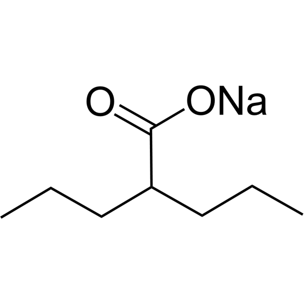
HY-10585A
Valproic acid sodiumHDAC Inhibitor -
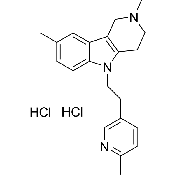
HY-14537
Latrepirdine dihydrochlorideAutophagy Inducer -
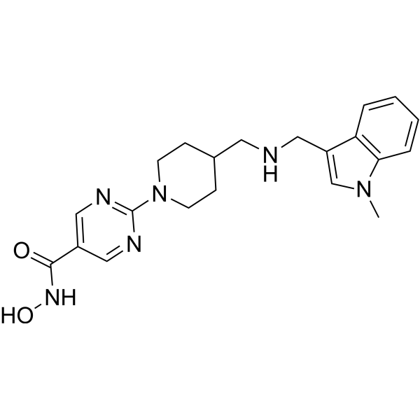
HY-15433
QuisinostatPan-HDAC Inhibitor -
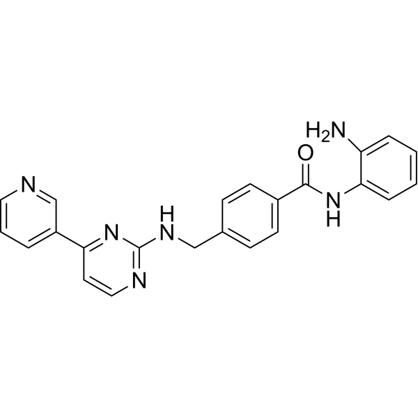
HY-12164
MocetinostatAutophagy Inducer
Publications Citing Use of MedChemExpress (MCE) Raddeanin A
More Customer Validation & Images

-
Cell Proliferation/Viability Assay

-
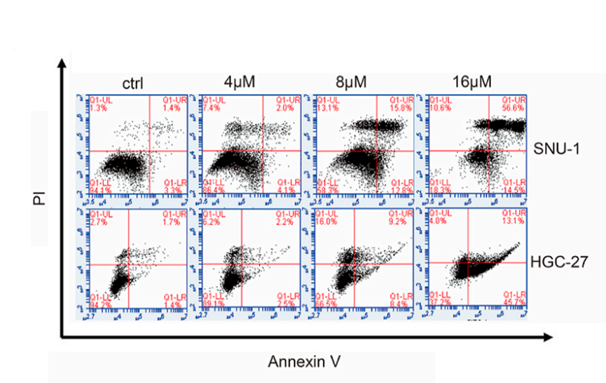
Apoptosis Analysis
-
IF

-
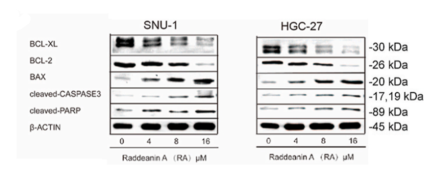
WB
-
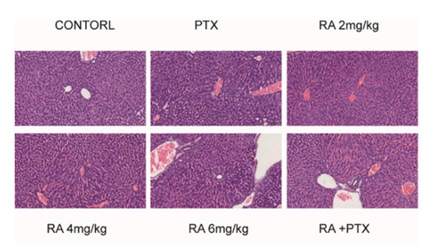
Histological Imaging/Staining
Voir tous les produits spécifiques à Isoform VEGFR
More

Activité biologique
Description
In Vitro
Raddeanin A (0.2-0.8 μM; 3-7 days) potently inhibits RANKL-induced osteoclast formation in BMMs, with a 72-hour cytotoxicity IC50 of 2.91 μM, and suppresses osteoclast survival in a concentration-dependent manner[1].
Raddeanin A (0.4 μM; 5-7 days) downregulates key osteoclastogenic markers CTSK and NFATc1 at both the gene and protein levels in RANKL-stimulated BMMs[1].
Raddeanin A (0.2-0.8 μM; 7-21 days), at concentrations up to 0.8 μM, does not inhibit osteoblast differentiation in MC3T3-E1 cells, and at 0.2 μM enhances mineralization and increases SPARC gene expression[1].
Raddeanin A (6.25-50 μM; 24-96 h) inhibits viability, proliferation, and invasion of MDA-MB-231 breast cancer cells, and induces apoptosis, with a 96-hour cytotoxicity IC50 of 15.77 μM[1].
Raddeanin A (3 μM; 6-12 h) inhibits AKT/mTOR signaling in MDA-MB-231 cells by reducing p-AKT and mTOR protein levels[1].
Raddeanin A (0.2-50 μM; 24-48 h) dose- and time-dependently inhibits the viability of human osteosarcoma MG-63, HOS, U-2 OS, Saos-2, and 143B cells, with MG-63 and HOS cells showing the highest sensitivity (IC50 values of 1.60 μM and 2.57 μM at 48 h, respectively)[2].
Raddeanin A (1-4 μM; 24 h) dose-dependently modulates mitochondrial apoptotic pathway proteins (reducing Bcl-2/Bax ratio, increasing cleaved caspase-3 and cleaved PARP) in human osteosarcoma MG-63 and HOS cells[2].
Raddeanin A (1-4 μM; 12 h) dose-dependently inhibits IκBα phosphorylation in human osteosarcoma MG-63 and HOS cells after 12 h of treatment[2].
Raddeanin A (1-4 μM; 2 μM, 6 h) dose-dependently suppresses p65 nuclear translocation in human osteosarcoma MG-63 and HOS cells, with 2 μM treatment for 6 h visibly reducing nuclear p65 localization[2].
Raddeanin A (100-800 nM; 48 h) dose-dependently reduces the viability of U87, U251, T98G, and LN299 human glioblastoma cells, with greater potency in U87 and U251 cells[4].
Raddeanin A (100-200 nM; 48 h) dose-dependently reduces the mRNA and protein expression of β-catenin and EMT-related biomarkers (N-cadherin, vimentin, snail) in U87 and U251 human glioblastoma cells[4].
Raddeanin A (100 nM) inhibits viability, migration, invasion, and EMT biomarker expression in β-catenin-overexpressing U87 and U251 human glioblastoma cells, but these effects are reversed by β-catenin overexpression[4].
Raddeanin A (0.125-0.5 μM; 24 h) inhibits Aβ1-42-induced activation of the NLRP3 inflammasome and secretion of inflammatory cytokines in MIO-M1 cells[5].
Raddeanin A (0-160 μg/mL; 24 h) reduces cell viability in a dose-dependent manner in RBE, LIPF155C, LIPF178C, and LICCF cholangiocarcinoma cell lines (EC50: 50.95-64.76 μg/mL; LC50: 34.65-49.47 μg/mL) with lower toxicity to normal HIBEpiC biliary epithelial cells[6].
Raddeanin A (1-5 μM; 20 h) dose-dependently increases HMGB1-Gluc activity, a marker of immunogenic cell death, in B16 and MC38 cells[7].
Raddeanin A (1-5 μM; 6-8 h) dose-dependently increases mitochondrial ROS production in MC38 cells[7].
Raddeanin A (1-4 μM; 24 h) dose-dependently inhibits the migration and invasion of H1299, A549, and PC-9 NSCLC cells after 24 h of treatment[8].
Raddeanin A (1-4 μM; 24 h) modulates EMT-related protein expression and specifically inhibits CDK6 expression and Rb phosphorylation in H1299, A549, and PC-9 NSCLC cells after 24 h of treatment[8].
Raddeanin A (1-4 μM; 24 h) dose-dependently induces G1 phase cell cycle arrest in H1299, A549, and PC-9 NSCLC cells after 24 h of treatment[8].
Raddeanin A (1-16 μM; 12 h) potently inhibits proliferation of human colorectal cancer HCT116 cells in a dose-dependent manner, with a 12 h IC50 of 2.61 μM[9].
Raddeanin A (2-4 μM; 12 h) dose-dependently downregulates the mRNA expression of apoptosis-related genes (caspase-3, PARP) and cell cycle-related genes (cyclinD1, cyclinE) in human colorectal cancer HCT116 cells[9].
Raddeanin A (2-4 μM; 12 h) modulates protein expression in human colorectal cancer HCT116 cells by increasing pro-apoptotic proteins, decreasing anti-apoptotic and cell cycle-related proteins, and suppressing the PI3K/AKT signaling pathway via reduced p-PI3K and p-AKT expression[9].
Raddeanin A (1.5-6 μM; 12-72 h) dose- and/or time-dependently inhibits the growth of AR-positive 22Rv1, C4-2, C4-2B, and LNCaP95 CRPC cells, with no effect on AR-null PC-3 and DU145 prostate cancer cells[10].
Raddeanin A (3 μM; 6-24 h) time-dependently downregulates mRNA levels of AR target genes PSA (in C4-2 and LNCaP95 cells) and UBE2C (in LNCaP95 cells)[10].
Raddeanin A (0.125-8 μM; 24-48 h) inhibits the proliferation of MM.1S, MM.1R, and RPMI 8226 multiple myeloma cells in a time-dependent and concentration-dependent manner, with IC50 values ranging from 1.058 μM (MM.1S, 48 h) to 6.091 μM (RPMI 8226, 24 h)[11].
MedChemExpress (MCE) has not independently confirmed the accuracy of these methods. They are for reference only. Further protocols information, click here.
-
Cell Line:RANKL-induced mouse bone marrow-derived macrophages (BMMs)
-
Concentration:0.4 μM
-
Incubation Time:5 days; 7 days
-
Result:Dramatically suppressed mRNA expression of cathepsin k (CTSK) and nuclear factor of activated T cells 1 (NFATc1). Reduced protein expression levels of CTSK and NFATc1.
-
Cell Line:MC3T3-E1 preosteoblast cells
-
Concentration:0.2-0.8 μM; 0.781 μM
-
Incubation Time:7, 14, 21 days
-
Result:Showed no significant difference in ALP activity between control and treated groups at day 7. Resulted in a larger total mineralized area compared to the control group at 0.2 μM at day 21. Significantly increased secreted protein acidic and rich in cysteine (SPARC) mRNA expression after 14 days of treatment. Showed no significant cytotoxic effect on MC3T3-E1 cells at doses below 0.781 μM.
-
Cell Line:MDA-MB-231 breast cancer cells
-
Concentration:3 μM
-
Incubation Time:6, 12 h
-
Result:Significantly downregulated both p-AKT phosphorylation and mTOR protein expression in a time-dependent manner.
-
Cell Line:human osteosarcoma MG-63 and HOS cells
-
Concentration:1-4 μM
-
Incubation Time:12 h
-
Result:Increased nuclear fragmentation and apoptotic body formation in a dose-dependent manner in both MG-63 and HOS cells.
-
Cell Line:human osteosarcoma MG-63 and HOS cells
-
Concentration:1-4 μM
-
Incubation Time:24 h
-
Result:Caused a dose-dependent increase in the percentage of both early and late apoptotic cells in MG-63 and HOS cells.
In Vivo
Raddeanin A (100 μg/kg; i.p.; every other day; 28 days) significantly inhibits breast cancer-induced osteolysis in female BALB/c nu/nu mice by preserving bone structure and increasing tumor cell apoptosis, as evidenced by higher BV/TV, reduced Tb. Sp, intact bone cortex, and increased TUNEL-positive cells[1].
Raddeanin A (1.25-5 mg/kg; i.p.; every other day; 20 consecutive days) dose-dependently inhibits the growth of HOS osteosarcoma xenografts in nude mice, while inducing tumor cell apoptosis and demonstrating low systemic toxicity[2].
Raddeanin A (0.4 μM; continuous immersion; 30 h) inhibits zebrafish intersegmental vessel formation by 67.64%[3].
Raddeanin A (5 mg/kg; i.p.; once every 2 days; 11 injections) reduces HCT-15 colorectal xenograft tumor volume and weight, increases tumor apoptosis and necrosis, and decreases intratumoral microvessel density without obvious toxicity[3].
Raddeanin A (100 mg/kg; i.p.; daily) inhibits glioblastoma tumor growth, reduces tumor vessel density, downregulates β-catenin-mediated EMT and angiogenesis, and increases survival rate to ~80% at day 30 in an intracranial U87 xenograft mouse model[4].
Raddeanin A (10 mg/kg; p.o.; daily; 9 weeks) protects the blood-retinal barrier and improves Alzheimer's disease-related retinopathy in 3×Tg-AD mice by inhibiting NLRP3-mediated inflammation, suppressing Wnt/β-catenin pathway-mediated apoptosis, and restoring retinal structural and vascular integrity[5].
Raddeanin A (1-4 mg/kg; i.p., i.t.; four times at indicated time points) inhibits MC38 colon adenocarcinoma growth in a DC and CD8+ T cell-dependent manner in C57BL/6J mice, and induces 60% tumor-free survival in a tumor rechallenge model[7].
Raddeanin A (4 mg/kg; i.t.; four times at indicated time points)'s antitumor activity against MC38 colon adenocarcinoma in C57BL/6J mice is dependent on CD8+ T cells and DCs, and combining it with anti-PD-1 antibody enhances therapeutic efficacy by reprogramming the tumor immune microenvironment[7].
Raddeanin A (0.5-1.0 mg/kg; i.p.; once every 2 days; 7 total doses over 30 days) exerts dose-dependent anti-NSCLC efficacy in BALB/c nude mouse xenografts, with the 1.0 mg/kg dose significantly reducing tumor volume and weight while showing no detectable organ toxicity[8].
Raddeanin A (4 mg/kg; injected; 2 weeks) significantly inhibits colorectal cancer xenograft tumor growth in BALB/c nude mice, reduces tumor volume and weight, induces tumor cell apoptosis at a rate of 43.6%, modulates apoptosis- and cell cycle-related proteins, and regulates the PI3K/AKT signaling pathway without causing liver toxicity[9].
MedChemExpress (MCE) has not independently confirmed the accuracy of these methods. They are for reference only.
-
Animal Model:C57BL/6 (8-week-old male; Ti-particle-induced calvarial osteolysis model)[1]
-
Dosage:50 μg/kg; 100 μg/kg
-
Administration:daily; 14 days
-
Result:Increased bone volume to total volume (BV/TV) ratio compared to vehicle group.
Decreased percentage of total porosity compared to vehicle group.
Reduced number of tartrate-resistant acid phosphatase (TRAP)-positive multinucleated osteoclasts compared to vehicle group.
Reduced number of cathepsin K (CTSK)-positive multinucleated osteoclasts compared to vehicle group.
-
Animal Model:BALB/c nu/nu (5-week-old female; breast cancer-induced osteolysis model)[1]
-
Dosage:100 μg/kg
-
Administration:i.p.; every other day; 28 days
-
Result:Increased trabecular bone volume to total volume (BV/TV) ratio compared to vehicle group.
Reduced trabecular separation (Tb. Sp) compared to vehicle group.
Preserved intact bone cortex (versus extensive trabecular bone resorption and discrete cortical bone in vehicle controls).
Increased apoptosis in the treated group, as shown by TUNEL assay.
-
Animal Model:BALB/c nude (male, 4 weeks old, 20 ± 2 g, subcutaneous osteosarcoma xenograft model)[2]
-
Dosage:1.25 mg/kg; 2.5 mg/kg; 5 mg/kg
-
Administration:i.p.; every other day; 20 consecutive days
-
Result:Significantly decelerated tumor growth in a dose-dependent manner.
Reduced tumor volumes in all treatment groups.
Induced significant apoptosis in tumor tissues via TUNEL staining.
Increased p-JNK protein expression and decreased p65 protein expression in tumor tissues via immunohistochemistry analysis. Caused no significant body weight loss during treatment.
-
Animal Model:BALB/c nude (5-week-old female, subcutaneous xenograft model)[3]
-
Dosage:5 mg/kg
-
Administration:i.p.; once every 2 days; 11 injections
-
Result:Reduced mean tumor volume to 765.3 mm3. Reduced mean tumor weight to 1.2 g. Decreased intratumoral microvessel density to ~20 vessels/mm2. Increased the percentage of TUNEL-positive apoptotic cells to ~60%. Increased tumor necrosis area to ~70%. Caused no significant body weight loss.
-
Animal Model:B6;129-APPswe/Psen1/tau-P301L (3×Tg-AD) (male, female, Alzheimer's disease transgenic model)[5]
-
Dosage:10 mg/kg
-
Administration:p.o.; daily; 9 weeks
-
Result:Significantly reduced the number of degenerated retinal capillaries compared to untreated 3×Tg-AD mice.
Increased retinal expression of tight junction proteins ZO-1, Occludin, and Claudin 5.
Ameliorated retinal structural abnormalities: restored total retinal thickness, ganglion cell layer + inner plexiform layer thickness, inner nuclear layer thickness, and outer nuclear layer thickness; improved disorganization of retinal cell layers.
Increased the Bcl-2/Bax protein expression ratio in retinal tissue.
Reduced retinal expression of NLRP3 inflammasome components (NLRP3, pro-Caspase-1, Caspase-1, ASC) and pro-inflammatory cytokines (IL-1β, IL-18).
Decreased retinal expression of β-catenin and phosphorylated LRP5/6, and increased retinal expression of GSK-3β, indicating inhibition of the Wnt/β-catenin pathway.
-
Animal Model:C57BL/6J (female, 6-8 weeks old, subcutaneous inoculation of 7×10⁶ MC38 tumor cells)[7]
-
Dosage:1 mg/kg; 2 mg/kg; 4 mg/kg
-
Administration:i.p., four times at indicated time points; i.t., four times at indicated time points
-
Result:Caused considerable inhibition of tumor volume and tumor weight.
Induced 60% tumor-free survival 30 days after rechallenge with live MC38 cells.
Markedly elevated the population of tumor-infiltrating CD8+ T cells and CD103+CD11c+ DCs, and increased levels of CD8+ T cell effector molecules GZMB and IFN-γ within the tumor microenvironment.
Upregulated CD40, CD80, CD86, and MHC-II expression on tumor-infiltrating CD103+CD11c+ DCs.
-
Animal Model:C57BL/6J (female, 6-8 weeks old, subcutaneous inoculation of 7×10⁶ MC38 tumor cells)[7]
-
Dosage:4 mg/kg
-
Administration:i.t., four times at indicated time points
-
Result:Had its MC38 tumor inhibition attenuated by anti-CD8 depletion antibody.
Had its MC38 tumor growth inhibition abolished by DC depletion via cytochrome c.
When combined with anti-PD-1 antibody, achieved greater tumor growth inhibition than either treatment alone, increased populations of tumor-infiltrating CD8+ T cells and CD103+CD11c+ DCs, increased cleaved caspase 3 levels, decreased populations of regulatory T cells and monocytic MDSCs, and upregulated CD40, CD80, CD86, and MHC-II expression on tumor-infiltrating CD103+CD11c+ DCs.


Chemical Information
-
CAS No. 89412-79-3
-
Appearance Solid
-
Masse moléculaire 897.10
-
Formule C47H76O16
-
Color White to off-white
-
SMILES
C[C@@]1(CC[C@]2(C(O)=O)[C@@]3([H])CC(C)(C)CC2)C3=CC[C@@]4([H])[C@]1(CC[C@]5([H])[C@@]4(CC[C@H](O[C@@](OC[C@H](O)[C@@H]6O)([H])[C@@H]6O[C@@](O[C@H](CO)[C@@H](O)[C@@H]7O)([H])[C@@H]7O[C@@](O[C@@H](C)[C@H](O)[C@H]8O)([H])[C@@H]8O)C5(C)C)C)C
-
Structure Classification
-
Initial Source
-
Livraison
Room temperature in continental US; may vary elsewhere.
-
Stockage
4°C, protect from light
* In solvent : -80°C, 6 months; -20°C, 1 month (protect from light)


Publications (1)
-
Journal Impact Factor
-
Most Recent
-
J Tradit Complement Med
Raddeanin A promotes the apoptosis of gastric cancer in conjunction with autophagy inhibitor Hydroxychloroquine via MAPK signaling pathway. [Abstract]2024 Jul 20;15(2):161-169. PMID: 40060151
Raddeanin A purchased from MedChemExpress. Usage Cited in: J Tradit Complement Med. 2024 Jul 20;15(2):161-169. [Abstract]
Effects of Raddeanin A (RA) (2, 4, 8, 16 μM) on cell proliferation.
Raddeanin A purchased from MedChemExpress. Usage Cited in: J Tradit Complement Med. 2024 Jul 20;15(2):161-169. [Abstract]
Effects of Raddeanin A (RA) (0, 4, 8, 16 μM) on cell apoptosis.
Raddeanin A purchased from MedChemExpress. Usage Cited in: J Tradit Complement Med. 2024 Jul 20;15(2):161-169. [Abstract]
Morphologic changes assessed by fluorescence microscopy treated with Raddeanin A (RA) (0, 4, 8, 16 μM).
Raddeanin A purchased from MedChemExpress. Usage Cited in: J Tradit Complement Med. 2024 Jul 20;15(2):161-169. [Abstract]
Levels of apoptosis-related proteins according to western blotting analysis treated with Raddeanin A (RA) (0, 4, 8, 16 μM).
Raddeanin A purchased from MedChemExpress. Usage Cited in: J Tradit Complement Med. 2024 Jul 20;15(2):161-169. [Abstract]
HE staining was performed on the livers of nude mice treated with Raddeanin A (RA) (2, 4, 6 mg/kg, i.p.).

Raddeanin A purchased from MedChemExpress. Usage Cited in: J Tradit Complement Med. 2024 Jul 20;15(2):161-169. [Abstract]
Tumor volume in mice that received subcutaneous injections treated with Raddeanin A (RA) (2, 4, 6 mg/kg, i.p.).


Solvant et solubilité
In Vitro:
DMSO : 50 mg/mL (55.74 mM; Need ultrasonic; Hygroscopic DMSO has a significant impact on the solubility of product, please use newly opened DMSO)
Please refer to the solubility information to select the appropriate solvent. Once prepared, please aliquot and store the solution to prevent product inactivation from repeated freeze-thaw cycles.
Storage method and period of stock solution: -80°C, 6 months; -20°C, 1 month (protect from light). When stored at -80°C, please use it within 6 months. When stored at -20°C, please use it within 1 month.
Please refer to the solubility information to select the appropriate solvent. Once prepared, please aliquot and store the solution to prevent product inactivation from repeated freeze-thaw cycles.
Storage method and period of stock solution: -80°C, 6 months; -20°C, 1 month (protect from light). When stored at -80°C, please use it within 6 months. When stored at -20°C, please use it within 1 month.
Concentration (start) × Volume (start) = Concentration (final) × Volume (final)
In Vivo:
Select the appropriate dissolution method based on your experimental animal and administration route.
- For the following dissolution methods, please ensure to first prepare a clear stock solution using an In Vitro approach and then sequentially add co-solvents:
- To ensure reliable experimental results, the clarified stock solution can be appropriately stored based on storage conditions. As for the working solution for In Vivo experiments, it is recommended to prepare freshly and use it on the same day.
- The percentages shown for the solvents indicate their volumetric ratio in the final prepared solution. If precipitation or phase separation occurs during preparation, heat and/or sonication can be used to aid dissolution.
Add each solvent one by one: 10% DMSO 40% PEG300 5% Tween-80 45% Saline
Solubility: ≥ 2.5 mg/mL (2.79 mM); Clear solution
This protocol yields a clear solution of ≥ 2.5 mg/mL (saturation unknown).
Taking 1 mL working solution as an example, add 100 μL DMSO stock solution (25.0 mg/mL) to 400 μL PEG300, and mix evenly; then add 50 μL Tween-80 and mix evenly; then add 450 μL Saline to adjust the volume to 1 mL.
Preparation of Saline: Dissolve 0.9 g sodium chloride in ddH₂O and dilute to 100 mL to obtain a clear Saline solution.
Add each solvent one by one: 10% DMSO 90% (20% SBE-β-CD in Saline)
Solubility: ≥ 2.5 mg/mL (2.79 mM); Clear solution
This protocol yields a clear solution of ≥ 2.5 mg/mL (saturation unknown).
Taking 1 mL working solution as an example, add 100 μL DMSO stock solution (25.0 mg/mL) to 900 μL 20% SBE-β-CD in Saline, and mix evenly.
Preparation of 20% SBE-β-CD in Saline (4°C, storage for one week): 2 g SBE-β-CD powder is dissolved in 10 mL Saline, completely dissolve until clear.
In Vivo Dissolution Calculator
Please enter the basic information of animal experiments:
-
-
-
-
Recommended: Prepare an additional quantity of animals to account for potential losses during experiments.
Please enter your animal formula composition:
-
%DMSO +
Recommended: Keep the proportion of DMSO in working solution below 2% if your animal is weak.
-
%+
-
+%Tween-80 + +
-
%Saline +
The co-solvents required include: DMSO, . All of co-solvents are available by MedChemExpress (MCE). , Tween 80. All of co-solvents are available by MedChemExpress (MCE).
Working solution concentration: 0.22 mg/mL
Method for preparing stock solution: mg drug dissolved in μL DMSO. Stock solution concentration: mg/mL. * In solvent : -80°C, 6 months; -20°C, 1 month (protect from light)
1. Take μL DMSO stock solution;
2. Add μL .
μL , mix evenly;
3. Then add μL Tween 80, mix evenly;
4. Then add μL
Please ensure that the stock solution in the first step is dissolved to a clear state, and add co-solvents in sequence. You can use ultrasonic heating (ultrasonic cleaner, recommended frequency 20-40 kHz), vortexing, etc. to assist dissolution.

Protocole
-
RNA extraction experimental
By lysing cells, releasing RNA, and removing impurities such as proteins and DNA, high-purity RNA products are finally obtained. The commonly used traditional method is the guanidine isothiocyanate/phenol/chloroform method (Trizol), which is suitable for a variety of animal materials including animal tissues, microorganisms, cultured cells, etc., and most plant materials.
-
Multiplex immunofluorescence IHC
Multiplex immunofluorescence IHC detects multiple protein biomarkers in one tissue section by sequential antibody staining, HRP-mediated tyramide fluorophore deposition, heat-mediated antibody stripping, nuclear counterstaining, multispectral imaging, spectral unmixing, and digital cell phenotyping; TSA deposits fluorophore near the antigen so the fluorescence signal remains after primary and secondary antibodies are removed, enabling repeated staining cycles, including with antibodies from the same host species. Classic FFPE tumor immune-profiling applications use panels such as CD3, CD8, CD68/CD163, FOXP3, PD-1, PD-L1, pancytokeratin, Ki67, and DAPI to identify tumor cells, immune-cell subsets, checkpoint-marker expression, co-expression phenotypes, cell density, and spatial relationships in the tumor microenvironment.
-
Human pluripotent stem cell endothelial-cell differentiation
Human pluripotent stem cell endothelial differentiation is based on stepwise developmental patterning: early activation of WNT/GSK3β inhibition promotes mesodermal or vascular progenitor entry, followed by endothelial specification using VEGF-related signaling, BMP4, FGF2, Notch modulation, or cAMP depending on the published protocol. Endothelial differentiation is read out by acquisition of CD31, CD34, VE-cadherin/CD144, KDR/VEGFR2, vWF, Tie2, NOS3, acetylated LDL uptake, tube/network formation, barrier function, and in vivo vessel-forming capacity where tested.
-
Cell migration
Cell migration is a method that plays an important role in wound healing, cell differentiation, embryonic development, etc.
-
Breast Cancer Modeling
Breast cancer is a heterogeneous cancer, and it has been distinguished into four subtypes: luminal A, luminal B, HER2-positive and basal-like. Molecular mutations, epigenetic alterations, hormone exposure and immune microenvironment are related to the progression of breast cancer.
-
Alzheimer’s Disease Modeling
Alzheimer’s Disease (AD) is a neurodegenerative disorder characterized by a progressive decline in cognitive functions and loss of specific types of neurons and synapses. Alzheimer's symptoms can be simulated in mice by injecting drugs (such as Aβ) or genetically modified.
-
Apoptosis
Apoptosis, also called programmed cell death, is generally characterized by distinct morphological characteristics.
-
TUNEL staining for apoptotic DNA fragmentation
TUNEL staining detects DNA strand breaks by using terminal deoxynucleotidyl transferase to add labeled nucleotides to exposed 3′-OH DNA termini, generating either microscopic staining in fixed cells or tissue sections, or fluorescence/cytometric signal in cell suspensions. TUNEL positivity reflects DNA fragmentation but should not be interpreted alone as definitive apoptosis, because TUNEL can also label necrotic, autolytic, mechanically damaged, or DNA-repair-associated DNA breaks.
-
Flow cytometric DNA-content cell-cycle staining
Flow cytometric DNA-content cell-cycle staining measures the fluorescence intensity of DNA-bound fluorochromes in single cells or nuclei to estimate DNA content distributions, allowing assignment of populations to G0/G1, S, and G2/M phases by DNA histogram deconvolution. Propidium iodide (PI) intercalates into DNA, and PI fluorescence is proportional to cellular DNA content when staining is performed under conditions that make DNA accessible and minimize non-DNA signal. Cells with G2/M DNA content are expected to show approximately twice the fluorescence intensity of G0/G1 cells, while S-phase cells occupy intermediate fluorescence values. PI-based DNA-content analysis can also detect cells with fractional DNA content, often reported as sub-G1, when DNA fragmentation and extraction during staining reduce retained DNA signal in apoptotic cells. DAPI is an alternative DNA fluorochrome for univariate DNA-content analysis, while bivariate approaches combining DNA content with proliferation
-
Cell Cytotoxicity Assay
Cytotoxicity assays are usually based on the assessment of cell membrane damage, which can also be indirectly detected by measuring cell viability. Detection methods include MTT assay, CKK-8 assay, LDH assay and ATP assay, etc.
-
Endothelial Tube Formation Assay
Endothelial tube formation assay evaluates the ability of endothelial cells to attach, migrate, align, and organize into capillary-like networks when cultured on gelled basement membrane extract or Matrigel; the readout is the morphology and quantity of tube-like networks, which reflects an in vitro endothelial morphogenesis step related to angiogenesis. Basement membrane extract/Matrigel provides laminin-rich extracellular matrix cues that support endothelial differentiation into capillary-like structures, but it can contain biologically active growth factors, so growth-factor-reduced matrix is preferred when testing defined angiogenic stimulators or inhibitors.
-
Organotypic 3D Invasion Assay
The organotypic 3D invasion assay measures carcinoma-cell invasion into a fibroblast-remodeled extracellular matrix, usually collagen I with or without basement-membrane matrix, under an air-liquid or grid-supported culture condition; the readout is invasion depth, invaded area, or an invasion index from histological or fluorescence images. This assay models stromal regulation of invasion because fibroblasts or CAFs remodel matrix, generate tracks, and can lead collective carcinoma-cell invasion; the resulting cancer-cell penetration into the gel reflects tumor-stroma-ECM interactions rather than migration on a rigid 2D substrate.
-
Apoptosis Solutions
Apoptosis is a regulated, generally non-lytic cell-death pathway that removes unwanted, damaged, infected, or abnormal cells through coordinated morphological changes, caspase activation, DNA fragmentation, and membrane remodeling. The intrinsic apoptosis pathway is controlled mainly by mitochondrial outer membrane permeabilization, BCL-2 family proteins, cytochrome c release, apoptosome formation, caspase-9 activation, and downstream executioner caspase-3/7 activation. The extrinsic apoptosis pathway is initiated by death receptors such as Fas, TNFR, and TRAIL receptors, which recruit adaptor proteins and activate caspase-8 before engaging executioner caspases or mitochondrial amplification through BID cleavage. Apoptosis is linked to many phenotypes, including cancer cell killing, tissue homeostasis, immune regulation, neurodegeneration, infection response, and treatment-induced cytotoxicity; unresolved questions include how apoptosis interacts with necroptosis, pyroptosis, ferroptos
-
Cell invasion
Cell invasion is the ability of cells to migrate from one area to another via the extracellular matrix. Cell invasion is the response of normal and cancer cells to chemical and mechanical stimuli. Before migrating to a new region, the extracellular matrix is degraded by proteases within the cell. Cell invasion often occurs during wound repair, vascularization and inflammation, abnormal tissue invasion, and tumor cell metastasis.
-
Matrigel/ECM Transwell Invasion Assay
The Matrigel/ECM Transwell invasion assay measures the ability of cells to move toward a chemoattractant while crossing an extracellular-matrix barrier placed on a porous membrane; therefore, the readout reflects both chemotactic motility and matrix invasion rather than migration alone. Matrigel is a basement-membrane-rich matrix derived from Engelbreth-Holm-Swarm mouse sarcoma and has been used as a reconstituted basement membrane barrier in chemoinvasion assays. The assay readout is generated by quantifying cells that reach the underside of the insert membrane or lower compartment after incubation, commonly by staining and counting invaded cells or by fluorescence-based quantification.
-
Endothelial Cell Migration/Angiogenic Sprouting Assay
Endothelial cell migration and angiogenic sprouting assays are in vitro (and partially ex vivo-adapted) functional models that quantify the ability of endothelial cells to undergo coordinated migration, extracellular matrix invasion, and multicellular organization into capillary-like sprouts in response to pro-angiogenic stimuli such as VEGF, bFGF, or conditioned microenvironments. These assays are used to model early angiogenic events including tip-cell formation, directional migration, and lumen-like sprout extension, which collectively reflect angiogenic activation and vascular morphogenesis processes observed in vivo.
-
Pyroptosis Solutions
Pyroptosis is a lytic inflammatory cell-death pathway executed by gasdermin pores, most classically through inflammasome-mediated activation of caspase-1, cleavage of gasdermin D, membrane pore formation, LDH release, and secretion of IL-1β and IL-18. The canonical pathway is commonly modeled by priming cells with an inflammatory signal such as LPS to induce pro-IL-1β and inflammasome components, followed by an activation signal such as ATP or nigericin to activate NLRP3, ASC speck formation, caspase-1 cleavage, GSDMD cleavage, cytokine release, and pyroptotic membrane rupture. The non-canonical pathway is triggered when cytosolic LPS activates mouse caspase-11 or human caspase-4/5, leading to GSDMD cleavage and pyroptosis, and this can secondarily activate NLRP3-dependent IL-1β release. Pyroptosis is linked to inflammatory injury, infection, cancer, liver disease, ocular disease, placental inflammation, and other disease phenotypes, but unresolved questions include which gasdermin fam
-
3D Collagen/Hydrogel Matrix Invasion Assay
The 3D collagen/hydrogel matrix invasion assay is based on embedding cells within or on top of a three-dimensional fibrillar extracellular matrix (typically type I collagen or collagen-rich hydrogels) to model cell migration through a physiologically relevant physical barrier. In this system, invasive behavior is quantified by measuring the ability of cells to degrade, remodel, and migrate through the 3D matrix architecture, which better reflects in vivo tissue invasion compared to 2D migration assays. Collagen-based 3D matrices provide structural cues such as fiber alignment and porosity that influence cell motility and integrin-mediated adhesion, enabling observation of collective or single-cell invasion modes depending on matrix density and organization.
-
Spheroid/Tumor Organoid Invasion Assay
The spheroid/tumor organoid invasion assay measures outward movement of cancer cells from a compact 3D aggregate into an extracellular matrix, usually collagen I, basement membrane matrix, or mixed collagen-Matrigel hydrogels; the readout is generated by bright-field, fluorescence, confocal, or time-lapse imaging of cell egress, invasion area, invasion distance, dispersion, protrusion formation, basement-membrane perforation, or cell trajectories. The assay reflects cell-cell cohesion, cell-matrix adhesion, matrix remodeling, protease-dependent invasion, contractility, and invasion behavior in a 3D microenvironment rather than migration on a flat 2D surface.
-
Ex Vivo Tissue Slice/Explant Invasion Assay
Ex vivo organotypic tissue slice cultures are based on maintaining thin, viable tissue sections at an air-liquid interface to preserve native cytoarchitecture and local cell-matrix interactions, enabling observation of cell behavior such as migration and tissue infiltration within a physiologically relevant 3D microenvironment. The method relies on maintaining tissue viability on porous membrane supports, allowing diffusion of nutrients and oxygen while preserving structural integrity for extended culture periods, which makes it suitable for studying dynamic cellular processes in intact tissue contexts such as cell movement and tissue remodeling. .
-
Patient-Derived Organoid Invasion Assay
Patient-derived organoid (PDO) invasion assays are based on the ability of epithelial tumor organoids to self-organize in three-dimensional extracellular matrix (ECM) hydrogels (commonly Matrigel) and to recapitulate key aspects of in vivo tissue architecture, including polarity, proliferation, and invasive outgrowth when exposed to permissive microenvironmental cues. In this system, invasion is operationally defined as the emergence of multicellular protrusions, collective budding, or single-cell dissemination from the organoid core into the surrounding ECM, reflecting epithelial-mesenchymal plasticity and matrix remodeling capacity. Organoid morphology and invasive behavior are typically monitored using brightfield or confocal microscopy over time, enabling quantitative assessment of invasion area, protrusion number, and structural disruption of the organoid spheroid architecture.
-
Vascular/Branching Fractal Analysis
Vascular/branching fractal analysis quantifies the geometric complexity of vessel trees or vascular networks from segmented 2D images, commonly by converting vessels into binary and/or skeletonized maps and estimating fractal dimension using box-counting or related approaches. Fractal dimension is interpreted as an image-derived readout of vascular branching complexity, space filling, or density, and has been applied to retinal photographs, fluorescein angiography, OCT angiography, capillary perfusion maps, and in vitro Matrigel angiogenesis networks. The assay readout is generated from vessel-positive pixels after image preprocessing, vessel segmentation, binarization, and optional skeletonization; reported outputs include fractal dimension, vessel density, branchpoint density, endpoint density, vessel length density, tortuosity, and generation-based branching metrics when VESGEN-style analysis is used. The biological interpretation is limited to quantitative vascular patterning and s
-
Mammalian live/dead viability and cytotoxicity staining
Live/dead viability and cytotoxicity staining assays are based on the simultaneous detection of intracellular esterase activity in metabolically active (viable) cells and membrane integrity loss in non-viable cells. In commonly used dual-staining approaches, membrane-permeant fluorogenic substrates are converted by intracellular esterases into fluorescent products in live cells, while impermeant DNA-binding dyes selectively enter cells with compromised plasma membranes and label nucleic acids in dead or dying cells, enabling discrimination between viable and non-viable populations by fluorescence microscopy or flow cytometry.
-
Annexin V plus membrane-impermeant dye apoptosis staining
Annexin V-based apoptosis assays rely on the detection of phosphatidylserine (PS) externalization from the inner leaflet of the plasma membrane to the outer leaflet, an early biochemical hallmark of apoptosis. Fluorescently labeled Annexin V binds PS in a calcium-dependent manner, enabling identification of early apoptotic cells by flow cytometry or fluorescence microscopy. When combined with a membrane-impermeant DNA-binding dye (e. g. , propidium iodide), this approach allows discrimination between viable (Annexin V−/dye−), early apoptotic (Annexin V+/dye−), and late apoptotic or necrotic (Annexin V+/dye+) cell populations by assessing membrane integrity and PS exposure.
-
BrdU Incorporation Assay
Bromodeoxyuridine (BrdU) incorporation assay is based on the principle that BrdU, a thymidine analog, is incorporated into newly synthesized DNA during the S phase of the cell cycle, thereby serving as a marker of DNA replication and cellular proliferation. Incorporated BrdU can be detected using anti-BrdU antibodies following DNA denaturation, enabling visualization or quantification of proliferating cells through immunochemical detection methods such as immunofluorescence or immunohistochemistry.
-
ROS/oxidative-stress fluorescent staining
ROS/oxidative-stress fluorescent staining uses cell-permeant fluorogenic probes that become fluorescent after oxidation inside cells or tissues; commonly used examples include DCFH-DA/DCFDA for broad cellular oxidant detection, DHE for superoxide-related signal detection, MitoSOX for mitochondrial superoxide-related signal detection, and CellROX probes for oxidative-stress-associated fluorescence readouts. The assay detects probe oxidation rather than a single ROS species unless the probe and analysis method have been chemically validated for that species. DCFH-DA enters cells, is deacetylated by intracellular esterases to DCFH, and produces fluorescent DCF after oxidation, so the readout is used as an operational measure of total cellular oxidative stress rather than a species-specific ROS measurement. DHE and MitoSOX can report superoxide-related oxidation, but red fluorescence alone can include non-specific ethidium-like oxidation products; HPLC or optimized spectral approaches are
-
Multiplex immunohistochemistry
Multiplex immunohistochemistry (mIHC), also known as tyramide dignal amplification (TSA), is an enzymatic detection method that uses horseradish peroxidase (HRP) to perform high-density in-situ labeling of target proteins or nucleic acids.
-
Protocol for Cell Cycle
Cell-cycle analysis by flow cytometry measures DNA content in single cells to estimate the fraction of cells in G0/G1, S, and G2/M phases. Propidium iodide intercalates into DNA, and after RNA removal with RNase, fluorescence intensity reflects cellular DNA content: 2N cells are assigned to G0/G1, cells between 2N and 4N to S phase, and 4N cells to G2/M. DNA-content analysis alone cannot reliably separate G0 from G1 or G2 from M. Ki-67 can distinguish quiescent G0 cells from cycling cells, EdU or BrdU incorporation marks active DNA synthesis in S phase, and phospho-histone H3 staining identifies mitotic cells within the 4N population.
-
Research Protocol for Inflammation-related Diseases
The NLRP3 inflammasome is a cytosolic innate immune signaling platform that integrates priming signals and danger-signal activation to promote caspase-1 activation, maturation of IL-1β and IL-18, and gasdermin D-mediated pyroptotic cell death. The core experimental logic is to determine whether inflammatory disease phenotypes are driven by increased NLRP3 expression, ASC-containing inflammasome assembly, caspase-1 cleavage, GSDMD cleavage, and extracellular release of IL-1β/IL-18 rather than by nonspecific cell injury alone. The pathway is strongly linked to inflammation-related disease phenotypes because monosodium urate crystals activate NALP3/NLRP3 inflammasome signaling in gout-like crystal inflammation, cholesterol crystals activate NLRP3 inflammasomes in atherogenesis models, and DSS-induced intestinal inflammation has been reported to involve NLRP3 inflammasome activity. However, experimental colitis studies also show context-dependent protective effects of NLRP3 inflammasome co
-
3D Tumor Spheroid Invasion Assay
3D tumor spheroid invasion assay measures outward tumor-cell movement from a compact multicellular spheroid into a surrounding extracellular matrix, producing image-based readouts such as invasion area, invasion distance, cell dispersion, or time-resolved cell movement. The method models tumor-cell interaction with matrix components in three dimensions and is used to study invasive phenotypes in cancer models including glioblastoma, squamous cell carcinoma, breast cancer, prostate cancer, ovarian cancer, and other solid tumor systems.
-
Research Protocol for Cancer Immunology
Cancer immunology studies how the immune system recognizes, suppresses, edits, or fails to eliminate malignant cells through tumor antigen release, antigen presentation, T-cell priming, immune trafficking, tumor-cell killing, and feedback inhibition in the tumor microenvironment. The cancer-immunity cycle links tumor antigenicity, dendritic-cell priming, CD8+ T-cell infiltration, cytotoxic function, and immune-checkpoint regulation to tumor rejection or immune escape. Immune-checkpoint pathways such as PD-1/PD-L1 and CTLA-4 suppress antitumor T-cell activity and can be therapeutically blocked, but many tumors remain resistant because of poor antigen presentation, weak T-cell infiltration, suppressive myeloid cells, regulatory T cells, and tumor-intrinsic immune-exclusion programs. Unresolved questions include which immune-cell states predict response, how tumor-intrinsic pathways exclude immune cells, how myeloid suppression limits checkpoint blockade, and which combination strategies
-
Matrigel Transwell/Boyden Chamber Invasion Assay
Matrigel Transwell/Boyden chamber invasion assay measures the ability of cells to degrade or traverse an extracellular matrix-coated porous membrane and move from an upper chamber toward a chemoattractant in a lower chamber. Invasion is distinguished from migration by coating the membrane with Matrigel or basement membrane matrix; uncoated inserts measure migration, while coated inserts require cells to cross an ECM barrier before reaching the underside of the membrane.
-
Cell Viability Determination by MTT Colorimetric Assay
The following protocol uses the MTT colorimetric assay as a classic literature-established method for assessing cell viability/metabolic activity in cultured mammalian cells. MTT[3-(4,5-dimethylthiazol-2-yl)-2,5-diphenyltetrazolium bromide] is reduced by metabolically active cells to a colored formazan product; the amount of formazan is quantified spectrophotometrically and provides an indirect measure of metabolically active viable cells. Importantly, MTT reduction reflects cellular oxidoreductase/metabolic activity rather than an absolute direct count of living cells, so changes in cellular metabolism can alter the signal independently of cell number.
-
Invadopodia/Fluorescent Gelatin Degradation Assay
Invadopodia/fluorescent gelatin degradation assay detects proteolytic extracellular matrix degradation by cancer-cell invadopodia, which are actin-rich protrusive structures associated with matrix remodeling, invasion, and metastasis. The readout is generated by culturing cells on fluorescent gelatin and measuring dark degraded areas where fluorescent substrate has been locally removed, often together with immunofluorescent detection of invadopodia markers such as F-actin, cortactin, and TKS5.
-
Osteoclast differentiation from monocyte/macrophage precursors
Osteoclast differentiation is an in vitro induction assay in which monocyte/macrophage-lineage precursors are exposed to macrophage colony-stimulating factor (M-CSF) and receptor activator of NF-κB ligand (RANKL), generating multinucleated osteoclasts that are commonly identified by tartrate-resistant acid phosphatase (TRAP) staining and functionally confirmed by resorption pits on dentin, bone, or mineralized substrates. M-CSF supports survival and expansion of osteoclast precursors, while RANKL binding to RANK drives osteoclast commitment, fusion, maturation, and resorptive function; osteoprotegerin inhibits this pathway by binding RANKL and preventing RANK activation. The main readouts are the number of TRAP-positive multinucleated cells, formation of F-actin rings, and resorbed surface area; TRAP-positive multinucleated cells indicate osteoclast differentiation, whereas pit formation on dentin, bone, or mineralized coating indicates functional bone-resorbing activity.


Pureté et documentation
-
Fiche technique (308 KB)
-
SDS (393 KB)
- English - EN (393 KB)
- Français - FR (393 KB)
- Deutsch - DE (393 KB)
- Norwegian - NO (393 KB)
- Español - ES (393 KB)
- Swedish - SV (393 KB)
- Italian - IT (393 KB)
- Korean - KR (393 KB)
- Portuguese - PT (393 KB)
-
Instruction de manipulation (2659 KB)
Références
[1]. Wang Q, et al. Raddeanin A suppresses breast cancer-associated osteolysis through inhibiting osteoclasts and breast cancer cells. Cell Death Dis. 2018 Mar 7;9(3):376. [Content Brief]
[2]. Ma B, et al. Raddeanin A, a natural triterpenoid saponin compound, exerts anticancer effect on human osteosarcoma via the ROS/JNK and NF-κB signal pathway. Toxicol Appl Pharmacol. 2018;353:87-101. [Content Brief]
[3]. Guan YY, et al. Raddeanin A, a triterpenoid saponin isolated from Anemone raddeana, suppresses the angiogenesis and growth of human colorectal tumor by inhibiting VEGFR2 signaling. Phytomedicine. 2015;22(1):103-110. [Content Brief]
[4]. Wu B, et al. Raddeanin A inhibited epithelial-mesenchymal transition (EMT) and angiogenesis in glioblastoma by downregulating β-catenin expression. Int J Med Sci. 2021;18(7):1609-1617. Published 2021 Feb 4. [Content Brief]
[5]. Wang XF, et al. Raddeanin A Protects the BRB Through Inhibiting Inflammation and Apoptosis in the Retina of Alzheimer's Disease. Neurochem Res. 2024;49(8):2197-2214. [Content Brief]
[6]. Guo SS, et al. Raddeanin A promotes apoptosis and ameliorates 5-fluorouracil resistance in cholangiocarcinoma cells. World J Gastroenterol. 2019;25(26):3380-3391. [Content Brief]
[7]. Yin M, et al. Raddeanin A Enhances Mitochondrial DNA-cGAS/STING Axis-Mediated Antitumor Immunity by Targeting Transactive Responsive DNA-Binding Protein 43. Adv Sci (Weinh). 2023;10(13):e2206737. [Content Brief]
[8]. Wang X, et al. Raddeanin A exerts potent efficacy against non-small cell lung cancer by inhibiting cyclin-dependent kinase 6. Transl Oncol. 2025;56:102382. [Content Brief]
[9]. Meng C, et al. Raddeanin A Induces Apoptosis and Cycle Arrest in Human HCT116 Cells through PI3K/AKT Pathway Regulation In Vitro and In Vivo. Evid Based Complement Alternat Med. 2019;2019:7457105. Published 2019 May 26. [Content Brief]
[10]. Xia H, et al. Raddeanin A down-regulates androgen receptor and its splice variants in prostate cancer. J Cell Mol Med. 2019;23(5):3656-3664. [Content Brief]
[11]. Jiang MZ, et al. The MAPK/ERK signaling pathway involved in Raddeanin A induces apoptosis via the mitochondrial pathway and G2 phase arrest in multiple myeloma. Sci Rep. 2024;14(1):29061. Published 2024 Nov 23. [Content Brief]


Complete Stock Solution Preparation Table
Please refer to the solubility information to select the appropriate solvent. Once prepared, please aliquot and store the solution to prevent product inactivation from repeated freeze-thaw cycles.
Storage method and period of stock solution: -80°C, 6 months; -20°C, 1 month (protect from light). When stored at -80°C, please use it within 6 months. When stored at -20°C, please use it within 1 month.
| Optional Solvent | Concentration Solvent Mass | 1 mg | 5 mg | 10 mg | 25 mg |
|---|---|---|---|---|---|
| DMSO | 1 mM | 1.1147 mL | 5.5735 mL | 11.1470 mL | 27.8676 mL |
| 5 mM | 0.2229 mL | 1.1147 mL | 2.2294 mL | 5.5735 mL | |
| 10 mM | 0.1115 mL | 0.5574 mL | 1.1147 mL | 2.7868 mL | |
| 15 mM | 0.0743 mL | 0.3716 mL | 0.7431 mL | 1.8578 mL | |
| 20 mM | 0.0557 mL | 0.2787 mL | 0.5574 mL | 1.3934 mL | |
| 25 mM | 0.0446 mL | 0.2229 mL | 0.4459 mL | 1.1147 mL | |
| 30 mM | 0.0372 mL | 0.1858 mL | 0.3716 mL | 0.9289 mL | |
| 40 mM | 0.0279 mL | 0.1393 mL | 0.2787 mL | 0.6967 mL | |
| 50 mM | 0.0223 mL | 0.1115 mL | 0.2229 mL | 0.5574 mL |


Raddeanin A Related Classifications
HY-N0819 Related Classifications
 Powered by Bioz