



D-GsMTx4
Based on 6 publication(s) in Google Scholar
D-GsMTx4 is a spider peptide and the D enantiomer of GsMTx4 (HY-P1410). D-GsMTx4 inhibits the mechanosensitive ion channel Piezo2. D-GsMTx4 inhibits [Ca2+]i elevation. D-GsMTx4 inhibits mTOR and PI3K-Akt signaling pathways. D-GsMTx4 inhibits mechanical allodynia and thermal hyperalgesia. D-GsMTx4 can be used in researches of mechanical stress, chronic pain and idiopathic pulmonary fibrosis.
For research use only. We do not sell to patients.
- Purity : 99.44%
- Formula: C185H273N49O45S6
- Molecular Weight:4095.84
-
Storage:
Sealed storage, away from moisture and light, under nitrogen.
Powder -80°C, 2 years , -20°C, 1 year* In solvent : -80°C, 6 months; -20°C, 1 month (sealed storage, away from moisture and light, under nitrogen)


To place orders, for customer services and technical support, please contact: MedChemExpress USA
Tel: 609-228-6898 E-mail: [email protected] [email protected]


-
 Biological Activity
-
Chemical Information
-
Solvent & Solubility
- Protocols
- Purity & Documentation
- References
-
 Help & FAQs


-

HY-P1410
GsMTx4MSCs Inhibitor -
HY-18723
Yoda 1Piezo1 Agonist -
HY-100208
HC-067047TRPV4 Antagonist -

HY-18739
Phorbol 12-myristate 13-acetatePKC Activator -

HY-Y0320
Dimethyl sulfoxide, meets analytical specification of Ch.P.Aprotic Solvent -
HY-B0490
Hygromycin BAntibiotic -

HY-10432
A 83-01ALK4/5/7 Inhibitor -

HY-101896
Fluo-4 AMCa2+ Indicator
Publications Citing Use of MedChemExpress (MCE) D-GsMTx4
More Customer Validation & Images

-
Cell Proliferation/Viability Assay
-
WB
-
WB
-
WB
-
Cell Proliferation/Viability Assay
All Calcium Channel Isoforms
More

Biological Activity
Description
In Vitro
D-GsMTx4 (5-10 µM) dose-dependently and reversibly inhibits Piezo2-mediated mechanosensitive inward currents in QGP-1 cells and human Piezo2-transfected HEK-293 cells[1].
D-GsMTx4 decreases phosphorylation levels of proteins in the mTOR and PI3K-Akt signaling pathways in primary human lung fibroblasts (HLFs)[2].
D-GsMTx4 (5 µM) inhibits mechanical stimulation-evoked [Ca2+]i increases in cultured DRG neurons from CYP-induced cystitis rats[3].
MedChemExpress (MCE) has not independently confirmed the accuracy of these methods. They are for reference only.
In Vivo
D-GsMTx4 (5-10 μg/50 μL; s.c. or intracisternally) significantly reduces mechanical allodynia in the orofacial neuropathic pain rat model induced by inferior alveolar nerve injury[4].
MedChemExpress (MCE) has not independently confirmed the accuracy of these methods. They are for reference only.
-
Animal Model:Male Sprague Dawley rats with IL-1β-induced inflammatory pain[4].
-
Dosage:10 μg/10 μL (normal saline)
-
Administration:Subcutaneous injection, 1 h after IL-1β injection
-
Result:Significantly blocked IL-1β-induced mechanical allodynia and thermal hyperalgesia, with antiallodynic effects lasting 3-7 h.


Chemical Information
-
Appearance Solid
-
Molecular Weight 4095.84
-
Formula C185H273N49O45S6
-
Color White to off-white
-
Sequence
d-(Gly-Cys-Leu-Glu-Phe-Trp-Trp-Lys-Cys-Asn-Pro-Asn-Asp-Asp-Lys-Cys-Cys-Arg-Pro-Lys-Leu-Lys-Cys-Ser-Lys-Leu-Phe-Lys-Leu-Cys-Asn-Phe-Ser-Phe)-NH2 (Disulfide bridge:Cys2-Cys17, Cys9-Cys23, Cys16-Cys30)
-
Sequence Shortening
d-(GCLEFWWKCNPNDDKCCRPKLKCSKLFKLCNFSF)-NH2 (Disulfide bridge:Cys2-Cys17, Cys9-Cys23, Cys16-Cys30)
-
Shipping
Room temperature in continental US; may vary elsewhere.
-
Storage
Sealed storage, away from moisture and light, under nitrogen
Powder -80°C 2 years -20°C 1 year * In solvent : -80°C, 6 months; -20°C, 1 month (sealed storage, away from moisture and light, under nitrogen)


Publications (6)
-
Journal Impact Factor
-
Most Recent
-
EMBO Mol Med
2025 Oct;17(10):2809-2826. PMID: 40921878 -
Curr Biol
Swimming motions evoke Piezo1-dependent Ca2+ events in vascular endothelial cells of larval zebrafish. [Abstract]2025 Dec 15;35(24):6137-6146.e5. PMID: 41237769 -
-
-
Am J Physiol Lung Cell Mol Physiol
Mechanical stretch promotes sustained proliferation and inflammation in developing human airway smooth muscle. [Abstract]2025 Aug 1;329(2):L296-L306. PMID: 40668642
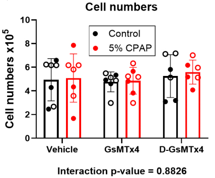
D-GsMTx4 purchased from MedChemExpress. Usage Cited in: Am J Physiol Lung Cell Mol Physiol. 2025 Aug 1;329(2):L296-L306. [Abstract]
D-GsMTx4 (1 μM). The short-term effect of stretch in fASM cell proliferation. fASM cells on Flexcell plates were pretreated with piezo inhibitors for 1h and then were exposed to 5% static stretch with 5% oscillatory stretch (5% CPAP) with continuous presence of the inhibitors. Control cells were exposed to 5% oscillatory stretch only. Cells were harvested after 24h treatment and counted.
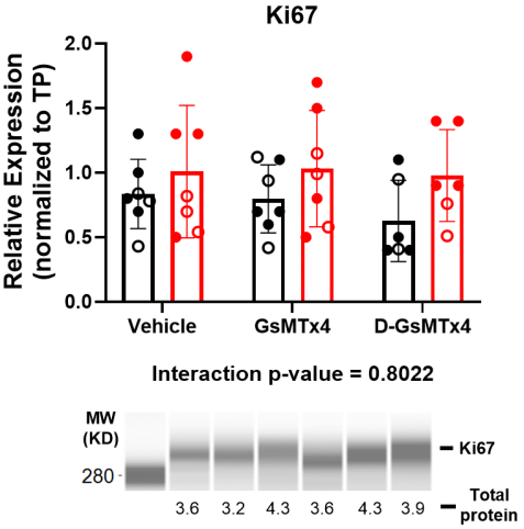
D-GsMTx4 purchased from MedChemExpress. Usage Cited in: Am J Physiol Lung Cell Mol Physiol. 2025 Aug 1;329(2):L296-L306. [Abstract]
D-GsMTx4 (1 μM). The protein expressions of Ki67 in fASM cells were determined by JESS.
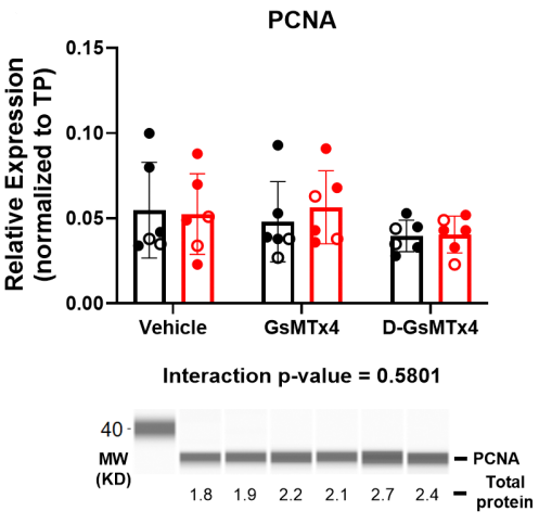
D-GsMTx4 purchased from MedChemExpress. Usage Cited in: Am J Physiol Lung Cell Mol Physiol. 2025 Aug 1;329(2):L296-L306. [Abstract]
D-GsMTx4 (1 μM). The protein expressions of PCNA in fASM cells were determined by JESS.
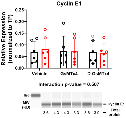
D-GsMTx4 purchased from MedChemExpress. Usage Cited in: Am J Physiol Lung Cell Mol Physiol. 2025 Aug 1;329(2):L296-L306. [Abstract]
D-GsMTx4 (1 μM). The protein expressions of Cyclin E1 in fASM cells were determined by JESS.
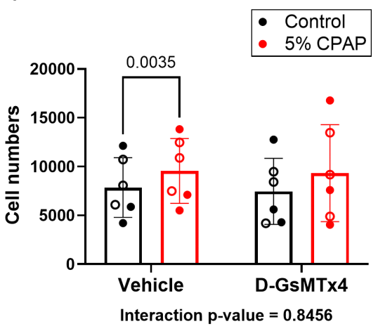
D-GsMTx4 purchased from MedChemExpress. Usage Cited in: Am J Physiol Lung Cell Mol Physiol. 2025 Aug 1;329(2):L296-L306. [Abstract]
The long-term effect of stretch in fASM cell proliferation. fASM cells on Flexcell plates were pretreated with D-GsMTx4 (1 μM) for 1h and then were exposed to 5% static stretch with 5% oscillatory stretch (5% CPAP) with or without the inhibitors. Control cells were exposed to 5% oscillatory stretch only. Cells were harvested after 24h treatment and seeded into 96-well plates. After 3-day culture without stretch, cell proliferation was measured by CyQUANT assay.
-
Pediatr Res
Stretch, Piezo channels, and store operated calcium entry in developing human airway smooth muscle. [Abstract]2026 Apr 24. PMID: 42032191


Solvent & Solubility
In Vitro:
DMSO : 100 mg/mL (24.42 mM; Need ultrasonic; Hygroscopic DMSO has a significant impact on the solubility of product, please use newly opened DMSO)
Please refer to the solubility information to select the appropriate solvent. Once prepared, please aliquot and store the solution to prevent product inactivation from repeated freeze-thaw cycles.
Storage method and period of stock solution: -80°C, 6 months; -20°C, 1 month (sealed storage, away from moisture and light, under nitrogen). When stored at -80°C, please use it within 6 months. When stored at -20°C, please use it within 1 month.
Please refer to the solubility information to select the appropriate solvent. Once prepared, please aliquot and store the solution to prevent product inactivation from repeated freeze-thaw cycles.
Storage method and period of stock solution: -80°C, 6 months; -20°C, 1 month (sealed storage, away from moisture and light, under nitrogen). When stored at -80°C, please use it within 6 months. When stored at -20°C, please use it within 1 month.
Concentration (start) × Volume (start) = Concentration (final) × Volume (final)


Purity & Documentation
-
Data Sheet (297 KB)
-
SDS (252 KB)
- English - EN (252 KB)
- Français - FR (252 KB)
- Deutsch - DE (252 KB)
- Norwegian - NO (252 KB)
- Español - ES (252 KB)
- Swedish - SV (252 KB)
- Italian - IT (252 KB)
- Korean - KR (252 KB)
- Portuguese - PT (252 KB)
-
Handling Instructions (2659 KB)
References
[1]. Alcaino C, Knutson K, Gottlieb PA, Farrugia G, Beyder A. Mechanosensitive ion channel Piezo2 is inhibited by D-GsMTx4. Channels (Austin). 2017 May 4;11(3):245-253. doi: 10.1080/19336950.2017.1279370. Epub 2017 Jan 13. Erratum for: Addendum to: Wang F, Knutson K, Alcaino C, Linden DR, Gibbons SJ, Kashyap PK, Grover M, Oeckler R, Gottlieb PA, Li HJ, et al. Mechanosensitive ion channel Piezo2 is important for enterochromaffin cell response to mechanical forces. J Physiol. 2017; 595(1):79-91 [Content Brief]
[3]. Liu L, et al. Piezo2 Channel Upregulation is Involved in Mechanical Allodynia in CYP-Induced Cystitis Rats. Mol Neurobiol. 2023 Sep;60(9):5000-5012. [Content Brief]
[4]. Jo MJ, et al. Blockade of Piezo2 Pathway Attenuates Inflammatory and Neuropathic Pain in the Orofacial Area. Pain Res Manag. 2024 Sep 27;2024:9179928. [Content Brief]


Complete Stock Solution Preparation Table
Please refer to the solubility information to select the appropriate solvent. Once prepared, please aliquot and store the solution to prevent product inactivation from repeated freeze-thaw cycles.
Storage method and period of stock solution: -80°C, 6 months; -20°C, 1 month (sealed storage, away from moisture and light, under nitrogen). When stored at -80°C, please use it within 6 months. When stored at -20°C, please use it within 1 month.
| Optional Solvent | Concentration Solvent Mass | 1 mg | 5 mg | 10 mg | 25 mg |
|---|---|---|---|---|---|
| DMSO | 1 mM | 0.2442 mL | 1.2208 mL | 2.4415 mL | 6.1038 mL |
| 5 mM | 0.0488 mL | 0.2442 mL | 0.4883 mL | 1.2208 mL | |
| 10 mM | 0.0244 mL | 0.1221 mL | 0.2442 mL | 0.6104 mL | |
| 15 mM | 0.0163 mL | 0.0814 mL | 0.1628 mL | 0.4069 mL | |
| 20 mM | 0.0122 mL | 0.0610 mL | 0.1221 mL | 0.3052 mL |


D-GsMTx4 Related Classifications
HY-P1410B Related Classifications
 Powered by Bioz