



4-DAMP
Based on 4 publication(s) in Google Scholar
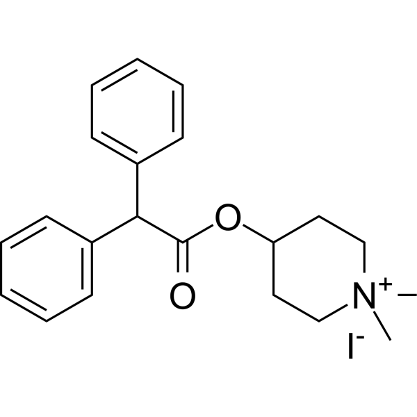
4-DAMP (4-DAMP methiodide) is a potent and selective antagonist of M3 receptors and also has a high affinity for the closely-related M5 receptors. 4-DAMP combined with 5-Fluorouracil (5-Fu) (HY-90006) could significantly reduce the cell viability and enhance apoptosis in MKN45 and BGC823 gastric cancer cells. 4-DAMP inhibits lipopolysaccharide (LPS)- and tobacco-induced pulmonary inflammation and reduces mucin 5AC (MUC5AC), oligomeric mucus/gel-forming secretion.
연구목적의 판매만을 진행합니다. 환자를 대상으로 한 판매는 하지 않습니다.
- Purity : 99.96%
- CAS No.: 1952-15-4
- 화학식: C21H26INO2
- 분자량:451.34
-
보관:
4°C, sealed storage, away from moisture and light
* In solvent : -80°C, 6 months; -20°C, 1 month (sealed storage, away from moisture and light)

To place orders, for customer services and technical support, please contact: MedChemExpress USA
Tel: 609-228-6898 E-mail: [email protected] [email protected]


-
 Biological Activity
-
Chemical Information
-
용액&용해도
- Protocol
- 순도&문서
- References
-
 Help & FAQs


-
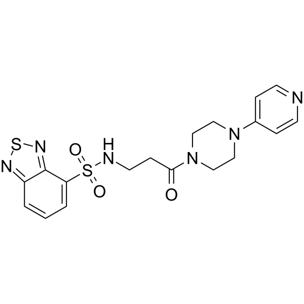
HY-108234
VU 0255035M1 Antagonist -
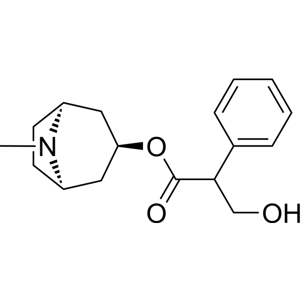
HY-B1205
AtropinemAChR Antagonist -
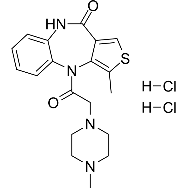
HY-B1789A
Telenzepine dihydrochlorideMuscarinic M1 Receptor Antagonist -
HY-107647
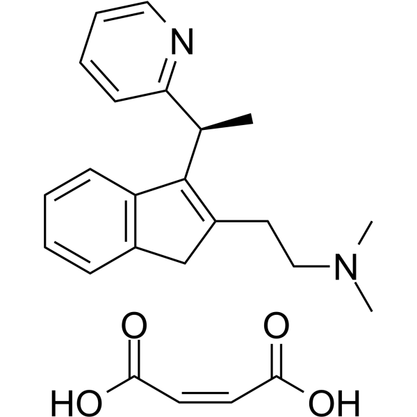
(S)-(+)-Dimethindene maleateM2 Receptor Antagonist -
HY-B0282
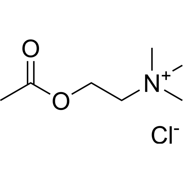
Acetylcholine chlorideCholinergic Agonist -
HY-101372A
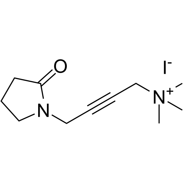
Oxotremorine M iodidemAChR Agonist -
HY-107654
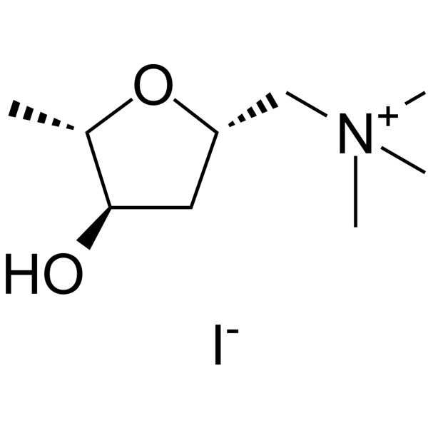
Muscarine iodidemAChR Agonist -
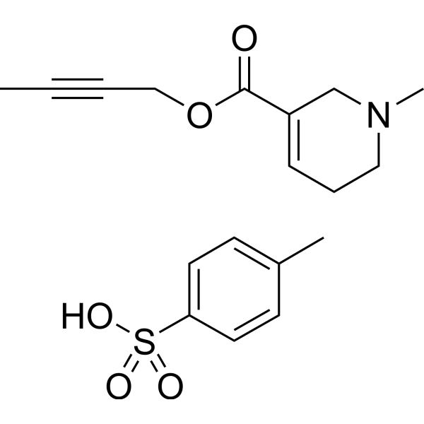
HY-107655
Arecaidine but-2-ynyl ester tosylatemAChR M2 Agonist -
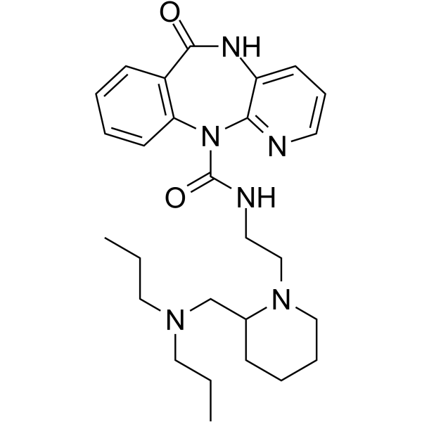
HY-107652
AF-DX 384 -
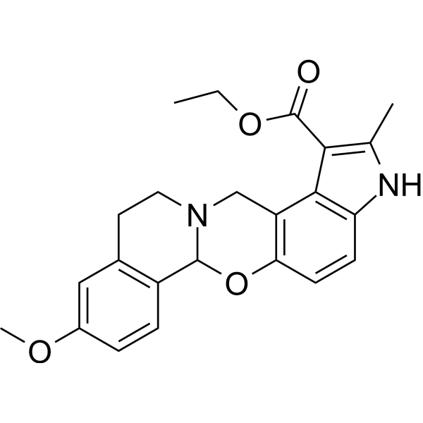
HY-107646
PD 102807M4 Receptor Antagonist -
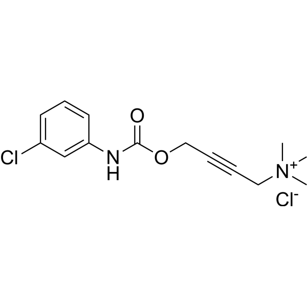
HY-107648
McN-A-343M1 Muscarinic Agonist -
HY-A0033
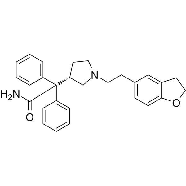
DarifenacinM3 Muscarinic Receptor Antagonist
Publications Citing Use of MedChemExpress (MCE) 4-DAMP
MoreAll EGFR Isoforms
More

Biological Activity
제품 설명
IC50 & Target
|
mAChR3 |
mAChR5 |
Cellular Effect
|
Cell Line

|
Type | Value | Description | References |
|---|---|---|---|---|
| CHO | IC50 |
3.2 x 10⁻¹⁰ M
Compound: 4-DAMP
|
Displacement of [3H]4-DAMP from human recombinant muscarinic M5 receptor expressed in CHO cells
Displacement of [3H]4-DAMP from human recombinant muscarinic M5 receptor expressed in CHO cells
|
[PMID: 26988801] |
| CHO | IC50 |
4.1 x 10⁻¹⁰ M
Compound: 4-DAMP
|
Displacement of [3H]4-DAMP from human recombinant muscarinic M3 receptor expressed in CHO cells
Displacement of [3H]4-DAMP from human recombinant muscarinic M3 receptor expressed in CHO cells
|
[PMID: 26988801] |
| CHO | IC50 |
4.9 x 10⁻¹⁰ M
Compound: 4-DAMP
|
Displacement of [3H]4-DAMP from human recombinant muscarinic M4 receptor expressed in CHO cells
Displacement of [3H]4-DAMP from human recombinant muscarinic M4 receptor expressed in CHO cells
|
[PMID: 26988801] |
| CHO | IC50 |
3.2 x 10⁻¹⁰ M
Compound: 4-DAMP
|
Displacement of [3H]4-DAMP from human recombinant M5 receptor expressed in CHO cells measured after 60 mins by scintillation counting method
Displacement of [3H]4-DAMP from human recombinant M5 receptor expressed in CHO cells measured after 60 mins by scintillation counting method
|
[PMID: 27876250] |
| CHO | IC50 |
4.1 x 10⁻¹⁰ M
Compound: 4-DAMP
|
Displacement of [3H]4-DAMP from human recombinant M3 receptor expressed in CHO cells measured after 60 mins by scintillation counting method
Displacement of [3H]4-DAMP from human recombinant M3 receptor expressed in CHO cells measured after 60 mins by scintillation counting method
|
[PMID: 27876250] |
| CHO | IC50 |
4.9 x 10⁻¹⁰ M
Compound: 4-DAMP
|
Displacement of [3H]4-DAMP from human recombinant M4 receptor expressed in CHO cells measured after 60 mins by scintillation counting method
Displacement of [3H]4-DAMP from human recombinant M4 receptor expressed in CHO cells measured after 60 mins by scintillation counting method
|
[PMID: 27876250] |
| CHO-K1 | IC50 |
0.055 μM
Compound: 4-DAMP
|
Displacement of [3H]N-methylscopolamine from human recombinant muscarinic M2 receptor expressed in CHOK1 cells after 2 hrs
Displacement of [3H]N-methylscopolamine from human recombinant muscarinic M2 receptor expressed in CHOK1 cells after 2 hrs
|
[PMID: 23403082] |
| CHO-K1 | IC50 |
4.5 nM
Compound: 4-DAMP
|
Displacement of [3H]N-methylscopolamine from human recombinant muscarinic M1 receptor expressed in CHOK1 cells after 2 hrs
Displacement of [3H]N-methylscopolamine from human recombinant muscarinic M1 receptor expressed in CHOK1 cells after 2 hrs
|
[PMID: 23403082] |
| CHO-K1 | IC50 |
5.1 nM
Compound: 4-DAMP
|
Displacement of [3H]N-methylscopolamine from human recombinant muscarinic M3 receptor expressed in CHOK1 cells after 2 hrs
Displacement of [3H]N-methylscopolamine from human recombinant muscarinic M3 receptor expressed in CHOK1 cells after 2 hrs
|
[PMID: 23403082] |
In Vitro
4-DAMP (10 μM, 5 days) combined with 5-Fu (5 μM, 5 days) can inhibit cell growth and promote apoptosis in MKN45 and BGC823 cells[2].
MedChemExpress (MCE) has not independently confirmed the accuracy of these methods. They are for reference only.
-
Cell Line:MKN45 and BGC823 cells
-
Concentration:10 μM
-
Incubation Time:5 days
-
Result:Reduced the cell viability to (30±4.0)% and (35±6.0)% combined with 5-Fu in MKN45 and BGC823 cells, respectively.
In Vivo
4-DAMP (0.6 or 0.12 mg/kg, intranasal instillation, daily for 35 days) reduces the allergic rhinitis (AR) symptom scores and nasal secretion weights, reverses goblet cell metaplasia, alleviates eosinophil infiltration, inhibits the levels of MUC5AC, histamine and cytokines, MMP9, EGFR in AR guinea pigs[3].
MedChemExpress (MCE) has not independently confirmed the accuracy of these methods. They are for reference only.
-
Animal Model:Mouse MKN45 and BGC823 tumor xenografts[2]
-
Dosage:1mg/kg
-
Administration:i.p., daily for 12 days
-
Result:Reduced the tumor sizes and the tumor weights in both MKN45 and BGC823 xenografts.
-
Animal Model:AR guinea pigs[3]
-
Dosage:0.6 or 0.12 mg/kg
-
Administration:intranasal instillation, daily for 35 days
-
Result:Reduced the weights of nasal secretions, reversed goblet cell metaplasia of the animal nasal mucosal epithelium, inhibited the levels of MUC5AC mRNA, IL-4, IL-13, and TNF-α in AR guinea pigs.
- Detection of Apoptosis
- TUNEL staining for apoptotic DNA fragmentation
- Cell Cytotoxicity Assay
- Mammalian live/dead viability and cytotoxicity staining
- Annexin V plus membrane-impermeant dye apoptosis staining
- Apoptosis Solutions
- CCK-8/WST-8 Cell Proliferation Assay
- Research Protocol for Inflammation-related Diseases
- MTT Cell Proliferation Assay
- Cell Viability Determination by MTT Colorimetric Assay


Chemical Information
-
CAS No. 1952-15-4
-
Appearance Solid
-
분자량 451.34
-
화학식 C21H26INO2
-
Color White to off-white
-
SMILES
C[N+]1(C)CCC(OC(C(C2=CC=CC=C2)C3=CC=CC=C3)=O)CC1.[I-]
-
Synonyms
4-DAMP methiodide
-
선적
Room temperature in continental US; may vary elsewhere.
-
보관
4°C, sealed storage, away from moisture and light
* In solvent : -80°C, 6 months; -20°C, 1 month (sealed storage, away from moisture and light)


Publications (4)
-
Journal Impact Factor
-
Most Recent
-
Cell Rep
Septo-subicular cholinergic circuit promotes seizure development via astrocytic inflammation. [Abstract]2025 May 14;44(5):115712. PMID: 40372911 -
Cell Biosci
Gut microbial metabolite deoxycholic acid facilitates Th17 differentiation through modulating cholesterol biosynthesis and participates in high-fat diet-associated colonic inflammation. [Abstract]2023 Oct 3;13(1):186. PMID: 37789469 -
Pharmaceuticals (Basel)
Ethanolic Extracts of Cupressaceae Species Conifers Provide Rapid Protection against Barium Chloride-Induced Cardiac Arrhythmia. [Abstract]2024 Jul 29;17(8):1003. PMID: 39204108 -
Mol Cell Endocrinol
Vagotomy suppresses food intake by increasing GLP-1 secretion via the M3 AChR-AMPKα pathway in mice. [Abstract]2025 Apr 1:599:112464. PMID: 39848433


용액&용해도
In Vitro:
DMSO : 100 mg/mL (221.56 mM; Need ultrasonic; Hygroscopic DMSO has a significant impact on the solubility of product, please use newly opened DMSO)
Please refer to the solubility information to select the appropriate solvent. Once prepared, please aliquot and store the solution to prevent product inactivation from repeated freeze-thaw cycles.
Storage method and period of stock solution: -80°C, 6 months; -20°C, 1 month (sealed storage, away from moisture and light). When stored at -80°C, please use it within 6 months. When stored at -20°C, please use it within 1 month.
Please refer to the solubility information to select the appropriate solvent. Once prepared, please aliquot and store the solution to prevent product inactivation from repeated freeze-thaw cycles.
Storage method and period of stock solution: -80°C, 6 months; -20°C, 1 month (sealed storage, away from moisture and light). When stored at -80°C, please use it within 6 months. When stored at -20°C, please use it within 1 month.
Concentration (start) × Volume (start) = Concentration (final) × Volume (final)
In Vivo:
Select the appropriate dissolution method based on your experimental animal and administration route.
- For the following dissolution methods, please ensure to first prepare a clear stock solution using an In Vitro approach and then sequentially add co-solvents:
- To ensure reliable experimental results, the clarified stock solution can be appropriately stored based on storage conditions. As for the working solution for In Vivo experiments, it is recommended to prepare freshly and use it on the same day.
- The percentages shown for the solvents indicate their volumetric ratio in the final prepared solution. If precipitation or phase separation occurs during preparation, heat and/or sonication can be used to aid dissolution.
Add each solvent one by one: 10% DMSO 40% PEG300 5% Tween-80 45% Saline
Solubility: ≥ 5 mg/mL (11.08 mM); Clear solution
This protocol yields a clear solution of ≥ 5 mg/mL (saturation unknown).
Taking 1 mL working solution as an example, add 100 μL DMSO stock solution (50.0 mg/mL) to 400 μL PEG300, and mix evenly; then add 50 μL Tween-80 and mix evenly; then add 450 μL Saline to adjust the volume to 1 mL.
Preparation of Saline: Dissolve 0.9 g sodium chloride in ddH₂O and dilute to 100 mL to obtain a clear Saline solution.
In Vivo Dissolution Calculator
Please enter the basic information of animal experiments:
-
-
-
-
Recommended: Prepare an additional quantity of animals to account for potential losses during experiments.
Please enter your animal formula composition:
-
%DMSO +
Recommended: Keep the proportion of DMSO in working solution below 2% if your animal is weak.
-
%+
-
+%Tween-80 + +
-
%Saline +
The co-solvents required include: DMSO, . All of co-solvents are available by MedChemExpress (MCE). , Tween 80. All of co-solvents are available by MedChemExpress (MCE).
Working solution concentration: 0.22 mg/mL
Method for preparing stock solution: mg drug dissolved in μL DMSO. Stock solution concentration: mg/mL. * In solvent : -80°C, 6 months; -20°C, 1 month (sealed storage, away from moisture and light)
1. Take μL DMSO stock solution;
2. Add μL .
μL , mix evenly;
3. Then add μL Tween 80, mix evenly;
4. Then add μL
Please ensure that the stock solution in the first step is dissolved to a clear state, and add co-solvents in sequence. You can use ultrasonic heating (ultrasonic cleaner, recommended frequency 20-40 kHz), vortexing, etc. to assist dissolution.


순도&문서
-
Data Sheet (279 KB)
-
SDS (394 KB)
- English - EN (394 KB)
- Français - FR (394 KB)
- Deutsch - DE (394 KB)
- Norwegian - NO (394 KB)
- Español - ES (394 KB)
- Swedish - SV (394 KB)
- Italian - IT (394 KB)
- Korean - KR (394 KB)
- Portuguese - PT (394 KB)
-
Handling Instructions (2659 KB)
References
[1]. Watson N, et al. Comparative pharmacology of recombinant human M3 and M5 muscarinic receptors expressed in CHO-K1 cells. Br J Pharmacol. 1999 May;127(2):590-6. [Content Brief]
[2]. Yu H, et al. Acetylcholine acts through M3 muscarinic receptor to activate the EGFR signaling and promotes gastric cancer cell proliferation. Sci Rep. 2017 Jan 19;7:40802. [Content Brief]
[3]. Huang Y, et al. Effects of 4-DAMP on allergic rhinitis in guinea pigs and its potential mechanism. Ann Transl Med. 2022 Jul;10(14):774. [Content Brief]


Complete Stock Solution Preparation Table
Please refer to the solubility information to select the appropriate solvent. Once prepared, please aliquot and store the solution to prevent product inactivation from repeated freeze-thaw cycles.
Storage method and period of stock solution: -80°C, 6 months; -20°C, 1 month (sealed storage, away from moisture and light). When stored at -80°C, please use it within 6 months. When stored at -20°C, please use it within 1 month.
| Optional Solvent | Concentration Solvent Mass | 1 mg | 5 mg | 10 mg | 25 mg |
|---|---|---|---|---|---|
| DMSO | 1 mM | 2.2156 mL | 11.0781 mL | 22.1562 mL | 55.3906 mL |
| 5 mM | 0.4431 mL | 2.2156 mL | 4.4312 mL | 11.0781 mL | |
| 10 mM | 0.2216 mL | 1.1078 mL | 2.2156 mL | 5.5391 mL | |
| 15 mM | 0.1477 mL | 0.7385 mL | 1.4771 mL | 3.6927 mL | |
| 20 mM | 0.1108 mL | 0.5539 mL | 1.1078 mL | 2.7695 mL | |
| 25 mM | 0.0886 mL | 0.4431 mL | 0.8862 mL | 2.2156 mL | |
| 30 mM | 0.0739 mL | 0.3693 mL | 0.7385 mL | 1.8464 mL | |
| 40 mM | 0.0554 mL | 0.2770 mL | 0.5539 mL | 1.3848 mL | |
| 50 mM | 0.0443 mL | 0.2216 mL | 0.4431 mL | 1.1078 mL | |
| 60 mM | 0.0369 mL | 0.1846 mL | 0.3693 mL | 0.9232 mL | |
| 80 mM | 0.0277 mL | 0.1385 mL | 0.2770 mL | 0.6924 mL | |
| 100 mM | 0.0222 mL | 0.1108 mL | 0.2216 mL | 0.5539 mL |


4-DAMP Related Classifications
HY-100958 Related Classifications
 Powered by Bioz