



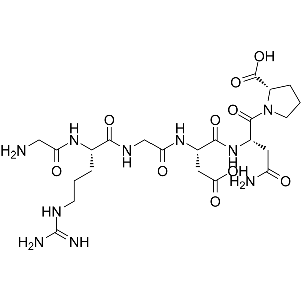
RGD peptide (GRGDNP)
Based on 10 publication(s) in Google Scholar
RGD peptide (GRGDNP) is an inhibitor of integrin-ligand interactions. RGD peptide (GRGDNP) competitively inhibits α5β1 binding with extracellular matrice (ECM). RGD peptide (GRGDNP) promotes apoptosis through activation of conformation changes that enhance pro-caspase-3 activation and autoprocessing. RGD peptide (GRGDNP) plays an important role in cell adhesion, migration, growth, and differentiation.
For research use only. We do not sell to patients.
- Purity : 99.83%
- CAS No.: 114681-65-1
- Formula: C23H38N10O10
- Molecular Weight:614.61
-
Storage:
Sealed storage, away from moisture.
Powder -80°C, 2 years , -20°C, 1 year* In solvent : -80°C, 6 months; -20°C, 1 month (sealed storage, away from moisture)

To place orders, for customer services and technical support, please contact: MedChemExpress USA
Tel: 609-228-6898 E-mail: [email protected] [email protected]


-
 Biological Activity
-
Chemical Information
-
Solvent & Solubility
- Protocols
- Purity & Documentation
- References
-
 Help & FAQs


-
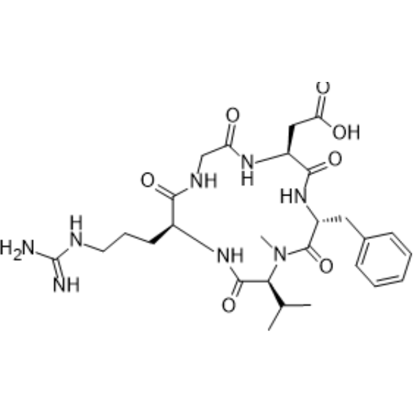
HY-16141
CilengitideIntegrin Inhibitor -
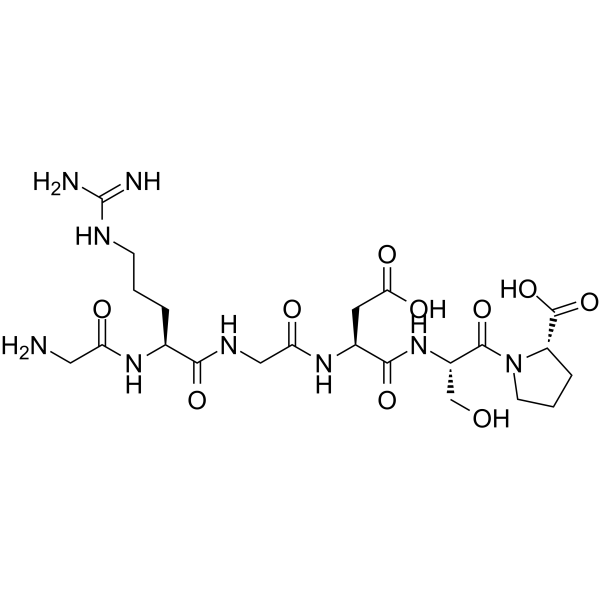
HY-P0290
GRGDSPIntegrin Inhibitor -
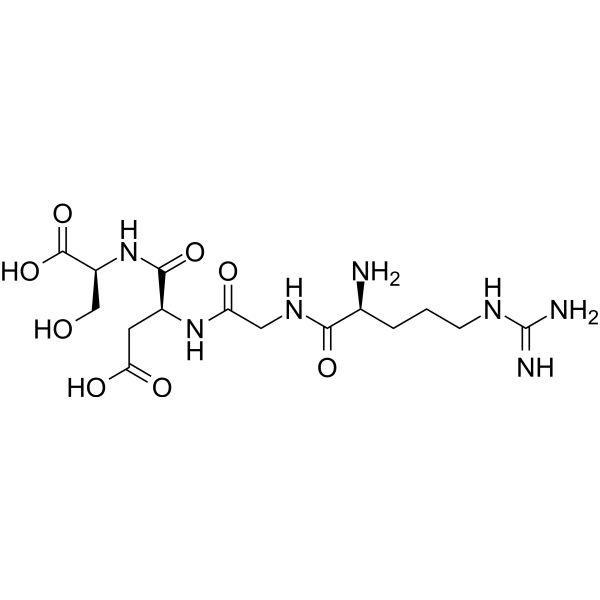
HY-12290
Arg-Gly-Asp-SerIntegrin Inhibitor -
HY-P0295
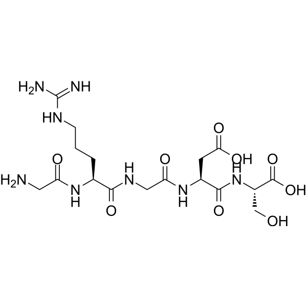
Gly-Arg-Gly-Asp-SerIntegrin Inhibitor -
HY-P0278
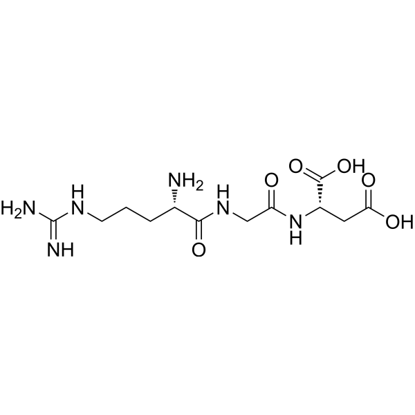
RGDTripeptide -

HY-15763
ErastinFerroptosis Inducer -
HY-P0023
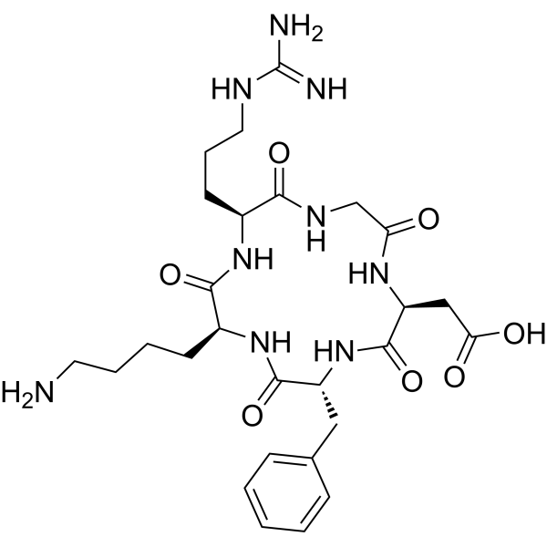
Cyclo(-RGDfK)αvβ3 Integrin Inhibitor -

HY-100579
Ferrostatin-1Ferroptosis Inhibitor -

HY-10431
SB-431542TGF-β Receptor Kinase Inhibitor -
HY-P0023A
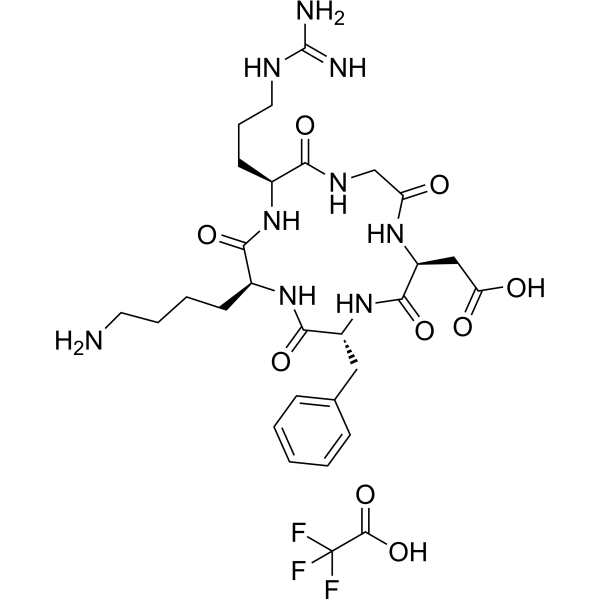
Cyclo(-RGDfK) TFAαvβ3 Integrin Inhibitor -
HY-A0070A
LiothyronineTIGIT/PVR Blocker -
HY-100563
Cyclo(RGDyK) trifluoroacetateαVβ3 Integrin Inhibitor
Publications Citing Use of MedChemExpress (MCE) RGD peptide (GRGDNP)
More- Adv Mater. 2024 Sep;36(36):e2406140. [Abstract]
- Nat Biomed Eng. 2025 Nov 3. [Abstract]
- Acta Pharm Sin B. 2026 Jun 8.
- Cell Death Dis. 2022 Jul 1;13(7):577. [Abstract]
- Int J Surg. 2026 Jul;112(7):p 13073-13091.
- Biofabrication. 2021 Apr 26;13(3). [Abstract]
- Br J Pharmacol. 2026 Aug;183(16):4989-5017. [Abstract]
- Biomacromolecules. 2023 Apr 10;24(4):1574-1584. [Abstract]
- J Cell Sci. 2025 Dec 1;138(23):jcs264412. [Abstract]
- bioRxiv. 2024 October 06.
 Customer Validation & Images

-
Bio/Physico-chemical Assay
All Caspase Isoforms
More

Biological Activity
Description
IC50 & Target
[2]|
α5β1 |
Caspase 3 |
Cellular Effect
|
Cell Line

|
Type | Value | Description | References |
|---|---|---|---|---|
| K562 | IC50 |
>100 μM
Compound: H-Gly-Arg-Gly-Asp-Asn-Pro-OH
|
Inhibition of alpha5beta1 integrin-mediated human K562 cell adhesion to fibronectin incubated for 30 mins before plating measured after 1 hr
Inhibition of alpha5beta1 integrin-mediated human K562 cell adhesion to fibronectin incubated for 30 mins before plating measured after 1 hr
|
[PMID: 23811088] |
In Vitro
RGD peptide (GRGDNP) (50 μM ; preincubation for 3 hours before stretch) abolishes stretch-induced IKK activation and IL-6 mRNA expression. It shows little effect on the IKK activity and IL-6 mRNA expression in unstretched HUVECs[2].
RGD peptide (GRGDNP) (300 μg/mL; for 6 hours) completely reverses enhanced FN1 expression in oxygen glucose deprivation(OGD) treated primary hippocampal neurons, HT22 cell lines and in its sEVs[3].
MedChemExpress (MCE) has not independently confirmed the accuracy of these methods. They are for reference only. Further protocols information, click here.


Chemical Information
-
CAS No. 114681-65-1
-
Appearance Solid
-
Molecular Weight 614.61
-
Formula C23H38N10O10
-
Color White to off-white
-
Sequence
Gly-Arg-Gly-Asp-Asn-Pro
-
Sequence Shortening
GRGDNP
-
Shipping
Room temperature in continental US; may vary elsewhere.
-
Storage
Sealed storage, away from moisture
Powder -80°C 2 years -20°C 1 year * In solvent : -80°C, 6 months; -20°C, 1 month (sealed storage, away from moisture)


Publications (10)
-
Journal Impact Factor
-
Most Recent
-
Adv Mater
2024 Sep;36(36):e2406140. PMID: 39023382
RGD peptide (GRGDNP) purchased from MedChemExpress. Usage Cited in: Adv Mater. 2024 Sep;36(36):e2406140. [Abstract]
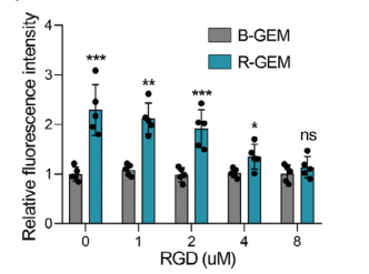
Comparison of the adhesion of B-GEM and R-GEM cells to 4T1 tumor cells after 10 min of coincubation in medium preadded with free RGD peptide (0-8 μM). Exogenous free RGD dose-dependently reversed the increase in adhesion of R-GEM cells to tumor cells.
-
Nat Biomed Eng
Nonexpansive biodegradable matrix promotes blood vessel organoid development for neurovascular repair and functional recovery in ischaemic stroke. [Abstract]2025 Nov 3. PMID: 41184604 -
-
Cell Death Dis
The kinase activity of integrin-linked kinase regulates cellular senescence in gastric cancer. [Abstract]2022 Jul 1;13(7):577. PMID: 35778385 -
-
Biofabrication
A novel 4D cell culture mimicking stomach peristalsis altered gastric cancer spheroids growth and malignance. [Abstract]2021 Apr 26;13(3). PMID: 33836517 -
Br J Pharmacol
Enzyme-responsive 612-AYR nanoparticles for targeted inhibition of B4GALT3-mediated ITGB1 glycosylation in hepatocellular carcinoma. [Abstract]2026 Aug;183(16):4989-5017. PMID: 42157415 -
Biomacromolecules
2023 Apr 10;24(4):1574-1584. PMID: 36943688 -
J Cell Sci
Bile canaliculi formation in primary hepatocytes requires α1β1 integrin-dependent adherens junction re-organization. [Abstract]2025 Dec 1;138(23):jcs264412. PMID: 41347643 -


Solvent & Solubility
In Vitro:
H2O : ≥ 100 mg/mL (162.70 mM)
DMSO : 3.12 mg/mL (5.08 mM; Need ultrasonic; Hygroscopic DMSO has a significant impact on the solubility of product, please use newly opened DMSO)
* "≥" means soluble, but saturation unknown.
Please refer to the solubility information to select the appropriate solvent. Once prepared, please aliquot and store the solution to prevent product inactivation from repeated freeze-thaw cycles.
Storage method and period of stock solution: -80°C, 6 months; -20°C, 1 month (sealed storage, away from moisture). When stored at -80°C, please use it within 6 months. When stored at -20°C, please use it within 1 month.
* Note: If you choose water as the stock solution, please dilute it to the working solution, then filter and sterilize it with a 0.22 μm filter before use.
Please refer to the solubility information to select the appropriate solvent. Once prepared, please aliquot and store the solution to prevent product inactivation from repeated freeze-thaw cycles.
Storage method and period of stock solution: -80°C, 6 months; -20°C, 1 month (sealed storage, away from moisture). When stored at -80°C, please use it within 6 months. When stored at -20°C, please use it within 1 month.
* Note: If you choose water as the stock solution, please dilute it to the working solution, then filter and sterilize it with a 0.22 μm filter before use.
Concentration (start) × Volume (start) = Concentration (final) × Volume (final)
In Vivo:
Select the appropriate dissolution method based on your experimental animal and administration route.
- For the following dissolution methods, please ensure to first prepare a clear stock solution using an In Vitro approach and then sequentially add co-solvents:
- To ensure reliable experimental results, the clarified stock solution can be appropriately stored based on storage conditions. As for the working solution for In Vivo experiments, it is recommended to prepare freshly and use it on the same day.
- The percentages shown for the solvents indicate their volumetric ratio in the final prepared solution. If precipitation or phase separation occurs during preparation, heat and/or sonication can be used to aid dissolution.
Add each solvent one by one: 10% DMSO 90% (20% SBE-β-CD in Saline)
Solubility: ≥ 0.67 mg/mL (1.09 mM); Clear solution
This protocol yields a clear solution of ≥ 0.67 mg/mL (saturation unknown).
Taking 1 mL working solution as an example, add 100 μL DMSO stock solution (6.7 mg/mL) to 900 μL 20% SBE-β-CD in Saline, and mix evenly.
Preparation of 20% SBE-β-CD in Saline (4°C, storage for one week): 2 g SBE-β-CD powder is dissolved in 10 mL Saline, completely dissolve until clear.
In Vivo Dissolution Calculator
Please enter the basic information of animal experiments:
-
-
-
-
Recommended: Prepare an additional quantity of animals to account for potential losses during experiments.
Working solution concentration: 0.22 mg/mL
This product has good water solubility, please refer to the measured solubility data in water/PBS/Saline for details.

Protocols
-
Apoptosis
Apoptosis, also called programmed cell death, is generally characterized by distinct morphological characteristics.
-
TUNEL staining for apoptotic DNA fragmentation
TUNEL staining detects DNA strand breaks by using terminal deoxynucleotidyl transferase to add labeled nucleotides to exposed 3′-OH DNA termini, generating either microscopic staining in fixed cells or tissue sections, or fluorescence/cytometric signal in cell suspensions. TUNEL positivity reflects DNA fragmentation but should not be interpreted alone as definitive apoptosis, because TUNEL can also label necrotic, autolytic, mechanically damaged, or DNA-repair-associated DNA breaks.
-
Annexin V plus membrane-impermeant dye apoptosis staining
Annexin V-based apoptosis assays rely on the detection of phosphatidylserine (PS) externalization from the inner leaflet of the plasma membrane to the outer leaflet, an early biochemical hallmark of apoptosis. Fluorescently labeled Annexin V binds PS in a calcium-dependent manner, enabling identification of early apoptotic cells by flow cytometry or fluorescence microscopy. When combined with a membrane-impermeant DNA-binding dye (e. g. , propidium iodide), this approach allows discrimination between viable (Annexin V−/dye−), early apoptotic (Annexin V+/dye−), and late apoptotic or necrotic (Annexin V+/dye+) cell populations by assessing membrane integrity and PS exposure.
-
Cell migration
Cell migration is a method that plays an important role in wound healing, cell differentiation, embryonic development, etc.
-
Apoptosis Solutions
Apoptosis is a regulated, generally non-lytic cell-death pathway that removes unwanted, damaged, infected, or abnormal cells through coordinated morphological changes, caspase activation, DNA fragmentation, and membrane remodeling. The intrinsic apoptosis pathway is controlled mainly by mitochondrial outer membrane permeabilization, BCL-2 family proteins, cytochrome c release, apoptosome formation, caspase-9 activation, and downstream executioner caspase-3/7 activation. The extrinsic apoptosis pathway is initiated by death receptors such as Fas, TNFR, and TRAIL receptors, which recruit adaptor proteins and activate caspase-8 before engaging executioner caspases or mitochondrial amplification through BID cleavage. Apoptosis is linked to many phenotypes, including cancer cell killing, tissue homeostasis, immune regulation, neurodegeneration, infection response, and treatment-induced cytotoxicity; unresolved questions include how apoptosis interacts with necroptosis, pyroptosis, ferroptos
-
Directly Induced Neuron Culture
Directly induced neuron culture converts somatic cells, most commonly fibroblasts, into induced neurons without passing through a pluripotent or neural progenitor stage; classic evidence shows that mouse fibroblasts can be converted by Ascl1, Brn2/Pou3f2, and Myt1l, human fibroblasts can be converted by defined neuronal transcription factors, and human fibroblasts can also be converted by miR-9/9-124 with neurogenic or subtype-specifying transcription factors. The readout is acquisition of neuronal identity and function, assessed by neuronal morphology, neuronal markers such as Tuj1/βIII-tubulin, MAP2, synapsin, and subtype markers when relevant, together with functional assays such as action-potential firing, synaptic activity, and electrophysiology.


Purity & Documentation
-
Data Sheet (278 KB)
-
SDS (394 KB)
- English - EN (394 KB)
- Français - FR (394 KB)
- Deutsch - DE (394 KB)
- Norwegian - NO (394 KB)
- Español - ES (394 KB)
- Swedish - SV (394 KB)
- Italian - IT (394 KB)
- Korean - KR (394 KB)
- Portuguese - PT (394 KB)
-
Handling Instructions (2659 KB)
References
[1]. C D Buckley, et al. RGD peptides induce apoptosis by direct caspase-3 activation. Nature. 1999 Feb 11;397(6719):534-9. [Content Brief]
[2]. Wei Xia, et al. Damaged brain accelerates bone healing by releasing small extracellular vesicles that target osteoprogenitors. Nat Commun. 2021 Oct 15;12(1):6043. [Content Brief]
[3]. Akitoshi Sasamoto, et al. Mechanotransduction by integrin is essential for IL-6 secretion from endothelial cells in response to uniaxial continuous stretch. Am J Physiol Cell Physiol. 2005 May;288(5):C1012-22. [Content Brief]


Complete Stock Solution Preparation Table
Please refer to the solubility information to select the appropriate solvent. Once prepared, please aliquot and store the solution to prevent product inactivation from repeated freeze-thaw cycles.
Storage method and period of stock solution: -80°C, 6 months; -20°C, 1 month (sealed storage, away from moisture). When stored at -80°C, please use it within 6 months. When stored at -20°C, please use it within 1 month.
| Optional Solvent | Concentration Solvent Mass | 1 mg | 5 mg | 10 mg | 25 mg |
|---|---|---|---|---|---|
| DMSO / H2O | 1 mM | 1.6270 mL | 8.1352 mL | 16.2705 mL | 40.6762 mL |
| 5 mM | 0.3254 mL | 1.6270 mL | 3.2541 mL | 8.1352 mL | |
| H2O | 10 mM | 0.1627 mL | 0.8135 mL | 1.6270 mL | 4.0676 mL |
| 15 mM | 0.1085 mL | 0.5423 mL | 1.0847 mL | 2.7117 mL | |
| 20 mM | 0.0814 mL | 0.4068 mL | 0.8135 mL | 2.0338 mL | |
| 25 mM | 0.0651 mL | 0.3254 mL | 0.6508 mL | 1.6270 mL | |
| 30 mM | 0.0542 mL | 0.2712 mL | 0.5423 mL | 1.3559 mL | |
| 40 mM | 0.0407 mL | 0.2034 mL | 0.4068 mL | 1.0169 mL | |
| 50 mM | 0.0325 mL | 0.1627 mL | 0.3254 mL | 0.8135 mL | |
| 60 mM | 0.0271 mL | 0.1356 mL | 0.2712 mL | 0.6779 mL | |
| 80 mM | 0.0203 mL | 0.1017 mL | 0.2034 mL | 0.5085 mL | |
| 100 mM | 0.0163 mL | 0.0814 mL | 0.1627 mL | 0.4068 mL |
* Note: If you choose water as the stock solution, please dilute it to the working solution, then filter and sterilize it with a 0.22 μm filter before use.


RGD peptide (GRGDNP) Related Classifications
HY-P1740 Related Classifications
 Powered by Bioz