



Chamaejasmine

Based on 1 Customer Validation
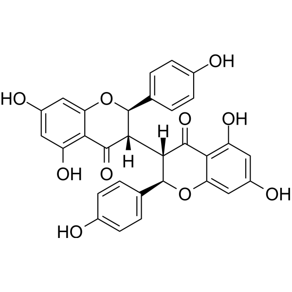
Chamaejasmine (Chamaejasmin) is a natural biflavonoid found in the roots of Stellera chamaejasme, exhibiting antitumor activity. Chamaejasmine inhibits Bcl-2, upregulates Bax, and induces cleavage of caspase-9/-3 and PARP. Chamaejasmine induces ROS production, Δψm loss, cytochrome c release, G2/M arrest, apoptosis, and autophagy. Chamaejasmine induces apoptosis and autophagy through activation of AMPK and inhibition of mTOR, and inhibits microtubule depolymerization by binding to β-tubulin. Chamaejasmine inhibits IL-4, IgE, β-hexosaminidase, and mast cell infiltration, and improves skin barrier function in AD models. Chamaejasmine exhibits cytotoxicity against various cancer cells. Chamaejasmine can be used in research related to various cancers such as lung adenocarcinoma and osteosarcoma, as well as atopic dermatitis.
For research use only. We do not sell to patients.
- Purity : 98.17%
- CAS No.: 69618-96-8
- Formula: C30H22O10
- Molecular Weight:542.49
-
Storage:
-20°C, protect from light
* In solvent : -80°C, 6 months; -20°C, 1 month (protect from light)

To place orders, for customer services and technical support, please contact: MedChemExpress USA
Tel: 609-228-6898 E-mail: [email protected] [email protected]


-
 Biological Activity
-
Chemical Information
-
Solvent & Solubility
- Purity & Documentation
- References
-
 Help & FAQs




Biological Activity
Description
|
Caspase-3 |
Caspase-9 |
Bcl-2 |
Bax |
IL-4 |
Cellular Effect
|
Cell Line

|
Type | Value | Description | References |
|---|---|---|---|---|
| A549 | IC50 |
7.72 μM
|
Cytotoxicity against human lung adenocarcinoma A549 cells assessed as reduction in cell viability by MTT assay.
Cytotoxicity against human lung adenocarcinoma A549 cells assessed as reduction in cell viability by MTT assay.
|
21952498 |
| H1975 | IC50 |
18.11 μM
|
Cytotoxicity against human non-small cell lung carcinoma H1975 cells assessed as reduction in cell viability by MTT assay.
Cytotoxicity against human non-small cell lung carcinoma H1975 cells assessed as reduction in cell viability by MTT assay.
|
21952498 |
| SMMC-7721 | IC50 |
14.04 μM
|
Cytotoxicity against human hepatoma SMMC-7721 cells assessed as reduction in cell viability by MTT assay.
Cytotoxicity against human hepatoma SMMC-7721 cells assessed as reduction in cell viability by MTT assay.
|
21952498 |
| SK-OV-3 | IC50 |
10.43 μM
|
Cytotoxicity against human adenocarcinoma SKOV-3 cells assessed as reduction in cell viability by MTT assay.
Cytotoxicity against human adenocarcinoma SKOV-3 cells assessed as reduction in cell viability by MTT assay.
|
21952498 |
| MCF7 | IC50 |
4.02 μM
|
Cytotoxicity against human MCF-7 cells assessed as inhibition of cell viability incubated for 72 hrs by MTT assay.
Cytotoxicity against human MCF-7 cells assessed as inhibition of cell viability incubated for 72 hrs by MTT assay.
|
21788932 |
| A549 | IC50 |
4.84 μM
|
Cytotoxicity against human A549 cells assessed as inhibition of cell viability incubated for 72 hrs by MTT assay.
Cytotoxicity against human A549 cells assessed as inhibition of cell viability incubated for 72 hrs by MTT assay.
|
21788932 |
| SGC-7901 | IC50 |
11.97 μM
|
Cytotoxicity against human SGC-7901 cells assessed as inhibition of cell viability incubated for 72 hrs by MTT assay.
Cytotoxicity against human SGC-7901 cells assessed as inhibition of cell viability incubated for 72 hrs by MTT assay.
|
21788932 |
| HCT-8 | IC50 |
12.45 μM
|
Cytotoxicity against human HCT-8 cells assessed as inhibition of cell viability incubated for 72 hrs by MTT assay.
Cytotoxicity against human HCT-8 cells assessed as inhibition of cell viability incubated for 72 hrs by MTT assay.
|
21788932 |
| HeLa | IC50 |
9.88 μM
|
Cytotoxicity against human Hela cells assessed as inhibition of cell viability incubated for 72 hrs by MTT assay.
Cytotoxicity against human Hela cells assessed as inhibition of cell viability incubated for 72 hrs by MTT assay.
|
21788932 |
| HepG2 | IC50 |
14.36 μM
|
Cytotoxicity against human HepG2 cells assessed as inhibition of cell viability incubated for 72 hrs by MTT assay.
Cytotoxicity against human HepG2 cells assessed as inhibition of cell viability incubated for 72 hrs by MTT assay.
|
21788932 |
| PC-3 | IC50 |
2.28 μM
|
Cytotoxicity against human PC-3 cells assessed as inhibition of cell viability incubated for 72 hrs by MTT assay.
Cytotoxicity against human PC-3 cells assessed as inhibition of cell viability incubated for 72 hrs by MTT assay.
|
21788932 |
| LNCaP | IC50 |
5.21 μM
|
Cytotoxicity against human LNCap cells assessed as inhibition of cell viability incubated for 72 hrs by MTT assay.
Cytotoxicity against human LNCap cells assessed as inhibition of cell viability incubated for 72 hrs by MTT assay.
|
21788932 |
| Vero | IC50 |
3.16 μM
|
Cytotoxicity against african green monkey Vero cells assessed as inhibition of cell viability incubated for 72 hrs by MTT assay.
Cytotoxicity against african green monkey Vero cells assessed as inhibition of cell viability incubated for 72 hrs by MTT assay.
|
21788932 |
| MDCK | IC50 |
4.57 μM
|
Cytotoxicity against Madin-Darby canine kidney MDCK cells assessed as inhibition of cell viability incubated for 72 hrs by MTT assay.
Cytotoxicity against Madin-Darby canine kidney MDCK cells assessed as inhibition of cell viability incubated for 72 hrs by MTT assay.
|
21788932 |
In Vitro
Chamaejasmine (2-64 μM; 24-72 h) exhibits the strongest cytotoxic activity against A549 human lung adenocarcinoma cells with an IC50 of 7.72 μM, compared with weaker effects on H1975, SMMC-7721, and SKOV-3 cell lines[1].
Chamaejasmine (0-8 μM; 48 h) induces the release of cytochrome c from mitochondria into the cytosol in A549 cells[1].
Chamaejasmine (0-8 μM; 48 h) upregulates Bax and downregulates Bcl-2 in A549 cells, increasing the Bax/Bcl-2 ratio[1].
Chamaejasmine (0-8 μM; 48 h) induces the cleavage of caspase-3, caspase-9, and PARP in a dose-dependent manner in A549 cells[1].
Chamaejasmine (40-160 μM; 24 h) inhibits the invasion of MG-63 cells in a concentration-dependent manner[2].
Chamaejasmine (0-100 μM; 72 h) exhibits potent in vitro cytotoxicity against multiple human cancer cell lines, with the strongest activity against PC-3 cells (IC50 = 2.28 μM)[4].
Chamaejasmine (24-72 h) inhibits PC-3 cell proliferation in a time-dependent manner, with the inhibitory effect gradually increasing over time[4].
Chamaejasmine (0.5-4 µM; 24-72 h) increases β-tubulin expression but not α-tubulin expression in PC-3 cells[4].
Chamaejasmine (0-8 μM; 48 h) induces G2/M phase cell cycle arrest in A549 cells in a concentration-dependent manner[1].
Chamaejasmine (0-8 μM; 48 h) induces apoptosis in A549 cells in a concentration-dependent manner[1].
Chamaejasmine (0-8 μM; 48 h) induces ROS generation in A549 cells in a concentration-dependent manner[1].
Chamaejasmine (0-8 μM; 48 h) induces dose-dependent disruption of mitochondrial membrane potential in A549 cells[1].
Chamaejasmine (0-160 μM; 24-48 h) inhibits the viability of MG-63, Saos-2, KHOS, and U2OS osteosarcoma cells in a time- and dose-dependent manner[2].
Chamaejasmine (0-160 μM; 48 h) induces apoptosis in MG-63 cells[2].
Chamaejasmine increases caspase-3 activity in MG-63 and KHOS cells, as well as caspase-9 activity in Saos-2 and U2OS cells[2].
Chamaejasmine (0-160 μM; 48 h) induces morphological features of apoptosis in MG-63 cells in a concentration-dependent manner[2].
Chamaejasmine (160 μM; 48 h) increases the expression of cleaved caspase 3, cleaved caspase 9, and bcl-2/bax in MG-63 cells[2].
Chamaejasmine (0-160 μM; 48 h) mediates autophagy in MG-63 cells through AMPK pathway signaling, as evidenced by an increase in acidic vesicular organelles[2].
Chamaejasmine (0-160 μM; 48 h) induces autophagy in MG-63 cells by increasing the levels of ATG-7, LC3B-II, and beclin-1 in a concentration-dependent manner[2].
Chamaejasmine (0-160 μM; 48 h) activates the AMPK/mTOR signaling pathway in MG-63 cells by increasing AMPK phosphorylation and decreasing mTOR phosphorylation[2].
Chamaejasmine induces ROS generation in MG-63 cells, and ROS production is required for Chamaejasmine-induced AMPK activation in MG-63 cells[2].
Chamaejasmine (30 μM; 1 h) most effectively inhibits DNP-specific IgE-induced degranulation in RBL-2H3 cells[3].
MedChemExpress (MCE) has not independently confirmed the accuracy of these methods. They are for reference only.
-
Cell Line:A549, H1975, SMMC-7721, SKOV-3
-
Concentration:2, 4, 8, 16, 32, 64 μM
-
Incubation Time:24, 48, 72 h
-
Result:Inhibited the growth of A549 cells in a time- and dose-dependent manner.
Showed more notable cytotoxicity against A549 than H1975, SMMC-7721 and SKOV-3, with IC50 values of 7.72, 18.11, 14.04 and 10.43 μM, respectively.
-
Cell Line:A549
-
Concentration:0, 2, 4, 8 μM
-
Incubation Time:48 h
-
Result:Increased the G2/M phase population from 13.06% to 34.53% at 2-8 μM, as compared to 9.36% of G2/M phase cells in untreated control samples.
Exerted growth-inhibitory effects via G2/M phase arrest in a concentration-dependent manner.
-
Cell Line:A549
-
Concentration:0, 2, 4, 8 μM
-
Incubation Time:48 h
-
Result:Increased the percentage of annexin V-FITC binding A549 cells from 13.06% to 76.46% at 2-8 µM in a concentration-dependent manner.
Shifted data points to the Q2 side in a dose-dependent manner, indicating that the cells moved to the late apoptotic stage.
-
Cell Line:A549
-
Concentration:0, 2, 4, 8 μM
-
Incubation Time:48 h
-
Result:Greatly increased cytochrome c in the cytosol of treated cells, indicating cytochrome c release from mitochondria to cytoplasm.\nRevealed a significant increase in the expression of Bax in treated cells and a significant decrease in Bcl-2 expression.
Increased the Bax/Bcl-2 ratio significantly.\nCleaved pro-caspase-9, pro-caspase-3, and pro-PARP to their active forms.
Increased the level of active protein with dose, demonstrating dose-dependent cleavage.
-
Cell Line:MG-63, Saos-2, KHOS, and U2OS
-
Concentration:0, 40, 80, and 160 μM
-
Incubation Time:24 or 48 h
-
Result:Inhibited cell growth of MG-63, Saos-2, KHOS, and U2OS cell lines effectively in a time- and dose-dependent manner.
-
Cell Line:MG-63
-
Concentration:40, 80, and 160 μM
-
Incubation Time:24 h
-
Result:Significantly inhibited cell invasion compared with control cells in a concentration-dependent manner.
-
Cell Line:MG-63
-
Concentration:0, 40, 80, and 160 μM
-
Incubation Time:48 h
-
Result:Induced apoptosis as analyzed by flow cytometry with Annexin V/PI kit.
-
Cell Line:MG-63
-
Concentration:0, 40, 80, and 160 μM
-
Incubation Time:48 h
-
Result:Caused condensed and fragmented nuclei, characteristic of apoptosis, in a concentration-dependent manner.
-
Cell Line:MG-63
-
Concentration:160 μM
-
Incubation Time:48 h
-
Result:Remarkably increased protein expression of cleaved caspase 3, cleaved caspase 9, and bcl-2/bax.
Decreased the ratio of Bcl-2/Bax.\nIncreased activity of p-AMPK and ATG-7.
Baf (100 nM) decreased the level of p-ampk/ampk and the expression of ATG-7 and markedly decreased the effects of chamaejasmine in the chamaejasmine + Baf co-treatment group.
-
Cell Line:MG-63
-
Concentration:0, 40, 80, and 160 μM
-
Incubation Time:48 h
-
Result:Exhibited more acidic vesicular organelles (AVOs) in the cytoplasm.
-
Cell Line:MG-63
-
Concentration:0, 40, 80, and 160 μM
-
Incubation Time:48 h
-
Result:Detected significant conversion of LC3-I to LC3-II.
Increased levels of ATG-7, LC3B-II and beclin-1 in a concentration-dependent manner.\nIncreased the phosphorylation of AMPK and decreased the phosphorylation of mTOR in a concentration-dependent manner.
-
Cell Line:MCF-7, A549, SGC-7901, HCT-8, HO-4980, Hela, HepG2, PC-3, LNCap, Vero, MDCK
-
Concentration:0, 3.13, 6.25, 12.5, 25, 50, 100 μM
-
Incubation Time:72 h
-
Result:Exhibited strong cytotoxicity against all nine cancer cell lines and normal cell lines with IC50 values of 4.02 μM for MCF-7, 4.84 μM for A549, 11.97 μM for SGC-7901, 12.45 μM for HCT-8, 5.31 μM for HO-4980, 9.88 μM for Hela, 14.36 μM for HepG2, 2.28 μM for PC-3, 5.21 μM for LNCap, 3.16 μM for Vero, and 4.57 μM for MDCK.
-
Cell Line:PC-3
-
Concentration:0, 0.5, 2, 4 µM (concentration-dependent); 2 µM (time-dependent)
-
Incubation Time:72 h (concentration-dependent); 0, 24, 48, 72 h (time-dependent)
-
Result:Increased the percentage of polymerized β-tubulin from 124.21% to 132.66% and 178.57% in a concentration-dependent manner.
After treatment with 2 µM, the percentage of polymerized β-tubulin increased to 120.64%, 143.12%, and 190.11%, respectively.
Largely unchanged the percentage of α-tubulin.
In Vivo
MedChemExpress (MCE) has not independently confirmed the accuracy of these methods. They are for reference only.
-
Animal Model:SKH-1 hairless mice (female, six weeks old, DNCB-induced AD-like skin lesions)[3]
-
Dosage:0.5%
-
Administration:topical; twice a day; 2 weeks
-
Result:Attenuated clinical symptoms of DNCB-induced dermatitis.
Reduced epidermal thickness by 81% compared to DNCB control.
Reduced mast cell number by 62% compared to DNCB control.
Decreased serum IgE concentration by 38% versus DNCB control.
Reduced mean total serum IL-4 level to 25.2 pg/mL compared to 42.5 pg/mL in DNCB controls.
Reduced TEWL to 43.5 g/m2h compared with 68.9 g/m2h in DNCB controls.
Increased skin hydration by 45% compared with DNCB controls.


Chemical Information
-
CAS No. 69618-96-8
-
Appearance Solid
-
Molecular Weight 542.49
-
Formula C30H22O10
-
Color White to off-white
-
SMILES
O=C1[C@]([C@@]2([H])[C@H](C3=CC=C(C=C3)O)OC4=CC(O)=CC(O)=C4C2=O)([H])[C@H](C5=CC=C(C=C5)O)OC6=CC(O)=CC(O)=C61
-
Synonyms
Chamaejasmin
-
Structure Classification
-
Shipping
Room temperature in continental US; may vary elsewhere.
-
Storage
-20°C, protect from light
* In solvent : -80°C, 6 months; -20°C, 1 month (protect from light)


Solvent & Solubility
In Vitro:
DMSO : 100 mg/mL (184.34 mM; Need ultrasonic; Hygroscopic DMSO has a significant impact on the solubility of product, please use newly opened DMSO)
Please refer to the solubility information to select the appropriate solvent. Once prepared, please aliquot and store the solution to prevent product inactivation from repeated freeze-thaw cycles.
Storage method and period of stock solution: -80°C, 6 months; -20°C, 1 month (protect from light). When stored at -80°C, please use it within 6 months. When stored at -20°C, please use it within 1 month.
Please refer to the solubility information to select the appropriate solvent. Once prepared, please aliquot and store the solution to prevent product inactivation from repeated freeze-thaw cycles.
Storage method and period of stock solution: -80°C, 6 months; -20°C, 1 month (protect from light). When stored at -80°C, please use it within 6 months. When stored at -20°C, please use it within 1 month.
Concentration (start) × Volume (start) = Concentration (final) × Volume (final)
In Vivo:
Select the appropriate dissolution method based on your experimental animal and administration route.
- For the following dissolution methods, please ensure to first prepare a clear stock solution using an In Vitro approach and then sequentially add co-solvents:
- To ensure reliable experimental results, the clarified stock solution can be appropriately stored based on storage conditions. As for the working solution for In Vivo experiments, it is recommended to prepare freshly and use it on the same day.
- The percentages shown for the solvents indicate their volumetric ratio in the final prepared solution. If precipitation or phase separation occurs during preparation, heat and/or sonication can be used to aid dissolution.
Add each solvent one by one: 10% DMSO 40% PEG300 5% Tween-80 45% Saline
Solubility: ≥ 2.5 mg/mL (4.61 mM); Clear solution
This protocol yields a clear solution of ≥ 2.5 mg/mL (saturation unknown).
Taking 1 mL working solution as an example, add 100 μL DMSO stock solution (25.0 mg/mL) to 400 μL PEG300, and mix evenly; then add 50 μL Tween-80 and mix evenly; then add 450 μL Saline to adjust the volume to 1 mL.
Preparation of Saline: Dissolve 0.9 g sodium chloride in ddH₂O and dilute to 100 mL to obtain a clear Saline solution.
Add each solvent one by one: 10% DMSO 90% (20% SBE-β-CD in Saline)
Solubility: ≥ 2.5 mg/mL (4.61 mM); Clear solution
This protocol yields a clear solution of ≥ 2.5 mg/mL (saturation unknown).
Taking 1 mL working solution as an example, add 100 μL DMSO stock solution (25.0 mg/mL) to 900 μL 20% SBE-β-CD in Saline, and mix evenly.
Preparation of 20% SBE-β-CD in Saline (4°C, storage for one week): 2 g SBE-β-CD powder is dissolved in 10 mL Saline, completely dissolve until clear.
In Vivo Dissolution Calculator
Please enter the basic information of animal experiments:
-
-
-
-
Recommended: Prepare an additional quantity of animals to account for potential losses during experiments.
Please enter your animal formula composition:
-
%DMSO +
Recommended: Keep the proportion of DMSO in working solution below 2% if your animal is weak.
-
%+
-
+%Tween-80 + +
-
%Saline +
The co-solvents required include: DMSO, . All of co-solvents are available by MedChemExpress (MCE). , Tween 80. All of co-solvents are available by MedChemExpress (MCE).
Working solution concentration: 0.22 mg/mL
Method for preparing stock solution: mg drug dissolved in μL DMSO. Stock solution concentration: mg/mL. * In solvent : -80°C, 6 months; -20°C, 1 month (protect from light)
1. Take μL DMSO stock solution;
2. Add μL .
μL , mix evenly;
3. Then add μL Tween 80, mix evenly;
4. Then add μL
Please ensure that the stock solution in the first step is dissolved to a clear state, and add co-solvents in sequence. You can use ultrasonic heating (ultrasonic cleaner, recommended frequency 20-40 kHz), vortexing, etc. to assist dissolution.


Purity & Documentation
-
Data Sheet (298 KB)
-
SDS (252 KB)
- English - EN (252 KB)
- Français - FR (252 KB)
- Deutsch - DE (252 KB)
- Norwegian - NO (252 KB)
- Español - ES (252 KB)
- Swedish - SV (252 KB)
- Italian - IT (252 KB)
- Korean - KR (252 KB)
- Portuguese - PT (252 KB)
-
Handling Instructions (2659 KB)
References


Complete Stock Solution Preparation Table
Please refer to the solubility information to select the appropriate solvent. Once prepared, please aliquot and store the solution to prevent product inactivation from repeated freeze-thaw cycles.
Storage method and period of stock solution: -80°C, 6 months; -20°C, 1 month (protect from light). When stored at -80°C, please use it within 6 months. When stored at -20°C, please use it within 1 month.
| Optional Solvent | Concentration Solvent Mass | 1 mg | 5 mg | 10 mg | 25 mg |
|---|---|---|---|---|---|
| DMSO | 1 mM | 1.8434 mL | 9.2168 mL | 18.4335 mL | 46.0838 mL |
| 5 mM | 0.3687 mL | 1.8434 mL | 3.6867 mL | 9.2168 mL | |
| 10 mM | 0.1843 mL | 0.9217 mL | 1.8434 mL | 4.6084 mL | |
| 15 mM | 0.1229 mL | 0.6145 mL | 1.2289 mL | 3.0723 mL | |
| 20 mM | 0.0922 mL | 0.4608 mL | 0.9217 mL | 2.3042 mL | |
| 25 mM | 0.0737 mL | 0.3687 mL | 0.7373 mL | 1.8434 mL | |
| 30 mM | 0.0614 mL | 0.3072 mL | 0.6145 mL | 1.5361 mL | |
| 40 mM | 0.0461 mL | 0.2304 mL | 0.4608 mL | 1.1521 mL | |
| 50 mM | 0.0369 mL | 0.1843 mL | 0.3687 mL | 0.9217 mL | |
| 60 mM | 0.0307 mL | 0.1536 mL | 0.3072 mL | 0.7681 mL | |
| 80 mM | 0.0230 mL | 0.1152 mL | 0.2304 mL | 0.5760 mL | |
| 100 mM | 0.0184 mL | 0.0922 mL | 0.1843 mL | 0.4608 mL |


Chamaejasmine Related Classifications
HY-N10093 Related Classifications
 Powered by Bioz