



Valproic acid-d4-1

Based on 1 Customer Validation
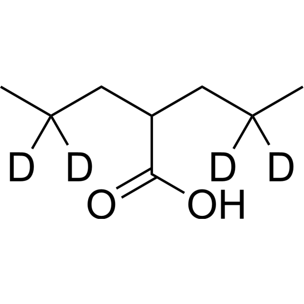
Valproic acid-d4-1 is the deuterium labeled Valproic acid. Valproic acid (VPA; 2-Propylpentanoic Acid) is an HDAC inhibitor, with IC50 in the range of 0.5 and 2 mM, also inhibits HDAC1 (IC50, 400 μM), and induces proteasomal degradation of HDAC2. Valproic acid activates Notch1 signaling and inhibits proliferation in small cell lung cancer (SCLC) cells. Valproic acid sodium salt is used in the treatment of epilepsy, bipolar disorder and prevention of migraine headaches.
For research use only. We do not sell to patients.
- Purity : 99.5%
- CAS No.: 345909-03-7
- Formula: C8H12D4O2
- Molecular Weight:148.24
-
Storage:Pure form -20°C, 3 years , 4°C, 2 years ; In solvent -80°C, 6 months , -20°C, 1 month

To place orders, for customer services and technical support, please contact: MedChemExpress USA
Tel: 609-228-6898 E-mail: [email protected] [email protected]


-
 Biological Activity
-
Chemical Information
- Protocols
- Purity & Documentation
- References
-
 Help & FAQs


All Endogenous Metabolite Isoforms
More

Biological Activity
Description
In Vitro
Stable heavy isotopes of hydrogen, carbon, and other elements have been incorporated into drug molecules, largely as tracers for quantitation during the drug development process. Deuteration has gained attention because of its potential to affect the pharmacokinetic and metabolic profiles of drugs[1].
MedChemExpress (MCE) has not independently confirmed the accuracy of these methods. They are for reference only.
Application
1. This compound can be used as a tracer
2. This compound can be used as an internal standard for quantitative analysis by NMR, GC-MS, or LC-MS.


Chemical Information
-
CAS No. 345909-03-7
-
Unlabeled CAS 99-66-1
-
Appearance Liquid
-
Molecular Weight 148.24
-
Formula C8H12D4O2
-
Color Colorless to light yellow
-
SMILES
CC([2H])([2H])CC(C(O)=O)CC([2H])([2H])C
-
Synonyms
VPA-d4-1; 2-Propylpentanoic acid-d4-1
-
Shipping
Room temperature in continental US; may vary elsewhere.
-
Storage
Pure form -20°C 3 years 4°C 2 years In solvent -80°C 6 months -20°C 1 month


Purity & Documentation
-
Data Sheet (274 KB)
-
SDS (557 KB)
- English - EN (557 KB)
- Français - FR (557 KB)
- Deutsch - DE (557 KB)
- Norwegian - NO (557 KB)
- Español - ES (557 KB)
- Swedish - SV (557 KB)
- Italian - IT (557 KB)
- Korean - KR (557 KB)
- Portuguese - PT (557 KB)
-
Handling Instructions (2659 KB)
References
[1]. Russak EM, et al. Impact of Deuterium Substitution on the Pharmacokinetics of Pharmaceuticals. Ann Pharmacother. 2019;53(2):211-216. [Content Brief]
[2]. Valproic acid, et al. Histone deacetylase is a direct target of valproic acid, a potent anticonvulsant, mood stabilizer, and teratogen. J Biol Chem. 2001 Sep 28;276(39):36734-41. [Content Brief]
[3]. Han BR, et al. Valproic acid inhibits the growth of HeLa cervical cancer cells via caspase-dependent apoptosis. Oncol Rep. 2013 Dec;30(6):2999-3005. [Content Brief]
[4]. Zhang ZH, et al. Valproic acid inhibits tumor angiogenesis in mice transplanted with Kasumi 1 leukemia cells. Mol Med Rep. 2013 Nov 28. [Content Brief]
[5]. Cohen OS, et al. Acute prenatal exposure to a moderate dose of valproic acid increases social behavior and alters gene expression in rats. Int J Dev Neurosci. 2013 Dec;31(8):740-50. [Content Brief]
[6]. Avery LB, et al. Valproic Acid Is a Novel Activator of AMP-Activated Protein Kinase and Decreases Liver Mass, Hepatic Fat Accumulation, and Serum Glucose in Obese Mice. Mol Pharmacol. 2014 Jan;85(1):1-10. [Content Brief]
[7]. Platta CS, et al. Valproic acid induces Notch1 signaling in small cell lung cancer cells. J Surg Res. 2008 Jul;148(1):31-7. [Content Brief]

Calculators
Concentration (start) × Volume (start) = Concentration (final) × Volume (final)


Valproic acid-d4-1 Related Classifications
HY-10585S4 Related Classifications
 Powered by Bioz