



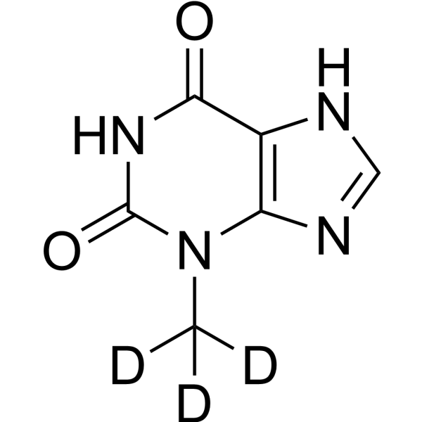
3-Methylxanthine-d3

Based on 1 Customer Validation
3-Methylxanthine-d3 is deuterated labeled 3-Methylxanthine (HY-50723). 3-Methylxanthine is a blood-brain barrier-permeable cyclic GMP phosphodiesterase inhibitor, with an IC50 of 920 μM against guinea pig cyclic GMP phosphodiesterase. 3-Methylxanthine relaxes the spontaneous tension of isolated guinea pig tracheal smooth muscle preparations. Through a DRD1-dependent pathway, 3-Methylxanthine upregulates the expression of γH2AX and activated caspase-3, downregulates the expression of Bcl-2, and enhances Cisplatin-induced apoptosis (apoptosis) in ovarian cancer cells both in vitro and in vivo. 3-Methylxanthine induces clonic convulsions in the central nervous system. 3-Methylxanthine is the major metabolite of Theophylline (HY-B0809). 3-Methylxanthine can be used in studies related to ovarian cancer and neurotoxic convulsions.
Nur für Forschungszwecke. Wir verkaufen nicht an Patienten.
- Reinheit : 99.61%
- Formel: C6H3D3N4O2
- Molecular Weight:169.16
-
Speicherung:
-20°C, protect from light, stored under nitrogen
* In solvent : -80°C, 6 months; -20°C, 1 month (protect from light, stored under nitrogen)

To place orders, for customer services and technical support, please contact: MedChemExpress USA
Tel: 609-228-6898 E-mail: [email protected] [email protected]


-
 Biologische Aktivität
-
Chemical Information
- Protokoll
- Reinheit & Dokumentation
- Verweise
-
 Help & FAQs


Alle Endogenous Metabolite Isoform-spezifische Produkte anzeigen
MoreAlle Caspase Isoform-spezifische Produkte anzeigen
More

Biologische Aktivität
Beschreibung
IC50 & Target
|
Caspase 3 |
Bcl-2 |
In Vitro
Stable heavy isotopes of hydrogen, carbon, and other elements have been incorporated into drug molecules, largely as tracers for quantitation during the drug development process. Deuteration has gained attention because of its potential to affect the pharmacokinetic and metabolic profiles of drugs[1].
MedChemExpress (MCE) has not independently confirmed the accuracy of these methods. They are for reference only.
Anwendung
1. This compound can be used as a tracer
2. This compound can be used as an internal standard for quantitative analysis by NMR, GC-MS, or LC-MS.


Chemical Information
-
Unlabeled CAS 1076-22-8
-
Appearance Solid
-
Molecular Weight 169.16
-
Formel C6H3D3N4O2
-
SMILES
O=C(N1)N(C([2H])([2H])[2H])C2=C(NC=N2)C1=O
-
Versand
Room temperature in continental US; may vary elsewhere.
-
Speicherung
-20°C, protect from light, stored under nitrogen
* In solvent : -80°C, 6 months; -20°C, 1 month (protect from light, stored under nitrogen)


Reinheit & Dokumentation
Verweise
[1]. Tanaka H, et al. Inhibition of cyclic GMP phosphodiesterase by xanthine derivatives relaxes guinea-pig trachealis smooth muscle. Clinical and experimental pharmacology & physiology. 1991 Mar;18(3):163-8. [Content Brief]
[2]. Mai Z, et al. Gut-derived metabolite 3-methylxanthine enhances cisplatin-induced apoptosis via dopamine receptor D1 in a mouse model of ovarian cancer. mSystems. 2024 Jul 23;9(7):e0130123. [Content Brief]
[3]. Yamamoto K, et al. Neurotoxic convulsions induced by theophylline and its metabolites in mice. Biological & pharmaceutical bulletin. 1996 Jun;19(6):869-72. [Content Brief]

Calculators
Konzentration (Stammlösung) × Volumen (Stammlösung) = Konzentration (Ziellösung) × Volumen (Ziellösung)


3-Methylxanthine-d3 Related Classifications
HY-50723S1 Related Classifications
 Powered by Bioz