



Fluazinam

Based on 1 Customer Validation
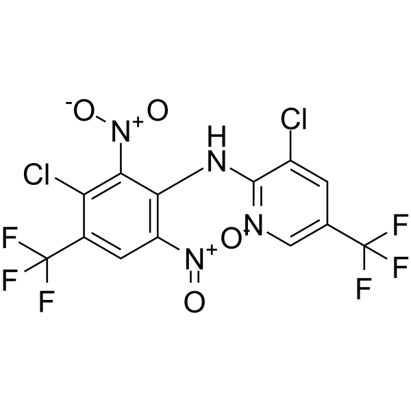
Fluazinam is an orally active dinitroaniline fungicide. Fluazinam induces phosphorylation of JNK, activates p38 pathway, decreases Bcl-2, activates caspase-3, decreases complex I activity, increases Autophagy and Apoptosis. Fluazinam has strong antifungal activity against F. fujikuroi and B. maydis. Fluazinam has a negative impact on Brachydanio rerio and worker bees.
For research use only. We do not sell to patients.
- Purity : 99.51%
- CAS No.: 79622-59-6
- Formula: C13H4Cl2F6N4O4
- Molecular Weight:465.09
-
Storage:Powder -20°C, 3 years , 4°C, 2 years ; In solvent -80°C, 2 years , -20°C, 1 year

To place orders, for customer services and technical support, please contact: MedChemExpress USA
Tel: 609-228-6898 E-mail: [email protected] [email protected]


-
 Biological Activity
-
Chemical Information
-
Solvent & Solubility
- Protocols
- Purity & Documentation
- References
-
 Help & FAQs


-
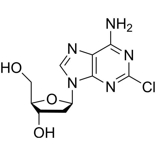
HY-13599
CladribineDNA Synthesis Inhibitor -
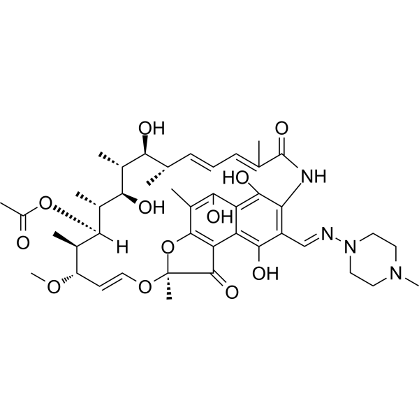
HY-B0272
RifampicinAntivirus Agent -
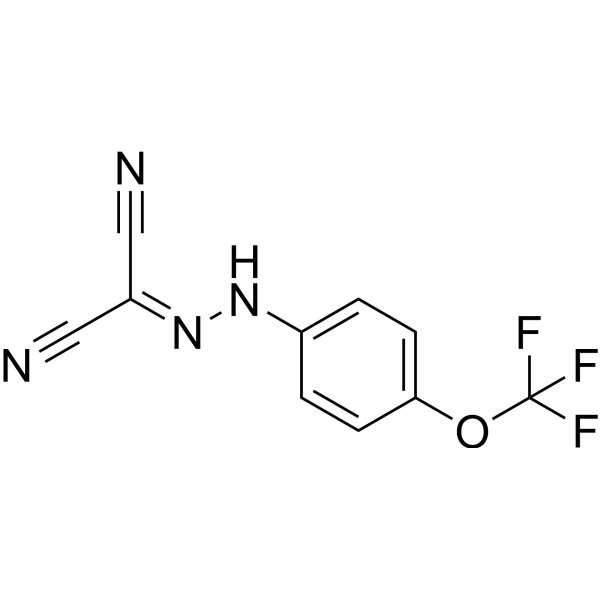
HY-100410
FCCPOXPHOS Uncoupler -
HY-14603
ClioquinolAnti-fungal Agent -
HY-B0850
DifenoconazoleTriazole Fungicide -
HY-114740
β-NaphthoflavoneAhR agonist -
HY-B0849
AzoxystrobinFungicide -
HY-B1664
Iopanoic acidIodinated Contrast Medium -
HY-B1425
EthoxyquinAntioxidant -
HY-N1405
Cucurbitacin ISTAT Inhibitor -

HY-B1839R
Fluazinam (Standard)Fungicide -
HY-124252
5,8,11-Eicosatriynoic acidLipoxygenase Inhibitor
All Caspase Isoforms
More

Biological Activity
Description
IC50 & Target
Fungal[1]
In Vitro
Fluazinam (0-5 μM; 24 h) induces SH-SY5Y cell death and nuclear damage[1].
Fluazinam (10-20 μg/mL) has great control efficiency against B. maydis on detached corn leaves[2].
Fluazinam has strong antifungal activity against F. fujikuroi, with EC50 values ranging from 0.0621 to 0.5446 μg/mL for mycelial growth inhibition of 103 F. fujikuroi isolates[3].
Fluazinam (0.01-0.5 μM) impairs oxidative phosphorylation and induces hyper/hypo-activity in a dose specific manner in zebrafish larvae[4].
Fluazinam (0.03-0.40 mg/L; 24-96 h) has acute toxicity to Brachydanio rerio[5].
MedChemExpress (MCE) has not independently confirmed the accuracy of these methods. They are for reference only.
-
Cell Line:SH-SY5Y
-
Concentration:1.25, 2.5, 5 μM
-
Incubation Time:10 min, 30 min, 1 h, 2 h, 4 h, 8 h, 16 h, 24 h
-
Result:Induced phosphorylation of JNK.
Activated the p38 pathway.
Induced a decrease in the level of Bcl-2 protein.
Activated caspase-3 in a dose-dependent manner.
Decreased complex I activity.
In Vivo
MedChemExpress (MCE) has not independently confirmed the accuracy of these methods. They are for reference only.
- Kinase activity and phosphorylation assays
- Western Blot
- Detection of Apoptosis
- TUNEL staining for apoptotic DNA fragmentation
- Annexin V plus membrane-impermeant dye apoptosis staining
- Autophagy
- Apoptosis Solutions
- Protocol for Kinase activity and phosphorylation assays
- Lysosome and acidic-vesicle live-cell staining
- Macroautophagy Solutions


Chemical Information
-
CAS No. 79622-59-6
-
Appearance Solid
-
Molecular Weight 465.09
-
Formula C13H4Cl2F6N4O4
-
Color Light yellow to yellow
-
SMILES
FC(C1=CN=C(NC2=C([N+]([O-])=O)C=C(C(F)(F)F)C(Cl)=C2[N+]([O-])=O)C(Cl)=C1)(F)F
-
Shipping
Room temperature in continental US; may vary elsewhere.
-
Storage
Powder -20°C 3 years 4°C 2 years In solvent -80°C 2 years -20°C 1 year


Solvent & Solubility
In Vitro:
DMSO : 125 mg/mL (268.77 mM; Need ultrasonic; Hygroscopic DMSO has a significant impact on the solubility of product, please use newly opened DMSO)
Please refer to the solubility information to select the appropriate solvent. Once prepared, please aliquot and store the solution to prevent product inactivation from repeated freeze-thaw cycles.
Storage method and period of stock solution: -80°C, 2 years; -20°C, 1 year. When stored at -80°C, please use it within 2 years. When stored at -20°C, please use it within 1 year.
Please refer to the solubility information to select the appropriate solvent. Once prepared, please aliquot and store the solution to prevent product inactivation from repeated freeze-thaw cycles.
Storage method and period of stock solution: -80°C, 2 years; -20°C, 1 year. When stored at -80°C, please use it within 2 years. When stored at -20°C, please use it within 1 year.
Concentration (start) × Volume (start) = Concentration (final) × Volume (final)
In Vivo:
Select the appropriate dissolution method based on your experimental animal and administration route.
- For the following dissolution methods, please ensure to first prepare a clear stock solution using an In Vitro approach and then sequentially add co-solvents:
- To ensure reliable experimental results, the clarified stock solution can be appropriately stored based on storage conditions. As for the working solution for In Vivo experiments, it is recommended to prepare freshly and use it on the same day.
- The percentages shown for the solvents indicate their volumetric ratio in the final prepared solution. If precipitation or phase separation occurs during preparation, heat and/or sonication can be used to aid dissolution.
Add each solvent one by one: 10% DMSO 40% PEG300 5% Tween-80 45% Saline
Solubility: ≥ 2.08 mg/mL (4.47 mM); Clear solution
This protocol yields a clear solution of ≥ 2.08 mg/mL (saturation unknown).
Taking 1 mL working solution as an example, add 100 μL DMSO stock solution (20.8 mg/mL) to 400 μL PEG300, and mix evenly; then add 50 μL Tween-80 and mix evenly; then add 450 μL Saline to adjust the volume to 1 mL.
Preparation of Saline: Dissolve 0.9 g sodium chloride in ddH₂O and dilute to 100 mL to obtain a clear Saline solution.
In Vivo Dissolution Calculator
Please enter the basic information of animal experiments:
-
-
-
-
Recommended: Prepare an additional quantity of animals to account for potential losses during experiments.
Please enter your animal formula composition:
-
%DMSO +
Recommended: Keep the proportion of DMSO in working solution below 2% if your animal is weak.
-
%+
-
+%Tween-80 + +
-
%Saline +
The co-solvents required include: DMSO, . All of co-solvents are available by MedChemExpress (MCE). , Tween 80. All of co-solvents are available by MedChemExpress (MCE).
Working solution concentration: 0.22 mg/mL
Method for preparing stock solution: mg drug dissolved in μL DMSO. Stock solution concentration: mg/mL.
1. Take μL DMSO stock solution;
2. Add μL .
μL , mix evenly;
3. Then add μL Tween 80, mix evenly;
4. Then add μL
Please ensure that the stock solution in the first step is dissolved to a clear state, and add co-solvents in sequence. You can use ultrasonic heating (ultrasonic cleaner, recommended frequency 20-40 kHz), vortexing, etc. to assist dissolution.


Purity & Documentation
-
Data Sheet (288 KB)
-
SDS (923 KB)
- English - EN (923 KB)
- Français - FR (923 KB)
- Deutsch - DE (923 KB)
- Norwegian - NO (923 KB)
- Español - ES (923 KB)
- Swedish - SV (923 KB)
- Italian - IT (923 KB)
- Korean - KR (923 KB)
- Portuguese - PT (923 KB)
-
Handling Instructions (2659 KB)
References
[1]. Lee JE, et al. Fluazinam targets mitochondrial complex I to induce reactive oxygen species-dependent cytotoxicity in SH-SY5Y cells. Neurochem Int. 2012 Jun;60(8):773-81. [Content Brief]
[2]. Chen YL, et al. Activity of the dinitroaniline fungicide fluazinam against Bipolaris maydis. Pestic Biochem Physiol. 2018 Jun;148:8-15. [Content Brief]
[3]. Qu XP, et al. Effects of the dinitroaniline fungicide fluazinam on Fusarium fujikuroi and rice. Pestic Biochem Physiol. 2018 Nov;152:98-105. [Content Brief]
[4]. Wang XH, et al. Fluazinam impairs oxidative phosphorylation and induces hyper/hypo-activity in a dose specific manner in zebrafish larvae. Chemosphere. 2018 Nov;210:633-644. [Content Brief]
[5]. Wu W, et al. Acute toxicity of fluazinam to aquatic organisms and its bioaccumulation in Brachydanio rerio. Environ Sci Pollut Res Int. 2020 Oct;27(28):35000-35007. [Content Brief]
[6]. Gomes DS, et al. Acute exposure to fungicide fluazinam induces cell death in the midgut, oxidative stress and alters behavior of the stingless bee Partamona helleri (Hymenoptera: Apidae). Ecotoxicol Environ Saf. 2024 Sep 1;282:116677. [Content Brief]
[7]. Hou YP, et al. Impact of fluazinam on morphological and physiological characteristics of Sclerotinia sclerotiorum. Pestic Biochem Physiol. 2019 Mar;155:81-89. [Content Brief]


Complete Stock Solution Preparation Table
Please refer to the solubility information to select the appropriate solvent. Once prepared, please aliquot and store the solution to prevent product inactivation from repeated freeze-thaw cycles.
Storage method and period of stock solution: -80°C, 2 years; -20°C, 1 year. When stored at -80°C, please use it within 2 years. When stored at -20°C, please use it within 1 year.
| Optional Solvent | Concentration Solvent Mass | 1 mg | 5 mg | 10 mg | 25 mg |
|---|---|---|---|---|---|
| DMSO | 1 mM | 2.1501 mL | 10.7506 mL | 21.5012 mL | 53.7530 mL |
| 5 mM | 0.4300 mL | 2.1501 mL | 4.3002 mL | 10.7506 mL | |
| 10 mM | 0.2150 mL | 1.0751 mL | 2.1501 mL | 5.3753 mL | |
| 15 mM | 0.1433 mL | 0.7167 mL | 1.4334 mL | 3.5835 mL | |
| 20 mM | 0.1075 mL | 0.5375 mL | 1.0751 mL | 2.6877 mL | |
| 25 mM | 0.0860 mL | 0.4300 mL | 0.8600 mL | 2.1501 mL | |
| 30 mM | 0.0717 mL | 0.3584 mL | 0.7167 mL | 1.7918 mL | |
| 40 mM | 0.0538 mL | 0.2688 mL | 0.5375 mL | 1.3438 mL | |
| 50 mM | 0.0430 mL | 0.2150 mL | 0.4300 mL | 1.0751 mL | |
| 60 mM | 0.0358 mL | 0.1792 mL | 0.3584 mL | 0.8959 mL | |
| 80 mM | 0.0269 mL | 0.1344 mL | 0.2688 mL | 0.6719 mL | |
| 100 mM | 0.0215 mL | 0.1075 mL | 0.2150 mL | 0.5375 mL |


Fluazinam Related Classifications
HY-B1839 Related Classifications
 Powered by Bioz