



Vaccarin
Based on 2 publication(s) in Google Scholar
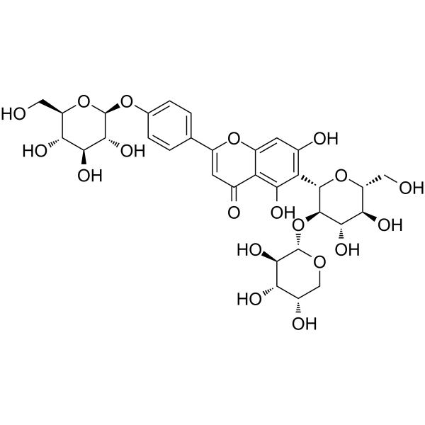
Vaccarin is an orally active flavonoid glycoside with multiple biological functions. Vaccarin promotes neovascularization by activating AKT and ERK. Vaccarin activates the AMPK signaling pathway to improve insulin resistance and steatosis. Vaccarin is a MAPK, NF-κB, and NFAT inhibitor, effectively blocking RANKL-induced osteoclastogenesis.
For research use only. We do not sell to patients.
- Purity : 99.05%
- CAS No.: 53452-16-7
- Formula: C32H38O19
- Molecular Weight:726.63
-
Storage:
4°C, protect from light
* In solvent : -80°C, 6 months; -20°C, 1 month (protect from light)

To place orders, for customer services and technical support, please contact: MedChemExpress USA
Tel: 609-228-6898 E-mail: [email protected] [email protected]


-
 Biological Activity
-
Chemical Information
-
Solvent & Solubility
- Protocols
- Purity & Documentation
- References
-
 Help & FAQs


-
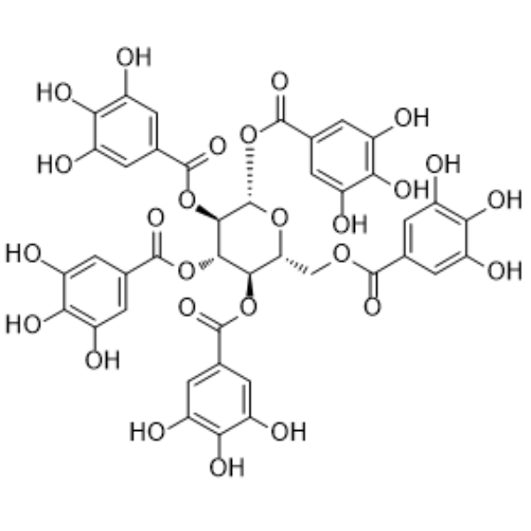
HY-N0527
PentagalloylglucoseAntioxidant -
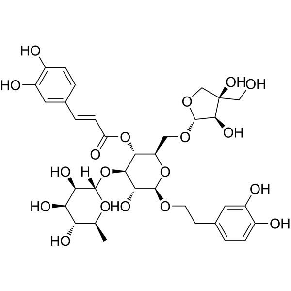
HY-N0029
Forsythoside BTNF Inhibitor -
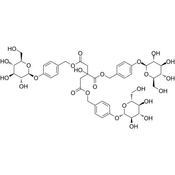
HY-N2031
ParishinSir2/Uth1/TOR Modulator -
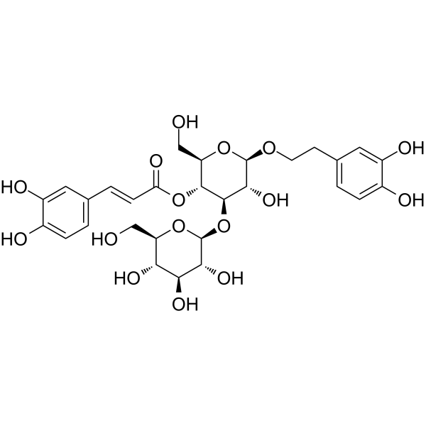
HY-N0031
PlantamajosideNF-κB Inhibitor -
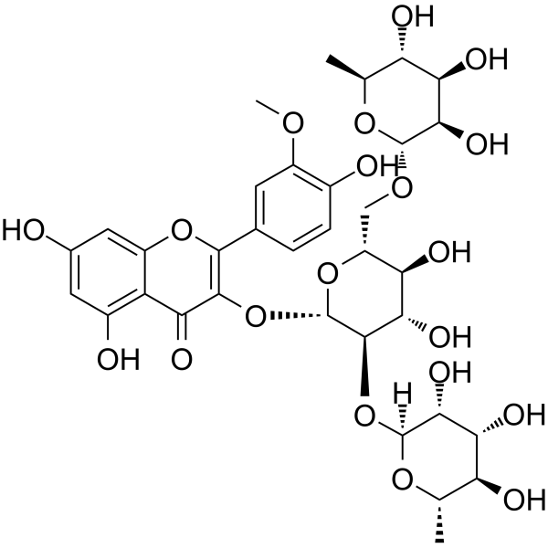
HY-N0712
TyphaneosidePI3K/Akt/mTOR Activator -
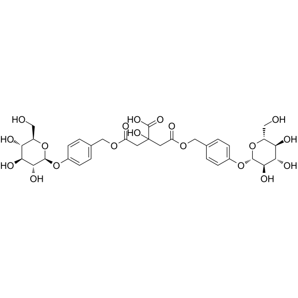
HY-N2125
Parishin C5-HT1A Receptor Agonist -
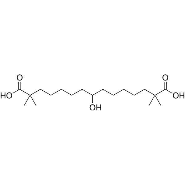
HY-12357
Bempedoic acidACL Inhibitor/AMPK Activator -
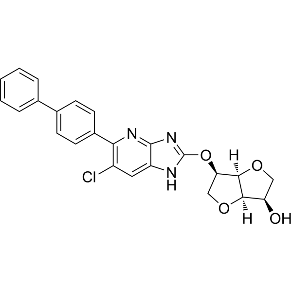
HY-111363
MK8722AMPK Activator -
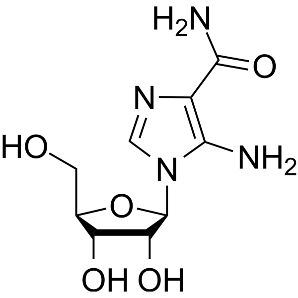
HY-13417
AICARAMPK Activator -
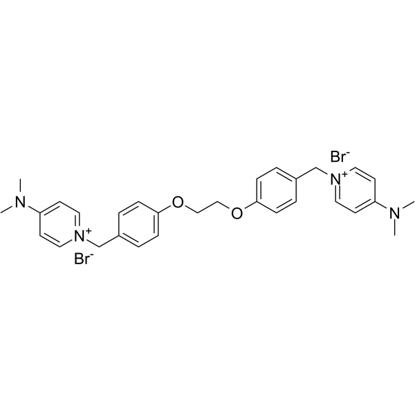
HY-115463
EB-3DChoKα Inhibitor -
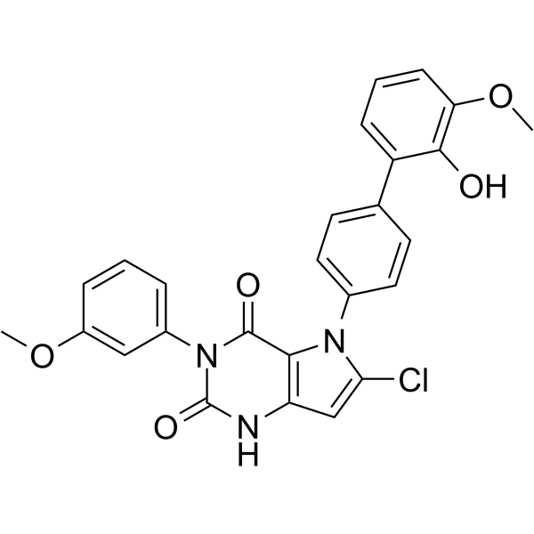
HY-100548
GSK621AMPK Activator -

HY-N1419R
Vaccarin (Standard)AMPK Activator
Publications Citing Use of MedChemExpress (MCE) Vaccarin
More Customer Validation & Images

-
Cell Proliferation/Viability Assay

-
WB

-
IF

-
IHC

-
RT-PCR

All AMPK Isoforms
More

Biological Activity
Description
In Vitro
Vaccarin (0.54-34.4 μM, 48 h) increases the proliferation activity of human microvascular endothelial cells-1 (HMEC-1)[1].
Vaccarin (1.08-2.15 μM, 24-48 h) promotes HMEC-1 migration and capillary lumen formation[1].
Vaccarin (5 μM, 24 h) activites insulin resistance in HepG2 cells by inhibiting gluconeogenesis and promoting glycogen synthesis[2].
Vaccarin (5 μM, 24 h) activates AMPK to increase insulin sensitivity and inhibit steatosis in HepG2 cells[2].
Vaccarin (4-64 μM, 48 h) inhibits RANKL-induced osteoclastogenesis in bone marrow macrophages (BMMs)[3].
Vaccarin (8 μM, 4 h) inhibits osteocalcin (OC) differentiation by inhibiting MAPK and NF-κB signaling pathways[3].
Vaccarin (4-16 μM) reduces the expression of OC marker genes by inhibiting the activation of NF-κB and NFATc1[3].
MedChemExpress (MCE) has not independently confirmed the accuracy of these methods. They are for reference only.
-
Cell Line:HepG2 cells
-
Concentration:5 μM
-
Incubation Time:24 h
-
Result:Down-regulated the protein levels of PEPCK, G6Pase, p-GS, SREBP-1 and FAS.
Up-regulated the protein levels of p-FOXO1, p-GSK3β, p-AMPK and p-ACC.
-
Cell Line:BMM
-
Concentration:8 μM
-
Incubation Time:4 h
-
Result:Down-regulated the protein expression levels of IκB-ɑ, p-NF-κBp65, p-p38, p-JNK, p-ERK, c-fos, and NFATc1.
In Vivo
Vaccarin (1 mg/kg, i.p., once per day for 4 weeks) inhibits insulin resistance and glucose/lipid metabolism disorders in the type 2 diabetes (T2DM) mouse model[2].
Vaccarin (2.5/5 mg/kg, once every 2 days for 14 days) inhibits osteolytic bone loss in the Ti particle-induced calvarial osteolysis mouse model[3].
MedChemExpress (MCE) has not independently confirmed the accuracy of these methods. They are for reference only.
-
Animal Model:Mouse matrigel plug model (Female ICR mice, aged 4–5 weeks old and weighing 17-20 g; subcutaneously injected with 500 μl of Matrigel containing heparin (113 units) and mouse EGF (120 ng) into the left hypogastric region)[1]
-
Dosage:5 and 10 mg/kg
-
Administration:Oral gavage (p.o.), once per day for 2 weeks
-
Result:Increased the number of endothelial cells.
Up-regulated the expression of p-Erk and p-Akt in blood vessels.
-
Animal Model:T2DM mouse model (Six-week-old male C57BL/6 J mice, intraperitoneal injection of STZ (HY-13753) and HFD)[2]
-
Dosage:1 mg/kg
-
Administration:Intraperitoneal injection (i.p.), once per day for 4 weeks
-
Result:Increased liver glycogen levels in mice.
Down-regulated the ratio of liver weight (LW) to body weight (BW), p-IRS-1 (Ser 307), TC, TG, ALT, AST, LDL-C and NEFA levels in the liver of mice.


Chemical Information
-
CAS No. 53452-16-7
-
Appearance Solid
-
Molecular Weight 726.63
-
Formula C32H38O19
-
Color Light yellow to yellow
-
SMILES
O=C1C=C(C2=CC=C(O[C@H]3[C@@H]([C@H]([C@@H]([C@@H](CO)O3)O)O)O)C=C2)OC4=CC(O)=C([C@H]5[C@@H]([C@H]([C@@H]([C@@H](CO)O5)O)O)O[C@H]6[C@@H]([C@H]([C@H](CO6)O)O)O)C(O)=C14
-
Structure Classification
-
Initial Source
-
Shipping
Room temperature in continental US; may vary elsewhere.
-
Storage
4°C, protect from light
* In solvent : -80°C, 6 months; -20°C, 1 month (protect from light)


Publications (2)
-
Journal Impact Factor
-
Most Recent
-
Phytomedicine
Vaccarin ameliorates osteoarthritis by suppressing the c-Jun N-terminal kinase (JNK)-serum amyloid A2 (SAA2) pathway mediating chondrocyte senescence. [Abstract]2025 Jun:141:156697. PMID: 40215820
Vaccarin purchased from MedChemExpress. Usage Cited in: Phytomedicine. 2025 Jun:141:156697. [Abstract]
CCK-8 assay showed no cytotoxicity up to 40 μM after 24 and 48 h treated with Vaccarin.
Vaccarin purchased from MedChemExpress. Usage Cited in: Phytomedicine. 2025 Jun:141:156697. [Abstract]
Western blot indicated Vaccarin dose-dependently restored ECM proteins including Aggrecan, Collagen II, and SOX9 suppressed by IL-1β treated with Vaccarin (5, 10, 20, 40 μM).
Vaccarin purchased from MedChemExpress. Usage Cited in: Phytomedicine. 2025 Jun:141:156697. [Abstract]
Immunofluorescence confirmed that Vaccarin (40 μM) restored IL-1β-reduced Collagen II and MMP13 levels.
Vaccarin purchased from MedChemExpress. Usage Cited in: Phytomedicine. 2025 Jun:141:156697. [Abstract]
Immunohistochemical staining Aggrecan and MMP13 treated with Vaccarin (25, 50 mg/kg, a intra-articular injection).
Vaccarin purchased from MedChemExpress. Usage Cited in: Phytomedicine. 2025 Jun:141:156697. [Abstract]
RT-qPCR analysis showing reduced SAA2 expression after Vaccarin (40 μM) or SP600125 treatment.
-
Kaohsiung J Med Sci
Vaccarin Improves Myocardial Ischemia-Reperfusion Injury by Attenuating Oxidative Stress and Ferroptosis Through Reducing NOX4-Modulated JAK2/STAT3 Pathway Activation. [Abstract]2026 Apr 29:e70226. PMID: 42053107


Solvent & Solubility
In Vitro:
DMSO : 125 mg/mL (172.03 mM; Need ultrasonic; Hygroscopic DMSO has a significant impact on the solubility of product, please use newly opened DMSO)
Please refer to the solubility information to select the appropriate solvent. Once prepared, please aliquot and store the solution to prevent product inactivation from repeated freeze-thaw cycles.
Storage method and period of stock solution: -80°C, 6 months; -20°C, 1 month (protect from light). When stored at -80°C, please use it within 6 months. When stored at -20°C, please use it within 1 month.
Please refer to the solubility information to select the appropriate solvent. Once prepared, please aliquot and store the solution to prevent product inactivation from repeated freeze-thaw cycles.
Storage method and period of stock solution: -80°C, 6 months; -20°C, 1 month (protect from light). When stored at -80°C, please use it within 6 months. When stored at -20°C, please use it within 1 month.
Concentration (start) × Volume (start) = Concentration (final) × Volume (final)
In Vivo:
Select the appropriate dissolution method based on your experimental animal and administration route.
- For the following dissolution methods, please ensure to first prepare a clear stock solution using an In Vitro approach and then sequentially add co-solvents:
- To ensure reliable experimental results, the clarified stock solution can be appropriately stored based on storage conditions. As for the working solution for In Vivo experiments, it is recommended to prepare freshly and use it on the same day.
- The percentages shown for the solvents indicate their volumetric ratio in the final prepared solution. If precipitation or phase separation occurs during preparation, heat and/or sonication can be used to aid dissolution.
Add each solvent one by one: 10% DMSO 40% PEG300 5% Tween-80 45% Saline
Solubility: ≥ 2.08 mg/mL (2.86 mM); Clear solution
This protocol yields a clear solution of ≥ 2.08 mg/mL (saturation unknown).
Taking 1 mL working solution as an example, add 100 μL DMSO stock solution (20.8 mg/mL) to 400 μL PEG300, and mix evenly; then add 50 μL Tween-80 and mix evenly; then add 450 μL Saline to adjust the volume to 1 mL.
Preparation of Saline: Dissolve 0.9 g sodium chloride in ddH₂O and dilute to 100 mL to obtain a clear Saline solution.
Add each solvent one by one: 10% DMSO 90% (20% SBE-β-CD in Saline)
Solubility: ≥ 2.08 mg/mL (2.86 mM); Clear solution
This protocol yields a clear solution of ≥ 2.08 mg/mL (saturation unknown).
Taking 1 mL working solution as an example, add 100 μL DMSO stock solution (20.8 mg/mL) to 900 μL 20% SBE-β-CD in Saline, and mix evenly.
Preparation of 20% SBE-β-CD in Saline (4°C, storage for one week): 2 g SBE-β-CD powder is dissolved in 10 mL Saline, completely dissolve until clear.
In Vivo Dissolution Calculator
Please enter the basic information of animal experiments:
-
-
-
-
Recommended: Prepare an additional quantity of animals to account for potential losses during experiments.
Please enter your animal formula composition:
-
%DMSO +
Recommended: Keep the proportion of DMSO in working solution below 2% if your animal is weak.
-
%+
-
+%Tween-80 + +
-
%Saline +
The co-solvents required include: DMSO, . All of co-solvents are available by MedChemExpress (MCE). , Tween 80. All of co-solvents are available by MedChemExpress (MCE).
Working solution concentration: 0.22 mg/mL
Method for preparing stock solution: mg drug dissolved in μL DMSO. Stock solution concentration: mg/mL. * In solvent : -80°C, 6 months; -20°C, 1 month (protect from light)
1. Take μL DMSO stock solution;
2. Add μL .
μL , mix evenly;
3. Then add μL Tween 80, mix evenly;
4. Then add μL
Please ensure that the stock solution in the first step is dissolved to a clear state, and add co-solvents in sequence. You can use ultrasonic heating (ultrasonic cleaner, recommended frequency 20-40 kHz), vortexing, etc. to assist dissolution.


Purity & Documentation
-
Data Sheet (282 KB)
-
SDS (393 KB)
- English - EN (393 KB)
- Français - FR (393 KB)
- Deutsch - DE (393 KB)
- Norwegian - NO (393 KB)
- Español - ES (393 KB)
- Swedish - SV (393 KB)
- Italian - IT (393 KB)
- Korean - KR (393 KB)
- Portuguese - PT (393 KB)
-
Handling Instructions (2659 KB)
References
[1]. Xie F, et al. Vaccarin promotes endothelial cell proliferation in association with neovascularization in vitro and in vivo. Mol Med Rep. 2015 Jul;12(1):1131-6. [Content Brief]
[2]. Lei Y, et al. Vaccarin ameliorates insulin resistance and steatosis by activating the AMPK signaling pathway. Eur J Pharmacol. 2019 May 15;851:13-24. [Content Brief]
[3]. Liu Y, et al. Vaccarin prevents titanium particle-induced osteolysis and inhibits RANKL-induced osteoclastogenesis by blocking NF-κB and MAPK signaling pathways. J Cell Physiol. 2019 Aug;234(8):13832-13842. [Content Brief]


Complete Stock Solution Preparation Table
Please refer to the solubility information to select the appropriate solvent. Once prepared, please aliquot and store the solution to prevent product inactivation from repeated freeze-thaw cycles.
Storage method and period of stock solution: -80°C, 6 months; -20°C, 1 month (protect from light). When stored at -80°C, please use it within 6 months. When stored at -20°C, please use it within 1 month.
| Optional Solvent | Concentration Solvent Mass | 1 mg | 5 mg | 10 mg | 25 mg |
|---|---|---|---|---|---|
| DMSO | 1 mM | 1.3762 mL | 6.8811 mL | 13.7622 mL | 34.4054 mL |
| 5 mM | 0.2752 mL | 1.3762 mL | 2.7524 mL | 6.8811 mL | |
| 10 mM | 0.1376 mL | 0.6881 mL | 1.3762 mL | 3.4405 mL | |
| 15 mM | 0.0917 mL | 0.4587 mL | 0.9175 mL | 2.2937 mL | |
| 20 mM | 0.0688 mL | 0.3441 mL | 0.6881 mL | 1.7203 mL | |
| 25 mM | 0.0550 mL | 0.2752 mL | 0.5505 mL | 1.3762 mL | |
| 30 mM | 0.0459 mL | 0.2294 mL | 0.4587 mL | 1.1468 mL | |
| 40 mM | 0.0344 mL | 0.1720 mL | 0.3441 mL | 0.8601 mL | |
| 50 mM | 0.0275 mL | 0.1376 mL | 0.2752 mL | 0.6881 mL | |
| 60 mM | 0.0229 mL | 0.1147 mL | 0.2294 mL | 0.5734 mL | |
| 80 mM | 0.0172 mL | 0.0860 mL | 0.1720 mL | 0.4301 mL | |
| 100 mM | 0.0138 mL | 0.0688 mL | 0.1376 mL | 0.3441 mL |


Vaccarin Related Classifications
HY-N1419 Related Classifications
 Powered by Bioz