



Gomisin D

Based on 1 Customer Validation
Gomisin D is an orally active lignan that binds to PDGFRβ with a Kd of 10 μM. By targeting PDGFRβ to regulate signaling pathways, Gomisin D inhibits the activation and proliferation of hepatic stellate cells and promotes their apoptosis, thereby ameliorating hepatic fibrosis. Gomisin D exhibits multiple activities such as photoprotection, antimelanogenesis, antioxidant effects, and hypoglycemic activity. Gomisin D can be used in studies related to diabetes, Alzheimer's disease, and hepatic fibrosis.
For research use only. We do not sell to patients.
- Purity : 99.86%
- CAS No.: 60546-10-3
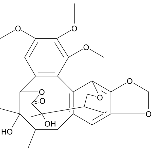
- Formula: C28H34O10
- Molecular Weight:530.56
-
Storage:
4°C, sealed storage, away from moisture and light
* In solvent : -80°C, 6 months; -20°C, 1 month (sealed storage, away from moisture and light)

To place orders, for customer services and technical support, please contact: MedChemExpress USA
Tel: 609-228-6898 E-mail: [email protected] [email protected]


-
 Biological Activity
-
Chemical Information
-
Solvent & Solubility
- Protocols
- Purity & Documentation
- References
-
 Help & FAQs


-
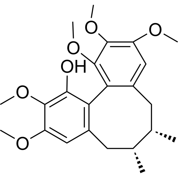
HY-N0859
SchisanhenolAntioxidant Agent -
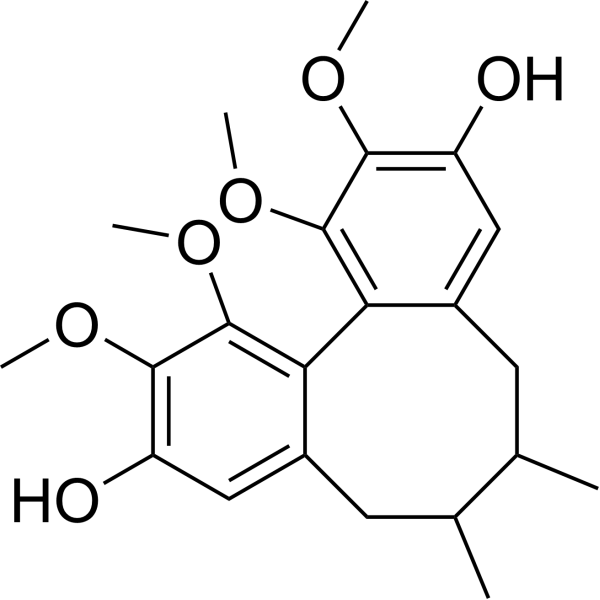
HY-N0385
Gomisin JCalcium Channel Activator -
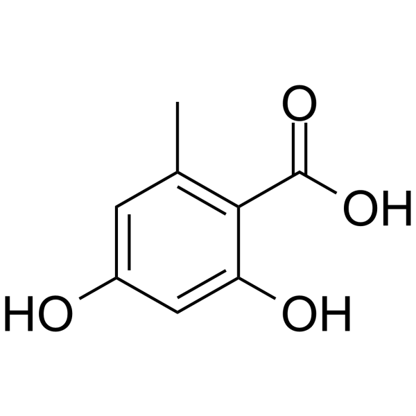
HY-N3126
Orsellinic acidBenzoic Acid Derivative -
HY-W040045
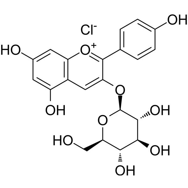
Callistephin chlorideROS/iNOS/TNF-α/COX-2 Inhibitor -
HY-N0701
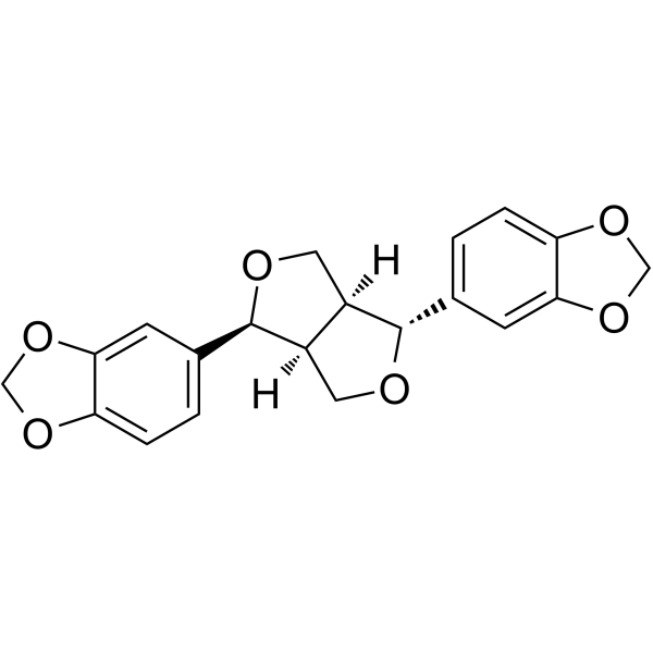
(-)-AsarininTetrahydrofurofurano Lignan -
HY-111253
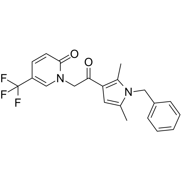
CYM-5478S1P2 Agonist -

HY-N0859R
Schisanhenol (Standard)Analytical Standard -
HY-N9926
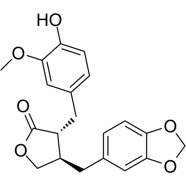
PluviatolideLignan -
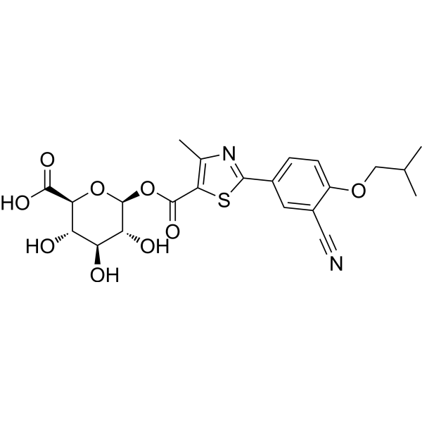
HY-129889
Febuxostat acyl glucuronideFebuxostat Metabolite -
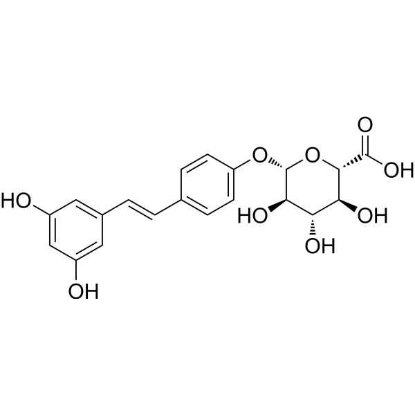
HY-118634
trans-Resveratrol-4'-O-D-GlucuronideAnti-fat Accumulation Agent -
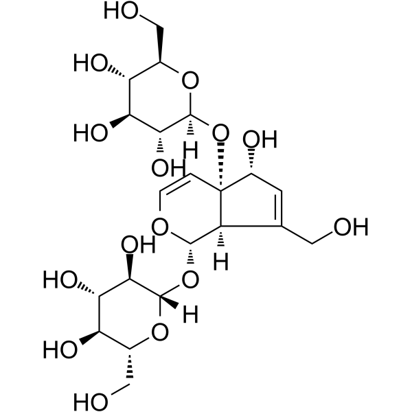
HY-N0915
MelittosideNatural Product -
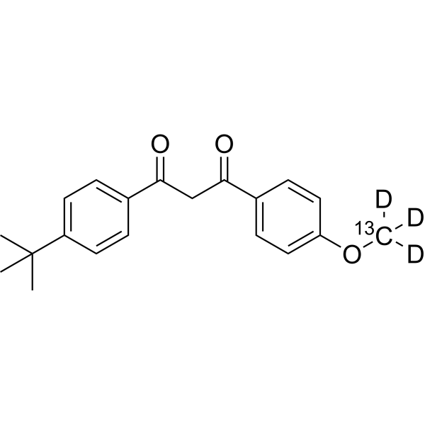
HY-B0316S
Avobenzone-13C,d3Stable Isotope


Biological Activity
Description
IC50 & Target
UDP-Glucuronosyltransferases[1]
Cellular Effect
|
Cell Line

|
Type | Value | Description | References |
|---|---|---|---|---|
| C8166 | CC50 |
185.7 μM
Compound: 23
|
Cytotoxicity against human C8166 cells after 3 days by MTT assay
Cytotoxicity against human C8166 cells after 3 days by MTT assay
|
[PMID: 23738539] |
| C8166 | EC50 |
9.8 μM
Compound: 23
|
Antiviral activity against HIV-1 3B infected in human C8166 cells assessed as inhibition of virus-induced cytopathic effect after 3 days by Syncytia Assay
Antiviral activity against HIV-1 3B infected in human C8166 cells assessed as inhibition of virus-induced cytopathic effect after 3 days by Syncytia Assay
|
[PMID: 23738539] |
In Vitro
Gomisin D (10-80 μM; 25 h) enhances the viability and reduces the cytotoxicity of HaCaT keratinocytes irradiated with UVA and UVB, and inhibits intracellular ROS generation[1].
Gomisin D (30 μM; 25 h) significantly inhibits apoptosis of UVA- and UVB-irradiated HaCaT keratinocytes[1].
Gomisin D (10-80 μM; 49 h) dose-dependently inhibits α-MSH-induced tyrosinase activity[1].
Gomisin D (30 μM; 49 h) significantly downregulates the mRNA and protein expression of α-MSH-induced melanogenesis-related factors MITF, tyrosinase, TRP-1 and TRP-2, as well as the phosphorylation of PKA and CREB, in B16F10 melanocytes[1].
Gomisin D (20-80 μM; 48 h) inhibits the activation of T6, LX-2 and primary mouse hepatic stellate cells by targeting PDGFRβ[3].
Gomisin D (40 μM) inhibits the downstream signaling pathways (p38, AKT, ERK pathways) of PDGFRβ in HSC-T6 cells by targeting PDGFRβ[3].
Gomisin D (5-80 μM) dose-dependently inhibits PDGF-BB-induced activation, proliferation, and PDGF-BB/PDGFRβ pathway signaling in LX-2 cells[3].
MedChemExpress (MCE) has not independently confirmed the accuracy of these methods. They are for reference only. Further protocols information, click here.
-
Cell Line:HaCaT keratinocytes (UVA or UVB irradiated)
-
Concentration:10, 20, 30, 40 and 80 μM
-
Incubation Time:1 h pre-incubation, 24 h post-irradiation incubation
-
Result:Improved viability of UVA- and UVB-irradiated HaCaT keratinocytes in a dose-dependent manner at 10, 20, 40, 80 μM.
Significantly increased viability and reduced LDH release in UVA- and UVB-irradiated HaCaT keratinocytes at 30 μM, compared to respective irradiated only groups.
Parmacokinetics
| Species | Dose | Route | AUC0-t | AUC0-∞ | MRT0-t | MRT0-∞ | T1/2 | Tmax | CL | V | Cmax | Bioavailability |
|---|---|---|---|---|---|---|---|---|---|---|---|---|
| Rat[2] | 5 mg/kg | i.v. | 3136.2 μg/L·h | 3261.1 μg/L·h | 6.0 h | 6.9 h | 5.0 h | 0.083 h | 1.6 L/h/kg | 11.2 L/kg | 539.5 μg/L | / |
| Rat[2] | 50 mg/kg | i.g. | 32795.6 μg/L·h | 35091.7 μg/L·h | 7.8 h | 10.6 h | 5.6 h | 3.0 h | 1.5 L/h/kg | 13.3 L/kg | 2575.2 μg/L | 107.6 % |
In Vivo
Gomisin D (50 mg/kg; p.o.; 30 days) causes no observable histotoxicity in male BALB/c mice[3].
MedChemExpress (MCE) has not independently confirmed the accuracy of these methods. They are for reference only.
-
Animal Model:BALB/c (male, 6-8 weeks old, 18-22 g, CCl4-induced hepatic fibrosis)[3]
-
Dosage:25 mg/kg; 50 mg/kg
-
Administration:i.g.; once daily; 4 weeks
-
Result:Improved survival status and liver morphology.
Reduced hepatocyte necrosis, preserved hepatic lobule structure, and decreased collagen deposition in both dose groups, with more significant improvements in the 50 mg/kg group.
Significantly reduced serum alanine aminotransferase (ALT) and aspartate aminotransferase (AST) levels compared to the model group in both dose groups.
Significantly decreased mRNA and protein levels of α-smooth muscle actin (α-SMA) and collagen I in liver tissue and isolated primary hepatic stellate cells (HSCs) from both dose groups, with the 50 mg/kg group showing greater reductions.
Significantly decreased protein levels of phosphorylated AKT (p-AKT), phosphorylated ERK (p-ERK), and phosphorylated p38 (p-p38) in liver tissue from both dose groups.
Significantly decreased mRNA levels of inflammatory factors IL-1β, IL-6, TNF-α, TGF-β, and COX-2 in liver tissue from both dose groups.


Chemical Information
-
CAS No. 60546-10-3
-
Appearance Solid
-
Molecular Weight 530.56
-
Formula C28H34O10
-
Color Off-white to light yellow
-
SMILES
COC1=C(OC)C(OC)=CC2=C1C3=C(CC(C)C(C)(O)C2O4)C=C(OCO5)C5=C3OCC(C)C(C)(O)C4=O
-
Structure Classification
-
Initial Source
-
Shipping
Room temperature in continental US; may vary elsewhere.
-
Storage
4°C, sealed storage, away from moisture and light
* In solvent : -80°C, 6 months; -20°C, 1 month (sealed storage, away from moisture and light)


Solvent & Solubility
In Vitro:
DMSO : 100 mg/mL (188.48 mM; Need ultrasonic; Hygroscopic DMSO has a significant impact on the solubility of product, please use newly opened DMSO)
Please refer to the solubility information to select the appropriate solvent. Once prepared, please aliquot and store the solution to prevent product inactivation from repeated freeze-thaw cycles.
Storage method and period of stock solution: -80°C, 6 months; -20°C, 1 month (sealed storage, away from moisture and light). When stored at -80°C, please use it within 6 months. When stored at -20°C, please use it within 1 month.
Please refer to the solubility information to select the appropriate solvent. Once prepared, please aliquot and store the solution to prevent product inactivation from repeated freeze-thaw cycles.
Storage method and period of stock solution: -80°C, 6 months; -20°C, 1 month (sealed storage, away from moisture and light). When stored at -80°C, please use it within 6 months. When stored at -20°C, please use it within 1 month.
Concentration (start) × Volume (start) = Concentration (final) × Volume (final)
In Vivo:
Select the appropriate dissolution method based on your experimental animal and administration route.
- For the following dissolution methods, please ensure to first prepare a clear stock solution using an In Vitro approach and then sequentially add co-solvents:
- To ensure reliable experimental results, the clarified stock solution can be appropriately stored based on storage conditions. As for the working solution for In Vivo experiments, it is recommended to prepare freshly and use it on the same day.
- The percentages shown for the solvents indicate their volumetric ratio in the final prepared solution. If precipitation or phase separation occurs during preparation, heat and/or sonication can be used to aid dissolution.
Add each solvent one by one: 10% DMSO 40% PEG300 5% Tween-80 45% Saline
Solubility: 2.5 mg/mL (4.71 mM); Suspended solution; Need ultrasonic
This protocol yields a suspended solution of 2.5 mg/mL. Suspended solution can be used for oral and intraperitoneal injection.
Taking 1 mL working solution as an example, add 100 μL DMSO stock solution (25.0 mg/mL) to 400 μL PEG300, and mix evenly; then add 50 μL Tween-80 and mix evenly; then add 450 μL Saline to adjust the volume to 1 mL.
Preparation of Saline: Dissolve 0.9 g sodium chloride in ddH₂O and dilute to 100 mL to obtain a clear Saline solution.
Add each solvent one by one: 10% DMSO 90% (20% SBE-β-CD in Saline)
Solubility: ≥ 2.5 mg/mL (4.71 mM); Clear solution
This protocol yields a clear solution of ≥ 2.5 mg/mL (saturation unknown).
Taking 1 mL working solution as an example, add 100 μL DMSO stock solution (25.0 mg/mL) to 900 μL 20% SBE-β-CD in Saline, and mix evenly.
Preparation of 20% SBE-β-CD in Saline (4°C, storage for one week): 2 g SBE-β-CD powder is dissolved in 10 mL Saline, completely dissolve until clear.
In Vivo Dissolution Calculator
Please enter the basic information of animal experiments:
-
-
-
-
Recommended: Prepare an additional quantity of animals to account for potential losses during experiments.
Please enter your animal formula composition:
-
%DMSO +
Recommended: Keep the proportion of DMSO in working solution below 2% if your animal is weak.
-
%+
-
+%Tween-80 + +
-
%Saline +
The co-solvents required include: DMSO, . All of co-solvents are available by MedChemExpress (MCE). , Tween 80. All of co-solvents are available by MedChemExpress (MCE).
Working solution concentration: 0.22 mg/mL
Method for preparing stock solution: mg drug dissolved in μL DMSO. Stock solution concentration: mg/mL. * In solvent : -80°C, 6 months; -20°C, 1 month (sealed storage, away from moisture and light)
1. Take μL DMSO stock solution;
2. Add μL .
μL , mix evenly;
3. Then add μL Tween 80, mix evenly;
4. Then add μL
Please ensure that the stock solution in the first step is dissolved to a clear state, and add co-solvents in sequence. You can use ultrasonic heating (ultrasonic cleaner, recommended frequency 20-40 kHz), vortexing, etc. to assist dissolution.

Protocols
-
Fibrosis/Collagen Morphometry
Fibrosis and collagen morphometry is based on the quantitative visualization of fibrillar collagen deposition in tissue sections using histochemical stains such as Sirius Red (Picrosirius Red) or Masson's trichrome, followed by image-based or polarization-enhanced analysis to estimate collagen proportional area as a surrogate of extracellular matrix accumulation during fibrotic remodeling. Sirius Red combined with polarized light microscopy enhances detection of collagen fibers due to birefringence properties, enabling more specific visualization of collagen type I and III fibrils compared to conventional bright-field histology, while whole-section or region-restricted digital morphometry reduces field-selection bias in fibrosis assessment. Alternative quantitative approaches include second harmonic generation (SHG) and two-photon excited fluorescence microscopy, which enable label-free detection of fibrillar collagen and have been validated against histological staining and biochemica
-
Collagen: Sirius Red Staining
Sirius Red or picrosirius red staining is a histochemical method for visualizing collagen-rich extracellular matrix in tissue sections, and collagen fibers are detected as red-stained structures under bright-field microscopy with enhanced birefringence under polarized light. Picrosirius red is useful for assessing total collagen organization, distribution, and fibrosis burden, but polarized color should not be interpreted as a definitive collagen type I versus type III readout because color is affected by fiber orientation, thickness, and packing.
-
Apoptosis
Apoptosis, also called programmed cell death, is generally characterized by distinct morphological characteristics.
-
TUNEL staining for apoptotic DNA fragmentation
TUNEL staining detects DNA strand breaks by using terminal deoxynucleotidyl transferase to add labeled nucleotides to exposed 3′-OH DNA termini, generating either microscopic staining in fixed cells or tissue sections, or fluorescence/cytometric signal in cell suspensions. TUNEL positivity reflects DNA fragmentation but should not be interpreted alone as definitive apoptosis, because TUNEL can also label necrotic, autolytic, mechanically damaged, or DNA-repair-associated DNA breaks.
-
Annexin V plus membrane-impermeant dye apoptosis staining
Annexin V-based apoptosis assays rely on the detection of phosphatidylserine (PS) externalization from the inner leaflet of the plasma membrane to the outer leaflet, an early biochemical hallmark of apoptosis. Fluorescently labeled Annexin V binds PS in a calcium-dependent manner, enabling identification of early apoptotic cells by flow cytometry or fluorescence microscopy. When combined with a membrane-impermeant DNA-binding dye (e. g. , propidium iodide), this approach allows discrimination between viable (Annexin V−/dye−), early apoptotic (Annexin V+/dye−), and late apoptotic or necrotic (Annexin V+/dye+) cell populations by assessing membrane integrity and PS exposure.
-
Connective Tissue: Masson's Trichrome/Collagen Trichrome Staining
Masson’s Trichrome (collagen/trichrome staining) is a histological technique that differentially stains tissue compartments using sequential acidic dyes to distinguish collagen from muscle and cytoplasmic components based on dye affinity and tissue permeability differences, enabling visualization of fibrosis and connective tissue architecture in histological sections. The classical formulation typically uses Weigert's iron hematoxylin for nuclear staining, Biebrich scarlet-acid fuchsin for cytoplasm and muscle, and aniline blue (or light green variants) for collagen, producing a characteristic blue/green collagen signal contrasted against red cytoplasm and dark nuclei. The staining principle relies on selective displacement of smaller dye molecules by larger anionic dyes in collagen-rich regions under controlled acidified conditions, which enhances collagen-specific dye retention. This property makes the method widely used for fibrosis assessment in organs such as heart, liver, lung, a
-
Apoptosis Solutions
Apoptosis is a regulated, generally non-lytic cell-death pathway that removes unwanted, damaged, infected, or abnormal cells through coordinated morphological changes, caspase activation, DNA fragmentation, and membrane remodeling. The intrinsic apoptosis pathway is controlled mainly by mitochondrial outer membrane permeabilization, BCL-2 family proteins, cytochrome c release, apoptosome formation, caspase-9 activation, and downstream executioner caspase-3/7 activation. The extrinsic apoptosis pathway is initiated by death receptors such as Fas, TNFR, and TRAIL receptors, which recruit adaptor proteins and activate caspase-8 before engaging executioner caspases or mitochondrial amplification through BID cleavage. Apoptosis is linked to many phenotypes, including cancer cell killing, tissue homeostasis, immune regulation, neurodegeneration, infection response, and treatment-induced cytotoxicity; unresolved questions include how apoptosis interacts with necroptosis, pyroptosis, ferroptos
-
Research Protocol for Metabolic Diseases
AMP-activated protein kinase, AMPK, is a conserved cellular energy sensor that responds to reduced cellular energy status and coordinates metabolism by increasing ATP-generating catabolic pathways while suppressing ATP-consuming anabolic processes. In metabolic disease research, the AMPK pathway is experimentally relevant because it regulates hepatic lipid synthesis, fatty acid oxidation, glucose production, skeletal-muscle glucose disposal, mTORC1-linked biosynthesis, autophagy, mitochondrial homeostasis, and whole-body energy balance. The central pathway logic is that energy stress, metformin, exercise-like stimulation, or direct AMPK activators increase AMPKα Thr172 phosphorylation and downstream substrate phosphorylation, including ACC and RAPTOR. Phosphorylation of ACC suppresses lipogenesis and supports fatty acid oxidation, whereas phosphorylation of RAPTOR suppresses mTORC1 signaling and links cellular energy status to growth and protein synthesis control. The pathway is linked
-
Cell Viability Determination by MTT Colorimetric Assay
The following protocol uses the MTT colorimetric assay as a classic literature-established method for assessing cell viability/metabolic activity in cultured mammalian cells. MTT[3-(4,5-dimethylthiazol-2-yl)-2,5-diphenyltetrazolium bromide] is reduced by metabolically active cells to a colored formazan product; the amount of formazan is quantified spectrophotometrically and provides an indirect measure of metabolically active viable cells. Importantly, MTT reduction reflects cellular oxidoreductase/metabolic activity rather than an absolute direct count of living cells, so changes in cellular metabolism can alter the signal independently of cell number.
-
Alzheimer’s Disease Modeling
Alzheimer’s Disease (AD) is a neurodegenerative disorder characterized by a progressive decline in cognitive functions and loss of specific types of neurons and synapses. Alzheimer's symptoms can be simulated in mice by injecting drugs (such as Aβ) or genetically modified.


Purity & Documentation
-
Data Sheet (290 KB)
-
SDS (394 KB)
- English - EN (394 KB)
- Français - FR (394 KB)
- Deutsch - DE (394 KB)
- Norwegian - NO (394 KB)
- Español - ES (394 KB)
- Swedish - SV (394 KB)
- Italian - IT (394 KB)
- Korean - KR (394 KB)
- Portuguese - PT (394 KB)
-
Handling Instructions (2659 KB)
References
[1]. Jeon JS, et al. Comparative study of the photo‑protective and anti‑melanogenic properties of gomisin D, J and O. Mol Med Rep. 2022;25(1):8. [Content Brief]
[2]. Zheng X, et al. A Rapid UPLC-MS Method for Quantification of Gomisin D in Rat Plasma and Its Application to a Pharmacokinetic and Bioavailability Study. Molecules. 2019;24(7):1403. Published 2019 Apr 10. [Content Brief]
[3]. Wang R, et al. Gomisin D alleviates liver fibrosis through targeting PDGFRβ in hepatic stellate cells. Int J Biol Macromol. 2023;235:123639. [Content Brief]


Complete Stock Solution Preparation Table
Please refer to the solubility information to select the appropriate solvent. Once prepared, please aliquot and store the solution to prevent product inactivation from repeated freeze-thaw cycles.
Storage method and period of stock solution: -80°C, 6 months; -20°C, 1 month (sealed storage, away from moisture and light). When stored at -80°C, please use it within 6 months. When stored at -20°C, please use it within 1 month.
| Optional Solvent | Concentration Solvent Mass | 1 mg | 5 mg | 10 mg | 25 mg |
|---|---|---|---|---|---|
| DMSO | 1 mM | 1.8848 mL | 9.4240 mL | 18.8480 mL | 47.1200 mL |
| 5 mM | 0.3770 mL | 1.8848 mL | 3.7696 mL | 9.4240 mL | |
| 10 mM | 0.1885 mL | 0.9424 mL | 1.8848 mL | 4.7120 mL | |
| 15 mM | 0.1257 mL | 0.6283 mL | 1.2565 mL | 3.1413 mL | |
| 20 mM | 0.0942 mL | 0.4712 mL | 0.9424 mL | 2.3560 mL | |
| 25 mM | 0.0754 mL | 0.3770 mL | 0.7539 mL | 1.8848 mL | |
| 30 mM | 0.0628 mL | 0.3141 mL | 0.6283 mL | 1.5707 mL | |
| 40 mM | 0.0471 mL | 0.2356 mL | 0.4712 mL | 1.1780 mL | |
| 50 mM | 0.0377 mL | 0.1885 mL | 0.3770 mL | 0.9424 mL | |
| 60 mM | 0.0314 mL | 0.1571 mL | 0.3141 mL | 0.7853 mL | |
| 80 mM | 0.0236 mL | 0.1178 mL | 0.2356 mL | 0.5890 mL | |
| 100 mM | 0.0188 mL | 0.0942 mL | 0.1885 mL | 0.4712 mL |


Gomisin D Related Classifications
HY-N2413 Related Classifications
 Powered by Bioz