



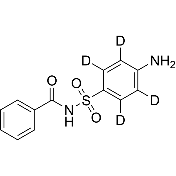
Sulfabenzamide-d4

Based on 1 Customer Validation
Sulfabenzamide (N-Sulfanilylbenzamide)-d4 is the deuterium labeled Sulfabenzamide (HY-B0960). Sulfabenzamide is a sulfonamide antibacterial agent. Sulfabenzamide exhibit antibacterial activity against Staphylococcus aureus (ATCC 25923) and Escherichia coli (ATCC 8739). Sulfabenzamide can promote autophagic cell autophagy in breast cancer cells through p53/ DRAM pathway. Sulfabenzamide increases caspase-3 activity, deactivates PARP1 and DNA-PK, downregulates AKT1 and AKT2. Sulfabenzamide can be used for the researches of breast cancer and bacterial infections.
연구목적의 판매만을 진행합니다. 환자를 대상으로 한 판매는 하지 않습니다.
- Purity : 99.74%
- CAS No.: 2732981-22-3
- 화학식: C13H8D4N2O3S
- 분자량:280.34
-
보관:Powder -20°C, 3 years ; In solvent -80°C, 6 months , -20°C, 1 month

To place orders, for customer services and technical support, please contact: MedChemExpress USA
Tel: 609-228-6898 E-mail: [email protected] [email protected]


-
 Biological Activity
-
Chemical Information
- Protocol
- 순도&문서
- References
-
 Help & FAQs


All Caspase Isoforms
More

Biological Activity
제품 설명
In Vitro
Stable heavy isotopes of hydrogen, carbon, and other elements have been incorporated into drug molecules, largely as tracers for quantitation during the drug development process. Deuteration has gained attention because of its potential to affect the pharmacokinetic and metabolic profiles of drugs[1].
MedChemExpress (MCE) has not independently confirmed the accuracy of these methods. They are for reference only. Further protocols information, click here.
신청
1. This compound can be used as a tracer
2. This compound can be used as an internal standard for quantitative analysis by NMR, GC-MS, or LC-MS.


Chemical Information
-
CAS No. 2732981-22-3
-
Unlabeled CAS 127-71-9
-
Appearance Solid
-
분자량 280.34
-
화학식 C13H8D4N2O3S
-
Color White to off-white
-
SMILES
O=C(NS(=O)(C1=C([2H])C([2H])=C(N)C([2H])=C1[2H])=O)C2=CC=CC=C2
-
Synonyms
N-Sulfanilylbenzamide-d4
-
선적
Room temperature in continental US; may vary elsewhere.
-
보관
Powder -20°C 3 years In solvent -80°C 6 months -20°C 1 month

Protocol
-
Research Protocol for Infectious Diseases
Infectious-disease experiments test how pathogens interact with host barriers, innate immune receptors, inflammatory signaling, pathogen replication, and tissue injury; pattern-recognition receptors such as TLRs, RIG-I-like receptors, NOD-like receptors, and inflammasomes detect microbial molecules and activate NF-κB, interferon, and cytokine responses. The central hypothesis is that infection severity reflects the balance between pathogen burden and host response: protective inflammation restricts pathogen growth, whereas excessive or mislocalized inflammation contributes to tissue damage and disease phenotype. Unresolved questions include which host pathways are protective versus pathogenic, why some infection models fail to translate to human disease, and which combined readouts best predict clinically relevant infection outcomes.
-
Autophagy
Autophagy is a process in which eukaryotic cells use lysosomes to degrade their own cytoplasmic proteins and damaged organelles under the regulation of autophagy related gene (Atg). Microtubule-associated proteins light chain 3 (LC3) is recognized as autophagy marker, which transfers from cytoplasmic LC3 (LC3-I) to membrane type (LC3-II). LC3-II/I ratio could be detected by Western Blot and fluorescence microscopy.
-
Apoptosis Solutions
Apoptosis is a regulated, generally non-lytic cell-death pathway that removes unwanted, damaged, infected, or abnormal cells through coordinated morphological changes, caspase activation, DNA fragmentation, and membrane remodeling. The intrinsic apoptosis pathway is controlled mainly by mitochondrial outer membrane permeabilization, BCL-2 family proteins, cytochrome c release, apoptosome formation, caspase-9 activation, and downstream executioner caspase-3/7 activation. The extrinsic apoptosis pathway is initiated by death receptors such as Fas, TNFR, and TRAIL receptors, which recruit adaptor proteins and activate caspase-8 before engaging executioner caspases or mitochondrial amplification through BID cleavage. Apoptosis is linked to many phenotypes, including cancer cell killing, tissue homeostasis, immune regulation, neurodegeneration, infection response, and treatment-induced cytotoxicity; unresolved questions include how apoptosis interacts with necroptosis, pyroptosis, ferroptos
-
Lysosome and acidic-vesicle live-cell staining
Lysosome and acidic-vesicle live-cell staining detects acidic intracellular compartments by using membrane-permeant acidotropic probes that accumulate in low-pH vesicles, including lysosomes, late endosomes, autolysosomes, and acidic phagosomes. LysoTracker staining is commonly used as an intensity-based readout of acidic lysosomal compartment abundance or enlargement, while acridine orange produces green fluorescence in less concentrated compartments and red fluorescence after concentration-dependent accumulation in acidic vesicular organelles. Loss or reduction of acridine-orange red signal can be used as a readout of lysosomal membrane permeabilization or reduced acidic-vesicle integrity. This protocol is designed for live cultured cells and can be adapted for fluorescence microscopy, high-content imaging, plate-reader readout, or flow cytometry when the selected literature supports the readout. Because these dyes report acidotropic accumulation rather than lysosome identity alone,
-
Bacterial live/dead nucleic-acid viability staining
The LIVE/DEAD bacterial viability staining method is based on differential permeability of nucleic-acid-binding fluorescent dyes, most commonly SYTO 9 and propidium iodide (PI), which enables discrimination of bacterial populations with intact versus compromised cytoplasmic membranes. SYTO 9 penetrates both intact and damaged bacterial membranes and binds nucleic acids to produce green fluorescence, whereas propidium iodide penetrates only cells with compromised membranes and fluoresces red while also reducing SYTO 9 signal through competitive binding and fluorescence interactions. The resulting fluorescence pattern is interpreted as a proxy for membrane integrity, which is widely used as an indicator of bacterial viability in microscopy, flow cytometry, and spectroscopic platforms. However, mechanistic studies show that SYTO 9 and PI interactions involve displacement and fluorescence resonance energy transfer effects, which can influence signal interpretation depending on dye ratios a
-
Macroautophagy Solutions
Macroautophagy is a conserved lysosome-dependent degradation pathway in which cytoplasmic material is sequestered into double-membrane autophagosomes and delivered to lysosomes for degradation and recycling. The pathway supports cellular homeostasis during nutrient limitation, organelle stress, protein-aggregate accumulation, infection, differentiation, and tissue remodeling by coupling cargo sequestration, autophagosome maturation, lysosomal fusion, and degradation of cargo-derived macromolecules. The core molecular sequence includes initiation by nutrient- and stress-regulated autophagy machinery, autophagosome nucleation, LC3/ATG8-family conjugation to autophagosomal membranes, cargo selection through receptors such as SQSTM1/p62, autophagosome-lysosome fusion, and lysosomal degradation. LC3 was identified as a mammalian homolog of yeast Atg8 that localizes to autophagosomal membranes after processing, and p62/SQSTM1 was shown to connect ubiquitinated cargo with autophagic degradati
-
Breast Cancer Modeling
Breast cancer is a heterogeneous cancer, and it has been distinguished into four subtypes: luminal A, luminal B, HER2-positive and basal-like. Molecular mutations, epigenetic alterations, hormone exposure and immune microenvironment are related to the progression of breast cancer.


순도&문서
-
Data Sheet (270 KB)
-
SDS (393 KB)
- English - EN (393 KB)
- Français - FR (393 KB)
- Deutsch - DE (393 KB)
- Norwegian - NO (393 KB)
- Español - ES (393 KB)
- Swedish - SV (393 KB)
- Italian - IT (393 KB)
- Korean - KR (393 KB)
- Portuguese - PT (393 KB)
-
Handling Instructions (2659 KB)
References

Calculators
Concentration (start) × Volume (start) = Concentration (final) × Volume (final)


Sulfabenzamide-d4 Related Classifications
HY-B0960S Related Classifications
 Powered by Bioz