
Licoflavanone
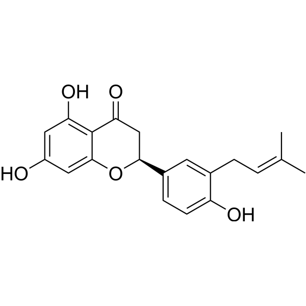
Licoflavanone (3′-Prenylnaringenin) is a flavanone with antioxidant, anti-inflammatory and anticancer activities. Licoflavanone can be isolated from the leaf extract of Glycyrrhiza glabra. Licoflavanone downregulates the mTOR/PI3K/AKT signaling pathway to inhibit the proliferation, migration and invasion of cancer cells, while activates Bax, Bad and multiple caspase enzymes to induce apoptosis. Its anti-inflammatory effect is manifested by reducing the nuclear translocation of NF-κB, decreasing the phosphorylation levels of p38, JNK and ERK1/2, thereby inhibiting the expression of nitric oxide, proinflammatory cytokines, COX-2 and iNOS. Licoflavanone is used in studies on nasopharyngeal carcinoma and related mechanisms.
For research use only. We do not sell to patients.
- CAS No.: 119240-82-3
- Formula: C20H20O5
- Molecular Weight:340.37
-
Storage:
Please store the product under the recommended conditions in the Certificate of Analysis.
All Caspase Isoforms
More
Biological Activity
Description
IC50 & Target
|
iNOS |
COX-2 |
In Vitro
Licoflavanone (12.5-100 μM; 24 h) reduces the viability of human nasopharyngeal carcinoma HK1 cells in a concentration-dependent manner[1].
Licoflavanone (12.5-100 μM; 72 h) induces destructive morphological changes and apoptosis in human nasopharyngeal HK1 cancer cells following 72 h of treatment at concentrations of 12.5, 25, and 100 μM[1].
Licoflavanone (12.5-100 μM; 24 h) regulates the expression of apoptosis-related proteins, upregulates pro-apoptotic proteins and downregulates anti-apoptotic proteins in human nasopharyngeal HK1 cancer cells following 24 h of treatment at concentrations of 12.5, 25 and 100 μM[1].
Licoflavanone (12.5-100 μM; 24 h) inhibits the mTOR/PI3K/AKT pathway in human nasopharyngeal carcinoma HK1 cells in a concentration-dependent manner by downregulating the expression of phosphorylated mTOR, PI3K and AKT (treatment concentrations: 12.5, 25 and 100 μM, treatment duration: 24 h)[1].
Licoflavanone (12.5-100 μM; 24 h) inhibits the migration and invasion of human nasopharyngeal HK1 cancer cells in a concentration-dependent manner[1].
Licoflavanone inhibits NO production in LPS-stimulated RAW 264.7 mouse macrophages with an IC50 of 37.68 μM, and suppresses the phosphorylation and activation of p38, JNK and ERK1/2 MAPKs[2].
MedChemExpress (MCE) has not independently confirmed the accuracy of these methods. They are for reference only. Further protocols information, click here.
-
Cell Line:human nasopharyngeal HK1 carcinoma cells
-
Concentration:12.5-100 μM
-
Incubation Time:24 h
-
Result:Reduced cell viability in a concentration-dependent manner, with viability dropping to almost 10% at the highest concentration tested.
Showed statistically significant effects in triplicate experiments.
-
Cell Line:human nasopharyngeal HK1 carcinoma cells
-
Concentration:12.5, 25, 100 μM
-
Incubation Time:24 h
-
Result:Induced pro-apoptotic changes including membrane damage, chromatin condensation, nuclear disintegration, formation of apoptotic bodies, and loss of normal cell morphology, while control cells showed normal structure and intact nuclear membranes.
-
Cell Line:human nasopharyngeal HK1 carcinoma cells
-
Concentration:12.5-100 μM
-
Incubation Time:24 h
-
Result:Significantly up-regulated protein expression of pro-apoptotic caspase-3, caspase-8, caspase-9, cleaved caspase-3, Bax, and Bad.
Markedly down-regulated protein expression of anti-apoptotic Bcl-2, XIAP, and Bcl-2-xL.\nMarkedly and concentration-dependently down-regulated the expression levels of phosphorylated mTOR, PI3K, and AKT.
Maintained almost constant protein expression levels of non-phosphorylated mTOR, PI3K, and AKT.
Showed statistically significant effects in triplicate experiments.
-
Cell Line:RAW 264.7 murine macrophages
-
Concentration:IC50 concentration
-
Incubation Time:6 h
-
Result:Significantly decreased mRNA levels of inducible nitric oxide synthase (iNOS), cyclooxygenase 2 (COX-2), tumor necrosis factor alpha (TNF-α), interleukin 1 beta (IL-1β), and interleukin 6 (IL-6) (p < 0.001 for all targets).
Chemical Information
-
CAS No. 119240-82-3
-
Molecular Weight 340.37
-
Formula C20H20O5
-
SMILES
O=C1C[C@@H](C2=CC=C(O)C(C/C=C(C)\C)=C2)OC3=CC(O)=CC(O)=C13
-
Synonyms
3′-Prenylnaringenin
-
Structure Classification
-
Initial Source
-
Shipping
Room temperature in continental US; may vary elsewhere.
-
Storage
Please store the product under the recommended conditions in the Certificate of Analysis.
Protocols
-
Western Blot
Western blotting (WB) is a commonly used experimental method in molecular biology, biochemistry, and immunogenetics for identifying and quantifying target proteins. It combines gel electrophoresis with immunoassay, enabling researchers to analyze protein expression, post-translational modifications, and molecular weight.
-
RNA extraction experimental
By lysing cells, releasing RNA, and removing impurities such as proteins and DNA, high-purity RNA products are finally obtained. The commonly used traditional method is the guanidine isothiocyanate/phenol/chloroform method (Trizol), which is suitable for a variety of animal materials including animal tissues, microorganisms, cultured cells, etc., and most plant materials.
-
Annexin V plus membrane-impermeant dye apoptosis staining
Annexin V-based apoptosis assays rely on the detection of phosphatidylserine (PS) externalization from the inner leaflet of the plasma membrane to the outer leaflet, an early biochemical hallmark of apoptosis. Fluorescently labeled Annexin V binds PS in a calcium-dependent manner, enabling identification of early apoptotic cells by flow cytometry or fluorescence microscopy. When combined with a membrane-impermeant DNA-binding dye (e. g. , propidium iodide), this approach allows discrimination between viable (Annexin V−/dye−), early apoptotic (Annexin V+/dye−), and late apoptotic or necrotic (Annexin V+/dye+) cell populations by assessing membrane integrity and PS exposure.
-
Cell migration
Cell migration is a method that plays an important role in wound healing, cell differentiation, embryonic development, etc.
-
Research Protocol for Inflammation-related Diseases
The NLRP3 inflammasome is a cytosolic innate immune signaling platform that integrates priming signals and danger-signal activation to promote caspase-1 activation, maturation of IL-1β and IL-18, and gasdermin D-mediated pyroptotic cell death. The core experimental logic is to determine whether inflammatory disease phenotypes are driven by increased NLRP3 expression, ASC-containing inflammasome assembly, caspase-1 cleavage, GSDMD cleavage, and extracellular release of IL-1β/IL-18 rather than by nonspecific cell injury alone. The pathway is strongly linked to inflammation-related disease phenotypes because monosodium urate crystals activate NALP3/NLRP3 inflammasome signaling in gout-like crystal inflammation, cholesterol crystals activate NLRP3 inflammasomes in atherogenesis models, and DSS-induced intestinal inflammation has been reported to involve NLRP3 inflammasome activity. However, experimental colitis studies also show context-dependent protective effects of NLRP3 inflammasome co
-
Cell Viability Determination by MTT Colorimetric Assay
The following protocol uses the MTT colorimetric assay as a classic literature-established method for assessing cell viability/metabolic activity in cultured mammalian cells. MTT[3-(4,5-dimethylthiazol-2-yl)-2,5-diphenyltetrazolium bromide] is reduced by metabolically active cells to a colored formazan product; the amount of formazan is quantified spectrophotometrically and provides an indirect measure of metabolically active viable cells. Importantly, MTT reduction reflects cellular oxidoreductase/metabolic activity rather than an absolute direct count of living cells, so changes in cellular metabolism can alter the signal independently of cell number.
-
Kinase activity and phosphorylation assays
Kinase activity assays measure the ability of kinases to transfer phosphate groups from ATP to specific substrates, while phosphorylation assays detect the presence and levels of phosphorylated proteins. Common methods include radiolabeled ATP incorporation (e. g. ,), ADP release detection via bioluminescence (e. g. ,[3]), enzyme-linked immunosorbent assays (ELISA) for phospho-specific epitopes (e. g. ,[6]), and microtiter-based formats for high-throughput screening (e. g. ,[8]). The ADP-Glo assay quantifies kinase activity by measuring ADP produced during phosphorylation using a luciferase-based system. Radiometric assays involve autoradiography or scintillation counting after incorporation of 32P-labeled ATP into substrate proteins. ELISA-based approaches rely on phospho-specific antibodies to detect activated kinases in cell lysates or purified samples.
-
Cytoplasmic-Nuclear Fractionated Protein Extraction
Cytoplasmic-nuclear fractionated protein extraction separates soluble cytoplasmic proteins from nuclear-enriched proteins by mild plasma-membrane permeabilization, differential centrifugation, washing of nuclei, and extraction of nuclear proteins for downstream immunoblotting or related molecular analysis. The readout is the relative abundance of a protein in cytoplasmic and nuclear fractions, commonly assessed by western blotting together with compartment markers such as tubulin or pyruvate kinase for cytoplasm and lamin, nucleoporin, hnRNP, H2AX, or Lamin B for nuclear fractions.
-
Apoptosis
Apoptosis, also called programmed cell death, is generally characterized by distinct morphological characteristics.
-
TUNEL staining for apoptotic DNA fragmentation
TUNEL staining detects DNA strand breaks by using terminal deoxynucleotidyl transferase to add labeled nucleotides to exposed 3′-OH DNA termini, generating either microscopic staining in fixed cells or tissue sections, or fluorescence/cytometric signal in cell suspensions. TUNEL positivity reflects DNA fragmentation but should not be interpreted alone as definitive apoptosis, because TUNEL can also label necrotic, autolytic, mechanically damaged, or DNA-repair-associated DNA breaks.
-
Apoptosis Solutions
Apoptosis is a regulated, generally non-lytic cell-death pathway that removes unwanted, damaged, infected, or abnormal cells through coordinated morphological changes, caspase activation, DNA fragmentation, and membrane remodeling. The intrinsic apoptosis pathway is controlled mainly by mitochondrial outer membrane permeabilization, BCL-2 family proteins, cytochrome c release, apoptosome formation, caspase-9 activation, and downstream executioner caspase-3/7 activation. The extrinsic apoptosis pathway is initiated by death receptors such as Fas, TNFR, and TRAIL receptors, which recruit adaptor proteins and activate caspase-8 before engaging executioner caspases or mitochondrial amplification through BID cleavage. Apoptosis is linked to many phenotypes, including cancer cell killing, tissue homeostasis, immune regulation, neurodegeneration, infection response, and treatment-induced cytotoxicity; unresolved questions include how apoptosis interacts with necroptosis, pyroptosis, ferroptos
-
Cell invasion
Cell invasion is the ability of cells to migrate from one area to another via the extracellular matrix. Cell invasion is the response of normal and cancer cells to chemical and mechanical stimuli. Before migrating to a new region, the extracellular matrix is degraded by proteases within the cell. Cell invasion often occurs during wound repair, vascularization and inflammation, abnormal tissue invasion, and tumor cell metastasis.
-
Protocol for Kinase activity and phosphorylation assays
Kinase activity assays measure transfer of phosphate from ATP to a protein or peptide substrate, generating phosphorylated substrate, ADP, or incorporated radiolabeled phosphate as the readout; phosphorylation assays measure site-specific phosphorylation in cells or tissues as a proxy for kinase-pathway activation, inhibition, or substrate regulation. Phosphorylation can be detected by phospho-specific Western blot, immunoprecipitation kinase assay, phospho-immunofluorescence, phospho-flow cytometry, luminescent ADP detection, radiolabeled ATP incorporation, or reporter-based pathway assays, and these readouts can be applied to cancer cells, primary neurons, mouse tumors, organoids, inflammatory macrophages, ferroptosis studies, and mitophagy studies when the kinase target is biologically relevant.
Purity & Documentation
References
Calculators
Concentration (start) × Volume (start) = Concentration (final) × Volume (final)

