



Lipoteichoic acid
Based on 2 publication(s) in Google Scholar
Lipoteichoic acid is an orally effect anti-inflammatory and antitumor agent. Lipoteichoic acid is a crucial immune molecule in Gram-positive bacteria that activates the complement system by inducing C3 and inhibiting CD55. Lipoteichoic acid regulates macrophage autophagy through the PI3K/Akt/mTOR pathway. Lipoteichoic acid induces lung damage in mice. Lipoteichoic acid inhibits the production of melanin.
Nur für Forschungszwecke. Wir verkaufen nicht an Patienten.
- CAS. Nr.: 56411-57-5
-
Speicherung:
4°C, protect from light
* In solvent : -80°C, 6 months; -20°C, 1 month (protect from light)

To place orders, for customer services and technical support, please contact: MedChemExpress USA
Tel: 609-228-6898 E-mail: [email protected] [email protected]


-
 Biologische Aktivität
-
Chemical Information
-
Lösungsmittel & Löslichkeit
- Protokoll
- Reinheit & Dokumentation
- Verweise
-
 Help & FAQs


-
HY-D1056
Lipopolysaccharides, from E. coli O55:B5Endotoxin -

HY-P75185
Flagellin Protein, Listeria monocytogenes (His) -
HY-127090
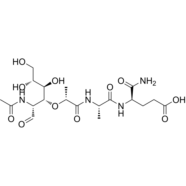
Muramyl dipeptideImmunoreactive Peptide -
HY-N6950
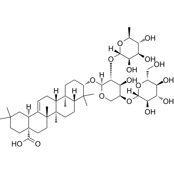
Hederacolchiside A1Antischistosomal Agent -
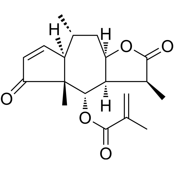
HY-N6843
Arnicolide DAntitumor Agent -
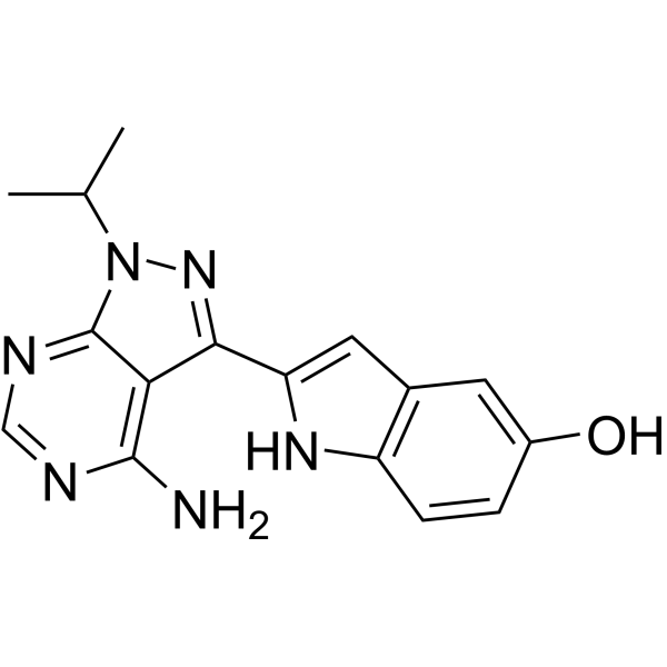
HY-10474
TorkinibmTOR Inhibitor -

HY-N6950R
Hederacolchiside A1 (Standard)Antischistosomal Agent -

HY-10474R
Torkinib (Standard)mTOR Inhibitor
Publications Citing Use of MedChemExpress (MCE) Lipoteichoic acid
More

Biologische Aktivität
Beschreibung
In Vitro
Lipoteichoic acid (0.1-20 μg/mL; 12-48 h) induces macrophage autophagy by inhibiting PI3K/AKT/mTOR pathway[2].
Lipoteichoic acid (0.1-100 μg/mL; 24 h) inhibits the production of melanin in B16F10 cells through MITF, ERK and PI3K/AKT signaling pathways[3].
Lipoteichoic acid (50-100 ng/mL; 2-3 h) shows no cytotoxicity to HT-29 cells, decreases the secretion of TNF-α and increases the secretion of IL-10 in HT-29 cells treated with LPS (HY-D1056)[4].
MedChemExpress (MCE) has not independently confirmed the accuracy of these methods. They are for reference only.
-
Cell Line:macrophage
-
Concentration:0.1, 1, 10, and 20 μg/mL
-
Incubation Time:12, 24 and 48 h
-
Result:Significantly decreased the expression levels of p-Akt and p-mTOR protein, but did not affect the expression of Akt and mTOR protein.
-
Cell Line:B16F10 cells
-
Concentration:0.1, 1, 10 and 100 μg/mL
-
Incubation Time:24 h
-
Result:Reduced the level of MITF in a dose-dependent manner.
Increased the phosphorylation levels of ERK, AKT and PI3K.
In Vivo
Lipoteichoic acid (0.1 mg; Oral administration; 20 days-34 weeks) has immunomodulatory and protective effects in UV-induced tumor models[5].
Lipoteichoic acid (5 mg/kg; Intratracheal injection; Single dose) can induce lung injury in mice[6].
MedChemExpress (MCE) has not independently confirmed the accuracy of these methods. They are for reference only.
-
Animal Model:DSS (HY-116282C) treated male swiss albino mice aged 6-8 weeks old[4]
-
Dosage:800 μg/100 μL
-
Administration:Oral gavage (i.g.); 7 days
-
Result:Significantly improved external colitis symptoms, disease activity scores, and weight gain in colitis mice.
Significantly improved key inflammatory markers such as the gut permeability, myeloperoxidase activity and histopathological damages in colon in colitis mice.
-
Animal Model:Female Crl:SKH-1-hrBR hairless mice aged 8-12 weeks old (20-25 g) with UV-induced tumors[5]
-
Dosage:100 μL (1 mg/mL)
-
Administration:Oral administration; 20 days and 34 weeks (3 times a week)
-
Result:Caused T cells in the mouse inguinal lymph nodes to produce higher levels of interferon-γ and a number of total, helper and cytotoxic T cells.
Significantly delayed the appearance of tumors.
-
Animal Model:Male C57BL/6 mice aged 6‑8 weeks old (22±3 g)[6]
-
Dosage:5 mg/kg
-
Administration:Intratracheal injection; Single dose
-
Result:Induced inflammatory cell infiltration, inter‑alveolar septal thickening and alveolar collapse.
Promoted the concentration of BALF total protein and the expression of inflammatory factors.
Increased lung neutrophil infiltration and MPO activity.
Clinical Trial
| NCT Number | Sponsor | Condition | Start Date |
Phase

|
|---|---|---|---|---|
| NCT01329991 | 2011-05 | PHASE1 |


Chemical Information
-
CAS. Nr. 56411-57-5
-
Appearance Solid
-
Color White to off-white
-
SMILES
[Lipoteichoic acid]
-
Structure Classification
-
Initial Source
Staphylococcus aureus
-
Versand
Room temperature in continental US; may vary elsewhere.
-
Speicherung
4°C, protect from light
* In solvent : -80°C, 6 months; -20°C, 1 month (protect from light)


Publications (2)
-
Journal Impact Factor
-
Most Recent
-
iScience
Newly isolated human-derived Lactobacillus fermentum strain stimulates IFN-γ-secreting CD8+ T cells for enhanced anti-cancer immunity. [Abstract]2026 Apr 8;29(5):115642. PMID: 42063558 -
Vet Res
2025 Jul 21;56(1):153. PMID: 40696454


Lösungsmittel & Löslichkeit
In Vitro:
H2O : 10 mg/mL (Need ultrasonic)


Reinheit & Dokumentation
-
Data Sheet (276 KB)
-
SDS (251 KB)
- English - EN (251 KB)
- Français - FR (251 KB)
- Deutsch - DE (251 KB)
- Norwegian - NO (251 KB)
- Español - ES (251 KB)
- Swedish - SV (251 KB)
- Italian - IT (251 KB)
- Korean - KR (251 KB)
- Portuguese - PT (251 KB)
-
Handling Instructions (2659 KB)
Verweise
[1]. Bong Jun Jung, et al. Lipoteichoic Acid from Staphylococcus aureus Activates the Complement System via C3 Induction and CD55 Inhibition. Microorganisms. 2021 May 24;9(6):1135. [Content Brief]
[2]. Lin D, et al. Enterococcus faecalis lipoteichoic acid regulates macrophages autophagy via PI3K/Akt/mTOR pathway. Biochem Biophys Res Commun. 2018 Apr 15;498(4):1028-1036. [Content Brief]
[3]. Kim HR, et al. Lipoteichoic acid isolated from Lactobacillus plantarum inhibits melanogenesis in B16F10 mouse melanoma cells. Mol Cells. 2015;38(2):163-70. [Content Brief]
[4]. Pradhan D, et al. Postbiotic Lipoteichoic acid of probiotic Lactobacillus origin ameliorates inflammation in HT-29 cells and colitis mice. Int J Biol Macromol. 2023 May 1;236:123962. [Content Brief]
[5]. Weill FS, et al. Lipoteichoic acid from Lactobacillus rhamnosus GG as an oral photoprotective agent against UV-induced carcinogenesis. Br J Nutr. 2013 Feb 14;109(3):457-66. [Content Brief]
[6]. Zhang Y, et al Shikonin ameliorates lipoteichoic acid induced acute lung injury via promotion of neutrophil apoptosis. Mol Med Rep. 2021 Feb;23(2):133. [Content Brief]
[7]. Hara H, et al. The NLRP6 Inflammasome Recognizes Lipoteichoic Acid and Regulates Gram-Positive Pathogen Infection. Cell. 2018 Nov 29;175(6):1651-1664.e14. [Content Brief]

Calculators
Konzentration (Stammlösung) × Volumen (Stammlösung) = Konzentration (Ziellösung) × Volumen (Ziellösung)


Lipoteichoic acid Related Classifications
HY-N9481 Related Classifications
 Powered by Bioz