



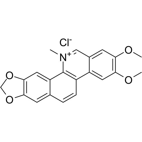
Nitidine chloride
Based on 4 publication(s) in Google Scholar
Nitidine chloride, a potential anti-malarial lead compound derived from Zanthoxylum nitidum (Roxb) DC, exerts potent anticancer activity through diverse pathways, including inducing apoptosis, inhibiting STAT3 signaling cascade, DNA topoisomerase 1 and 2A, ERK and c-Src/FAK associated signaling pathway, also has anti-inflammatory activity. Nitidine chloride inhibits LPS-induced inflammatory cytokines production via MAPK and NF-kB pathway.
For research use only. We do not sell to patients.
- Purity : 99.82%
- CAS No.: 13063-04-2
- Formula: C21H18ClNO4
- Molecular Weight:383.82
-
Storage:
4°C, sealed storage, away from moisture and light
* In solvent : -80°C, 6 months; -20°C, 1 month (sealed storage, away from moisture and light)

To place orders, for customer services and technical support, please contact: MedChemExpress USA
Tel: 609-228-6898 E-mail: [email protected] [email protected]


-
 Biological Activity
-
Chemical Information
-
Solvent & Solubility
- Protocols
- Purity & Documentation
- References
-
 Help & FAQs


-
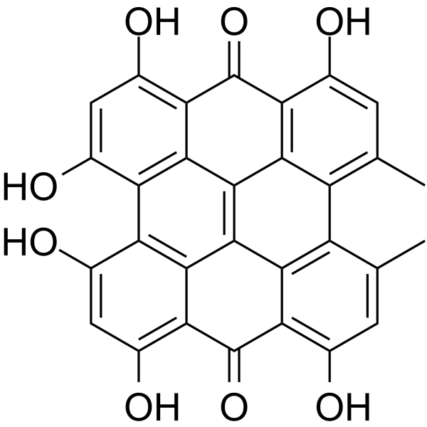
HY-N0453
HypericinPKC Inhibitor -
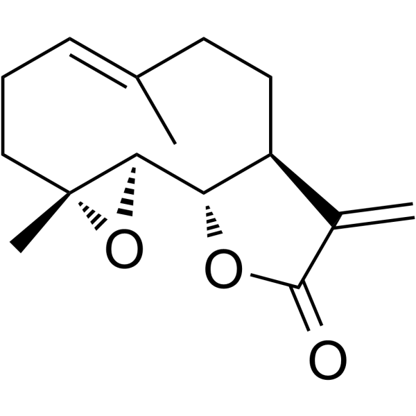
HY-N0141
ParthenolideNF-κB Inhibitor -
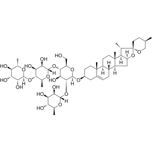
HY-N2389
Formosanin CApoptosis/Autophagy Inducer -
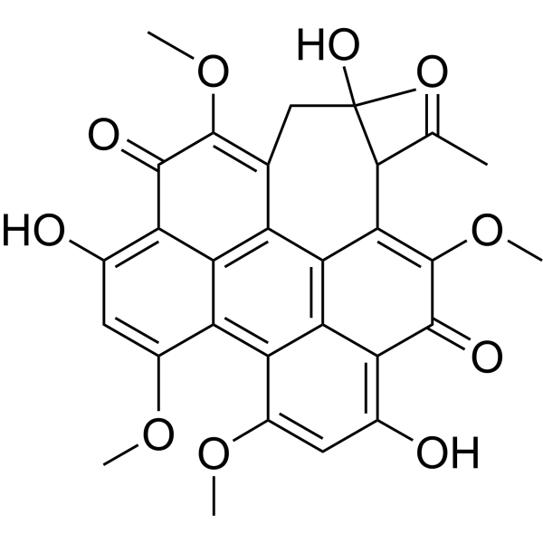
HY-N2575
Hypocrellin APhotosensitizer -
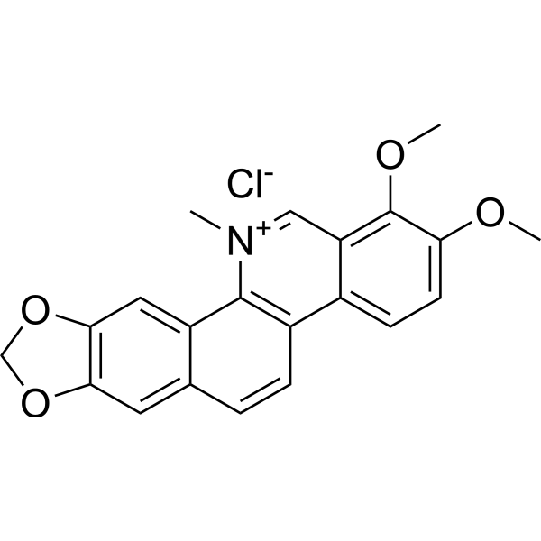
HY-12048
Chelerythrine chlorideBcl-2 Family Inhibitor -
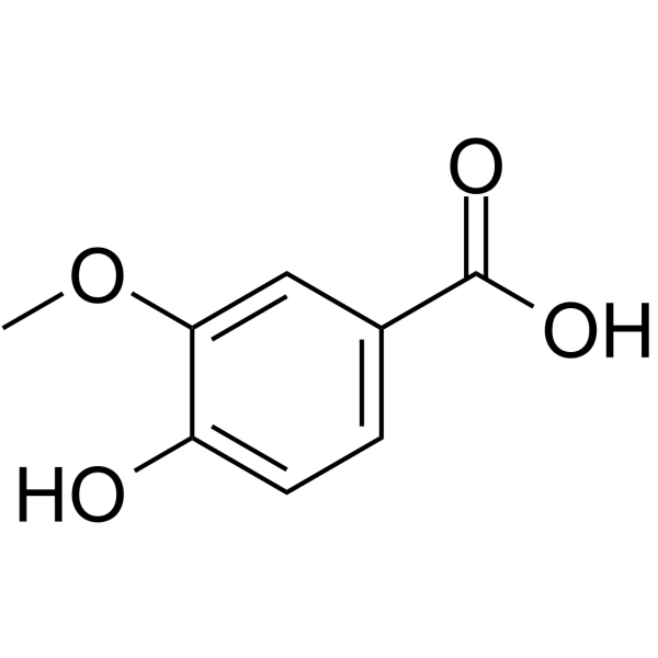
HY-N0708
Vanillic acidFlavouring Agent -

HY-N0498R
Nitidine chloride (Standard)Anti-malarial Agent -
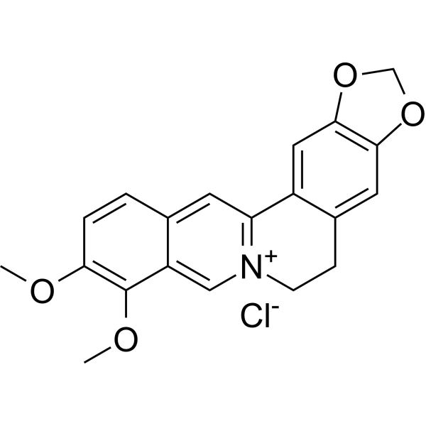
HY-18258
Berberine chlorideAntibiotic -
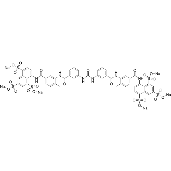
HY-B0879A
Suramin sodium saltPTPases Inhibitor -
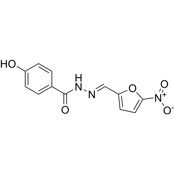
HY-B1436
NifuroxazideBacterial Inhibitor -
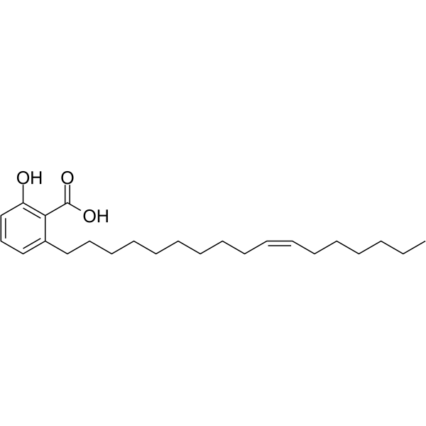
HY-N2116
Ginkgolic acid C17:1Antitumor Agent -
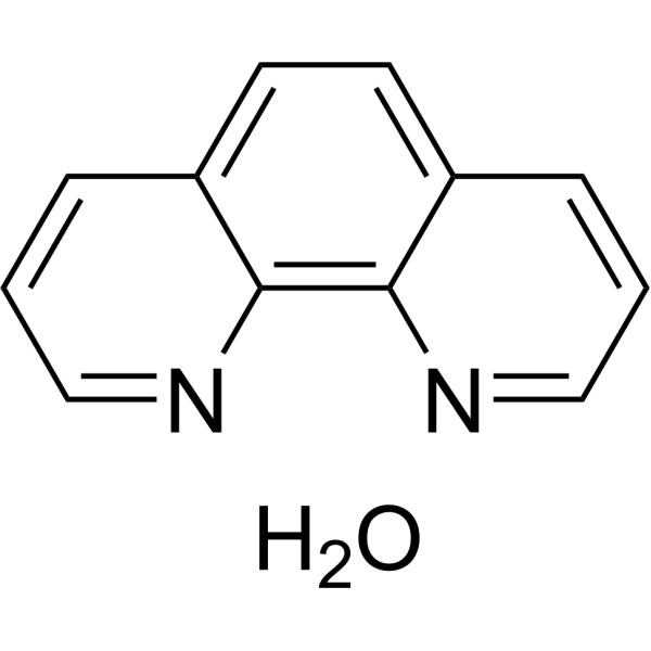
HY-Y1841
o-Phenanthroline monohydrateMMP Inhibitor
Publications Citing Use of MedChemExpress (MCE) Nitidine chloride
MoreAll Parasite Isoforms
MoreAll Topoisomerase Isoforms
More

Biological Activity
Description
IC50 & Target
|
Plasmodium |
Cellular Effect
|
Cell Line

|
Type | Value | Description | References |
|---|---|---|---|---|
| CEM-SS | EC50 |
14 μM
Compound: Nitidine
|
Antiviral activity against HIV1 RF in human CEM-SS cells assessed as inhibition of virus-induced cytopathic effect
Antiviral activity against HIV1 RF in human CEM-SS cells assessed as inhibition of virus-induced cytopathic effect
|
[PMID: 11430019] |
| HCT-116 | IC50 |
4.42 μM
Compound: 1c
|
Antiproliferative activity against human HCT116 cells assessed as inhibition of cell viability after 48 hrs by MTT assay
Antiproliferative activity against human HCT116 cells assessed as inhibition of cell viability after 48 hrs by MTT assay
|
[PMID: 26026362] |
| HeLa | IC50 |
0.05 μg/mL
Compound: 1
|
Growth inhibition of human HeLa S3 cells after 72 hrs
Growth inhibition of human HeLa S3 cells after 72 hrs
|
[PMID: 10395504] |
| HepG2 | IC50 |
3.52 μM
Compound: 1c
|
Cytotoxicity against human HepG2 cells
Cytotoxicity against human HepG2 cells
|
[PMID: 26026362] |
| HL-60 | IC50 |
3.41 μM
Compound: 1c
|
Antiproliferative activity against human HL60 cells assessed as inhibition of cell viability after 48 hrs by WST-1 assay
Antiproliferative activity against human HL60 cells assessed as inhibition of cell viability after 48 hrs by WST-1 assay
|
[PMID: 26026362] |
| HT-29 | IC50 |
2.75 μM
Compound: 1, 1-k6932
|
Cytotoxicity against human HT-29 cells after 72 hrs by MTS assay
Cytotoxicity against human HT-29 cells after 72 hrs by MTS assay
|
[PMID: 21435753] |
| KB | ED50 |
4.3 μg/mL
Compound: Nitidine
|
Cytotoxicity against human KB cells
Cytotoxicity against human KB cells
|
[PMID: 7130986] |
| Panel NCI-60 (60 carcinoma cell lines) | GI50 |
125 nM
Compound: Nitidine
|
Growth inhibitory activity against human cancer cell line in the NCI's anticancer drug screening program
Growth inhibitory activity against human cancer cell line in the NCI's anticancer drug screening program
|
[PMID: 15743190] |
In Vitro
Nitidine chloride (0-32 μM, 24-48 h) inhibits the proliferation of A498 and 768-O cells[1].
Nitidine chloride (8-16 μM, 24 h) induces apoptosis in A498 and 768-O cells[1].
Nitidine chloride (0-16 μM, 48 h) induces apoptosis in A498 cells, causes cell cycle arrest, and inhibits the ERK pathway[1].
Nitidine chloride (0-10 μM, 24 h) induces apoptosis in HSC3 and HSC4 cells, inducing apoptosis in OSCC cells through dephosphorylation of STAT3[2].
MedChemExpress (MCE) has not independently confirmed the accuracy of these methods. They are for reference only. Further protocols information, click here.
-
Cell Line:768-O, A498
-
Concentration:0, 4, 6, 8, 16, 32 μM
-
Incubation Time:24, 48 h
-
Result:Inhibited cell proliferation in a time- and dose-dependent manner.
-
Cell Line:768-O, A498
-
Concentration:8, 16 μM
-
Incubation Time:24 h
-
Result:Increased apoptosis rate in a dose-dependent manner.
-
Cell Line:A498
-
Concentration:0, 8, 16 μM
-
Incubation Time:48 h
-
Result:Increased Bax expression, decreased Bcl-2 expression, increased p53 expression, decreased pro-caspase-3 expression, increased cleaved caspase-3 expression and PARP cleavage level, inhibited ERK1/2, p38 and Akt phosphorylation.
-
Cell Line:HSC3, HSC4
-
Concentration:0, 0.1, 5, 10 μM
-
Incubation Time:24 h
-
Result:Reduced cell viability.
-
Cell Line:HSC3, HSC4, OSCC
-
Concentration:0, 0.1, 5, 10 μM
-
Incubation Time:24 h
-
Result:Dephosphorylated STAT3 and induced the cleavage of PARP and caspase-3 in a concentration- and time-dependent manner.
In Vivo
Nitidine chloride (0.375-10 mg/kg, intraperitoneal injection, once daily, 3 weeks) downregulates hypertrophy in mouse myocardium through AGT4B-mediated autophagy[3].
Nitidine chloride (5-10 mg/kg, intraperitoneal injection, single dose) exerts anti-inflammatory effects in endotoxin-treated mice through Akt-mediated IL-10 production[4].
MedChemExpress (MCE) has not independently confirmed the accuracy of these methods. They are for reference only.
-
Animal Model:A549 nude mouse xenograft model[1]
-
Dosage:5 mg/kg; daily; 5 weeks
-
Administration:Intraperitoneal injection (i.p.)
-
Result:Showed non-toxic effects, reduced tumor growth, and induced apoptosis in renal cancer cells in vivo.
-
Animal Model:Mice, beagles[3]
-
Dosage:2.5, 5, 10 mg/kg; 0.375, 0.75, 1.5 mg/kg; daily; 4 weeks
-
Administration:Intraperitoneal injection (i.p.)
-
Result:Both induced dose-dependent cardiac hypertrophy and dysfunction, while medium and high doses led to death in beagle dogs. Cardiac autophagy levels in cardiomyocytes were reduced. Overexpression of ATG4B reversed cardiac hypertrophy and reduced autophagy levels.
-
Animal Model:LPS induced endotoxemic mice[4]
-
Dosage:5, 10 mg/kg; single dose
-
Administration:Intraperitoneal injection (i.p.)
-
Result:Increased IL-10 production, inhibited inflammatory response, reduced mortality, and enhanced Akt activation.


Chemical Information
-
CAS No. 13063-04-2
-
Appearance Solid
-
Molecular Weight 383.82
-
Formula C21H18ClNO4
-
Color Light yellow to yellow
-
SMILES
C[N+]1=CC2=CC(OC)=C(OC)C=C2C(C=CC3=C4)=C1C3=CC5=C4OCO5.[Cl-]
-
Structure Classification
-
Initial Source
-
Shipping
Room temperature in continental US; may vary elsewhere.
-
Storage
4°C, sealed storage, away from moisture and light
* In solvent : -80°C, 6 months; -20°C, 1 month (sealed storage, away from moisture and light)


Publications (4)
-
Journal Impact Factor
-
Most Recent
-
Free Radic Biol Med
Targeting ABCB6 with nitidine chloride inhibits PI3K/AKT signaling pathway to promote ferroptosis in multiple myeloma. [Abstract]2023 Jul:203:86-101. PMID: 37044150 -
Life Sci
Nitidine chloride inhibits mTORC1 signaling through ATF4-mediated Sestrin2 induction and targets IGF2R for lysosomal degradation. [Abstract]2024 Sep 15:353:122918. PMID: 39034027 -
PLoS Pathog
G-quadruplex in the TMV Genome Regulates Viral Proliferation and Acts as Antiviral Target of Photodynamic Therapy. [Abstract]2023 Dec 7;19(12):e1011796. PMID: 38060599 -
J Pharm Biomed Anal
Integrated network pharmacology and metabolomics to investigate the effect and mechanism of nitidine chloride against cholangiocarcinoma. [Abstract]2025 Nov 15:265:117063. PMID: 40652810


Solvent & Solubility
In Vitro:
DMSO : 2 mg/mL (5.21 mM; Need ultrasonic; Hygroscopic DMSO has a significant impact on the solubility of product, please use newly opened DMSO)
* Nitidine chloride is usually formulated as a suspension.
Please refer to the solubility information to select the appropriate solvent. Once prepared, please aliquot and store the solution to prevent product inactivation from repeated freeze-thaw cycles.
Storage method and period of stock solution: -80°C, 6 months; -20°C, 1 month (sealed storage, away from moisture and light). When stored at -80°C, please use it within 6 months. When stored at -20°C, please use it within 1 month.
Please refer to the solubility information to select the appropriate solvent. Once prepared, please aliquot and store the solution to prevent product inactivation from repeated freeze-thaw cycles.
Storage method and period of stock solution: -80°C, 6 months; -20°C, 1 month (sealed storage, away from moisture and light). When stored at -80°C, please use it within 6 months. When stored at -20°C, please use it within 1 month.
Concentration (start) × Volume (start) = Concentration (final) × Volume (final)
In Vivo:
For the following dissolution methods, please prepare the working solution directly:
It is recommended to prepare fresh solutions and use them promptly within a short period of time.
The percentages shown for the solvents indicate their volumetric ratio in the final prepared solution. If precipitation or phase separation occurs during preparation, heat and/or sonication can be used to aid dissolution.
Add each solvent one by one: 15% Cremophor EL 85% Saline
Solubility: 2 mg/mL (5.21 mM); Suspended solution; Need ultrasonic

Protocols
-
Apoptosis
Apoptosis, also called programmed cell death, is generally characterized by distinct morphological characteristics.
-
TUNEL staining for apoptotic DNA fragmentation
TUNEL staining detects DNA strand breaks by using terminal deoxynucleotidyl transferase to add labeled nucleotides to exposed 3′-OH DNA termini, generating either microscopic staining in fixed cells or tissue sections, or fluorescence/cytometric signal in cell suspensions. TUNEL positivity reflects DNA fragmentation but should not be interpreted alone as definitive apoptosis, because TUNEL can also label necrotic, autolytic, mechanically damaged, or DNA-repair-associated DNA breaks.
-
LPS-Induced Endotoxemia/Systemic Inflammation
Lipopolysaccharide (LPS)-induced endotoxemia is a widely used in vivo model of acute systemic inflammation in which LPS, a Gram-negative bacterial endotoxin, activates innate immune signaling primarily through TLR4, leading to rapid and transient induction of pro-inflammatory cytokines such as TNF-α, IL-6, and IL-1β in circulation and tissues. This cytokine surge is commonly used as a measurable readout of systemic inflammatory activation and immune dysregulation, and is typically assessed within hours after intraperitoneal LPS administration in mouse models of endotoxemia. The model captures key features of systemic inflammatory response syndrome, including cytokine release, immune cell activation, and downstream tissue responses, and has been used to evaluate anti-inflammatory interventions such as cytokine modulation, lipid mediators, and immune cell-targeting therapies.
-
Annexin V plus membrane-impermeant dye apoptosis staining
Annexin V-based apoptosis assays rely on the detection of phosphatidylserine (PS) externalization from the inner leaflet of the plasma membrane to the outer leaflet, an early biochemical hallmark of apoptosis. Fluorescently labeled Annexin V binds PS in a calcium-dependent manner, enabling identification of early apoptotic cells by flow cytometry or fluorescence microscopy. When combined with a membrane-impermeant DNA-binding dye (e. g. , propidium iodide), this approach allows discrimination between viable (Annexin V−/dye−), early apoptotic (Annexin V+/dye−), and late apoptotic or necrotic (Annexin V+/dye+) cell populations by assessing membrane integrity and PS exposure.
-
Cotton Pellet Granuloma
Cotton pellet granuloma is a classical in vivo chronic inflammation model used to evaluate the anti-inflammatory potential of test substances by measuring their ability to inhibit granuloma tissue formation around an implanted foreign body (cotton pellet) in rodents. The method is based on the biological response to a sterile implanted material, which induces proliferative phase inflammation characterized by fibroblast proliferation and collagen-rich granuloma formation, and the final readout reflects the extent of chronic inflammatory tissue growth surrounding the pellet. In multiple preclinical pharmacological evaluations, inhibition of cotton pellet-induced granuloma formation has been used as an indicator of anti-inflammatory activity in both synthetic and natural product screening contexts.
-
Carrageenan-Induced Paw Edema
Carrageenan-induced paw edema is an acute inflammation model in which intraplantar injection of carrageenan induces localized inflammatory swelling characterized by vascular permeability, leukocyte infiltration, and production of inflammatory mediators such as prostaglandins and cytokines, making it widely used to evaluate anti-inflammatory agents in vivo. The resulting paw volume or thickness increase is quantified over time as a direct readout of inflammatory intensity and drug efficacy, typically reflecting cyclooxygenase-mediated prostaglandin-driven edema formation and immune cell recruitment in peripheral tissue[20].
-
Apoptosis Solutions
Apoptosis is a regulated, generally non-lytic cell-death pathway that removes unwanted, damaged, infected, or abnormal cells through coordinated morphological changes, caspase activation, DNA fragmentation, and membrane remodeling. The intrinsic apoptosis pathway is controlled mainly by mitochondrial outer membrane permeabilization, BCL-2 family proteins, cytochrome c release, apoptosome formation, caspase-9 activation, and downstream executioner caspase-3/7 activation. The extrinsic apoptosis pathway is initiated by death receptors such as Fas, TNFR, and TRAIL receptors, which recruit adaptor proteins and activate caspase-8 before engaging executioner caspases or mitochondrial amplification through BID cleavage. Apoptosis is linked to many phenotypes, including cancer cell killing, tissue homeostasis, immune regulation, neurodegeneration, infection response, and treatment-induced cytotoxicity; unresolved questions include how apoptosis interacts with necroptosis, pyroptosis, ferroptos
-
Research Protocol for Inflammation-related Diseases
The NLRP3 inflammasome is a cytosolic innate immune signaling platform that integrates priming signals and danger-signal activation to promote caspase-1 activation, maturation of IL-1β and IL-18, and gasdermin D-mediated pyroptotic cell death. The core experimental logic is to determine whether inflammatory disease phenotypes are driven by increased NLRP3 expression, ASC-containing inflammasome assembly, caspase-1 cleavage, GSDMD cleavage, and extracellular release of IL-1β/IL-18 rather than by nonspecific cell injury alone. The pathway is strongly linked to inflammation-related disease phenotypes because monosodium urate crystals activate NALP3/NLRP3 inflammasome signaling in gout-like crystal inflammation, cholesterol crystals activate NLRP3 inflammasomes in atherogenesis models, and DSS-induced intestinal inflammation has been reported to involve NLRP3 inflammasome activity. However, experimental colitis studies also show context-dependent protective effects of NLRP3 inflammasome co


Purity & Documentation
-
Data Sheet (284 KB)
-
SDS (480 KB)
- English - EN (480 KB)
- Français - FR (480 KB)
- Deutsch - DE (480 KB)
- Norwegian - NO (480 KB)
- Español - ES (480 KB)
- Swedish - SV (480 KB)
- Italian - IT (480 KB)
- Korean - KR (480 KB)
- Portuguese - PT (480 KB)
-
Handling Instructions (2659 KB)
References
[1]. Fang Z, et al. Nitidine chloride induces apoptosis and inhibits tumor cell proliferation via suppressing ERK signaling pathway in renal cancer. Food Chem Toxicol. 2014 Apr;66:210-6. [Content Brief]
[2]. Lee-Han Kim, et al. Nitidine chloride acts as an apoptosis inducer in human oral cancer cells and a nude mouse xenograft model via inhibition of STAT3. Oncotarget. 2017 Aug 24;8(53):91306-91315. [Content Brief]
[3]. Yang Hong, et al. Nitidine chloride induces cardiac hypertrophy in mice by targeting autophagy-related 4B cysteine peptidase. Acta Pharmacol Sin. 2023 Mar;44(3):561-572. [Content Brief]
[4]. Niao Yang, et al. Nitidine chloride exerts anti-inflammatory action by targeting Topoisomerase I and enhancing IL-10 production. Pharmacol Res. 2019 Oct:148:104368. [Content Brief]
[5]. Wang Z, et al. Nitidine chloride inhibits LPS-induced inflammatory cytokines production via MAPK and NF-kappaB pathway in RAW 264.7 cells. J Ethnopharmacol. 2012 Oct 31;144(1):145-50. [Content Brief]


Complete Stock Solution Preparation Table
Please refer to the solubility information to select the appropriate solvent. Once prepared, please aliquot and store the solution to prevent product inactivation from repeated freeze-thaw cycles.
Storage method and period of stock solution: -80°C, 6 months; -20°C, 1 month (sealed storage, away from moisture and light). When stored at -80°C, please use it within 6 months. When stored at -20°C, please use it within 1 month.
| Optional Solvent | Concentration Solvent Mass | 1 mg | 5 mg | 10 mg | 25 mg |
|---|---|---|---|---|---|
| DMSO | 1 mM | 2.6054 mL | 13.0269 mL | 26.0539 mL | 65.1347 mL |
| 5 mM | 0.5211 mL | 2.6054 mL | 5.2108 mL | 13.0269 mL |


Nitidine chloride Related Classifications
HY-N0498 Related Classifications
 Powered by Bioz