



Oosporein

Based on 1 Customer Validation
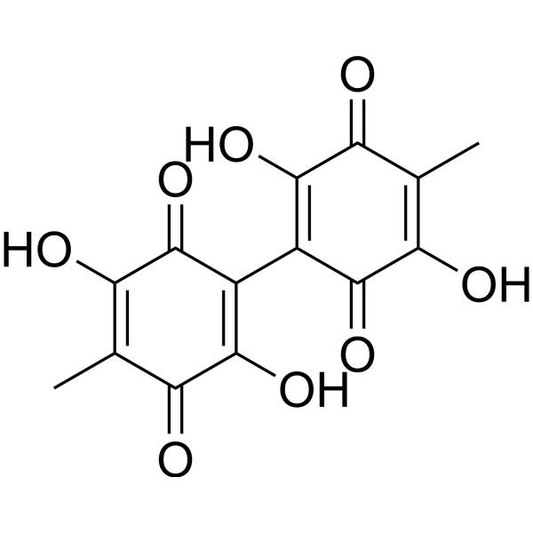
Oosporein is a microbial metabolite and a red crystalline toxin produced by various fungi. Oosporein can promote the reproduction of fungi in host bodies by inhibiting insect immunity, and possesses multiple activities such as antibacterial, antiviral (HSV), and insecticidal effects. Oosporein can inhibit plant growth. In addition, Oosporein can also induce apoptosis, cell membrane damage, oxidative stress, and mitochondrial damage. Oosporein has certain antitumor activity.
For research use only. We do not sell to patients.
- Purity : 99.16%
- CAS No.: 475-54-7
- Formula: C14H10O8
- Molecular Weight:306.22
-
Storage:Powder -20°C, 3 years ; In solvent -80°C, 6 months , -20°C, 1 month

To place orders, for customer services and technical support, please contact: MedChemExpress USA
Tel: 609-228-6898 E-mail: [email protected] [email protected]


-
 Biological Activity
-
Chemical Information
-
Solvent & Solubility
- Protocols
- Purity & Documentation
- References
-
 Help & FAQs


-

HY-N7719R
Oosporein (Standard)Standard of Oosporein -
HY-N0662
AmentoflavoneGABA(A) Negative Modulator -
HY-A0070A
LiothyronineTIGIT/PVR Blocker -
HY-N1401
20(R)-Ginsenoside Rh2Anti-tumor/Anti-viral/Anti-inflammatory Agent -
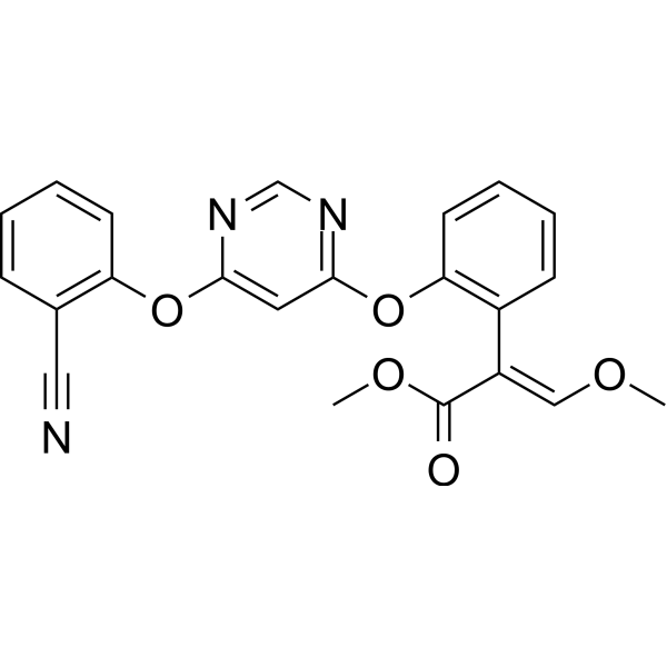
HY-B0849
AzoxystrobinFungicide -

HY-N10544
δ-CadineneApoptosis Inducer -
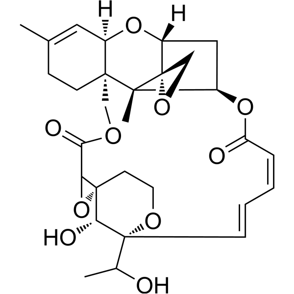
HY-130476
Satratoxin GLarge Cyclic Mucor Toxin. -
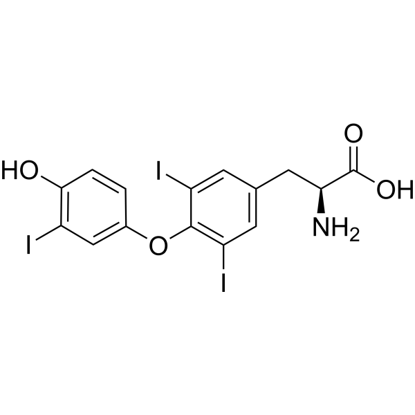
HY-A0070AG
Liothyronine (GMP)TRα/TRβ Agonist (GMP) -
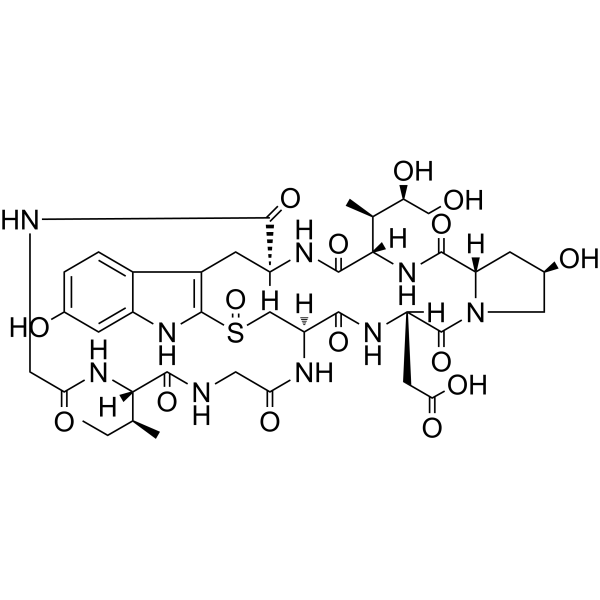
HY-125586
β-AmanitinRNA Polymerase II/III Inhibitor -
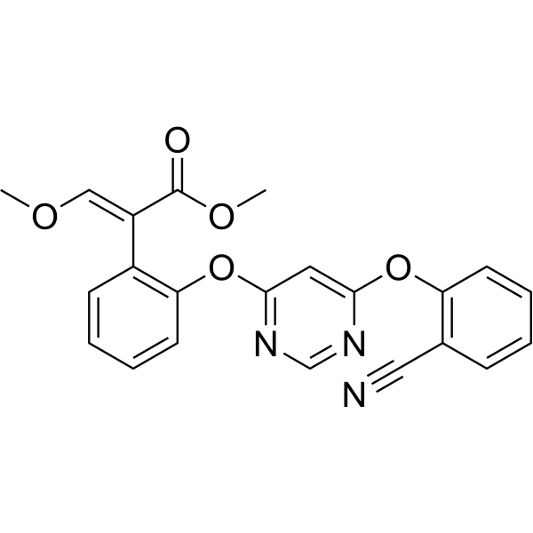
HY-B0849R
Azoxystrobin (Standard)Fungicide -
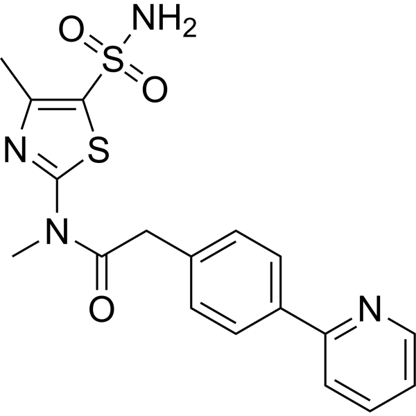
HY-15303
PritelivirHSV Inhibitor -
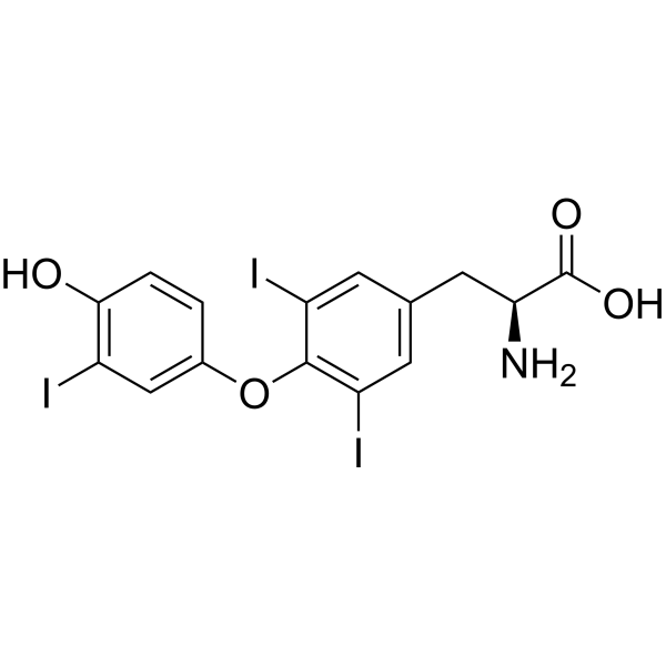
HY-A0070AR
Liothyronine (Standard)TRα/TRβ Agonist


Biological Activity
Description
IC50 & Target
[4]|
Microbial Metabolite |
Caspase 3 |
Caspase-6 |
Caspase-9 |
In Vitro
Oosporein exhibits antitumor activity in HL-60 and A549 cells, with an IC50 of 28.66 μM[1].
Oosporein shows moderate antibacterial activity against Gram-positive bacteria (such as Staphylococcus aureus, Bacillus subtilis, and Micrococcus lysodeikticus), with MIC values of 12.5-50 μg/mL[2].
Oosporein (10 μM-10 mM; 1-14 days) can inhibit the growth of plants like oats and wheat coleoptiles, and also cause abnormal plant morphology, such as dwarfing of tobacco and kidney bean plants and leaf shrinkage[3].
Oosporein (0-200 µM; 2-24 h) inhibits cell viability, induces apoptosis, causes cell membrane damage, oxidative stress, and mitochondrial damage in MDCK and RAW 264.7 cells, such as increasing intracellular ROS production, decreasing mitochondrial membrane potential, and enhancing glutathione hydroxylase generation[4].
MedChemExpress (MCE) has not independently confirmed the accuracy of these methods. They are for reference only. Further protocols information, click here.
-
Cell Line:Madin-Darby canine kidney (MDCK) and RAW 264.7 cells
-
Concentration:0, 5, 10, 25, 50, 100, 150 and 200 µM
-
Incubation Time:3 and 24 h
-
Result:Inhibited the cell viability, with IC50 values of 86 µM and 78 µM for MDCK and RAW 264.7 cells for 24 h exposure, respectively.
-
Cell Line:Madin-Darby canine kidney (MDCK) and RAW 264.7 cells
-
Concentration:25, 50 and 100 μM
-
Incubation Time:2 h
-
Result:Increased the mRNA levels of HSP70, CAS3, CAS6, and CAS9.
In Vivo
Oosporein (20-200 μM; intraperitoneal injection; single dose) causes histopathological damage in the kidneys and spleens of Balb/c mice, with the degree of damage being proportional to the dose[4].
MedChemExpress (MCE) has not independently confirmed the accuracy of these methods. They are for reference only.


Chemical Information
-
CAS No. 475-54-7
-
Appearance Solid
-
Molecular Weight 306.22
-
Formula C14H10O8
-
Color Brown to reddish brown
-
SMILES
O=C(C(C)=C1O)C(O)=C(C2=C(O)C(C(C)=C(O)C2=O)=O)C1=O
-
Structure Classification
-
Initial Source
Chaetomium
-
Shipping
Room temperature in continental US; may vary elsewhere.
-
Storage
Powder -20°C 3 years In solvent -80°C 6 months -20°C 1 month


Solvent & Solubility
In Vitro:
DMSO : ≥ 50 mg/mL (163.28 mM; Hygroscopic DMSO has a significant impact on the solubility of product, please use newly opened DMSO)
* "≥" means soluble, but saturation unknown.
Please refer to the solubility information to select the appropriate solvent. Once prepared, please aliquot and store the solution to prevent product inactivation from repeated freeze-thaw cycles.
Storage method and period of stock solution: -80°C, 6 months; -20°C, 1 month. When stored at -80°C, please use it within 6 months. When stored at -20°C, please use it within 1 month.
Please refer to the solubility information to select the appropriate solvent. Once prepared, please aliquot and store the solution to prevent product inactivation from repeated freeze-thaw cycles.
Storage method and period of stock solution: -80°C, 6 months; -20°C, 1 month. When stored at -80°C, please use it within 6 months. When stored at -20°C, please use it within 1 month.
Concentration (start) × Volume (start) = Concentration (final) × Volume (final)

Protocols
-
Research Protocol for Infectious Diseases
Infectious-disease experiments test how pathogens interact with host barriers, innate immune receptors, inflammatory signaling, pathogen replication, and tissue injury; pattern-recognition receptors such as TLRs, RIG-I-like receptors, NOD-like receptors, and inflammasomes detect microbial molecules and activate NF-κB, interferon, and cytokine responses. The central hypothesis is that infection severity reflects the balance between pathogen burden and host response: protective inflammation restricts pathogen growth, whereas excessive or mislocalized inflammation contributes to tissue damage and disease phenotype. Unresolved questions include which host pathways are protective versus pathogenic, why some infection models fail to translate to human disease, and which combined readouts best predict clinically relevant infection outcomes.
-
Apoptosis
Apoptosis, also called programmed cell death, is generally characterized by distinct morphological characteristics.
-
TUNEL staining for apoptotic DNA fragmentation
TUNEL staining detects DNA strand breaks by using terminal deoxynucleotidyl transferase to add labeled nucleotides to exposed 3′-OH DNA termini, generating either microscopic staining in fixed cells or tissue sections, or fluorescence/cytometric signal in cell suspensions. TUNEL positivity reflects DNA fragmentation but should not be interpreted alone as definitive apoptosis, because TUNEL can also label necrotic, autolytic, mechanically damaged, or DNA-repair-associated DNA breaks.
-
Annexin V plus membrane-impermeant dye apoptosis staining
Annexin V-based apoptosis assays rely on the detection of phosphatidylserine (PS) externalization from the inner leaflet of the plasma membrane to the outer leaflet, an early biochemical hallmark of apoptosis. Fluorescently labeled Annexin V binds PS in a calcium-dependent manner, enabling identification of early apoptotic cells by flow cytometry or fluorescence microscopy. When combined with a membrane-impermeant DNA-binding dye (e. g. , propidium iodide), this approach allows discrimination between viable (Annexin V−/dye−), early apoptotic (Annexin V+/dye−), and late apoptotic or necrotic (Annexin V+/dye+) cell populations by assessing membrane integrity and PS exposure.
-
ROS/oxidative-stress fluorescent staining
ROS/oxidative-stress fluorescent staining uses cell-permeant fluorogenic probes that become fluorescent after oxidation inside cells or tissues; commonly used examples include DCFH-DA/DCFDA for broad cellular oxidant detection, DHE for superoxide-related signal detection, MitoSOX for mitochondrial superoxide-related signal detection, and CellROX probes for oxidative-stress-associated fluorescence readouts. The assay detects probe oxidation rather than a single ROS species unless the probe and analysis method have been chemically validated for that species. DCFH-DA enters cells, is deacetylated by intracellular esterases to DCFH, and produces fluorescent DCF after oxidation, so the readout is used as an operational measure of total cellular oxidative stress rather than a species-specific ROS measurement. DHE and MitoSOX can report superoxide-related oxidation, but red fluorescence alone can include non-specific ethidium-like oxidation products; HPLC or optimized spectral approaches are
-
Apoptosis Solutions
Apoptosis is a regulated, generally non-lytic cell-death pathway that removes unwanted, damaged, infected, or abnormal cells through coordinated morphological changes, caspase activation, DNA fragmentation, and membrane remodeling. The intrinsic apoptosis pathway is controlled mainly by mitochondrial outer membrane permeabilization, BCL-2 family proteins, cytochrome c release, apoptosome formation, caspase-9 activation, and downstream executioner caspase-3/7 activation. The extrinsic apoptosis pathway is initiated by death receptors such as Fas, TNFR, and TRAIL receptors, which recruit adaptor proteins and activate caspase-8 before engaging executioner caspases or mitochondrial amplification through BID cleavage. Apoptosis is linked to many phenotypes, including cancer cell killing, tissue homeostasis, immune regulation, neurodegeneration, infection response, and treatment-induced cytotoxicity; unresolved questions include how apoptosis interacts with necroptosis, pyroptosis, ferroptos
-
Subcutaneous Cell-Line-Derived Xenograft
Subcutaneous cell-line-derived xenograft (CDX) models are established by implanting cultured human cancer cell lines into immunodeficient mice, where the injected cells form localized tumors that can be monitored in vivo as a measure of tumorigenic potential, growth kinetics, and treatment response. These models are widely used in oncology research because they allow reproducible tumor formation and enable comparative assessment of tumor growth between different cell lines or genetic manipulations in a controlled in vivo microenvironment. Subcutaneous implantation of cancer cells in immunodeficient mice is a standard approach for evaluating tumor growth behavior and therapeutic response across multiple cancer types, including prostate, esophageal, pancreatic, and colon cancer models.


Purity & Documentation
-
Data Sheet (278 KB)
-
SDS (251 KB)
- English - EN (251 KB)
- Français - FR (251 KB)
- Deutsch - DE (251 KB)
- Norwegian - NO (251 KB)
- Español - ES (251 KB)
- Swedish - SV (251 KB)
- Italian - IT (251 KB)
- Korean - KR (251 KB)
- Portuguese - PT (251 KB)
-
Handling Instructions (2659 KB)
References
[3]. Cole RJ, et al. Toxic effects of oosporein from Chaetomium trilaterale. J Agric Food Chem. 1974;22(3):517-520. [Content Brief]
[4]. Ramesha A, et al. Cytotoxic effects of oosporein isolated from endophytic fungus Cochliobolus kusanoi. Front Microbiol. 2015 Sep 1;6:870. [Content Brief]
[5]. Feng P, et al. C. Fungal biosynthesis of the bibenzoquinone oosporein to evade insect immunity. Proc Natl Acad Sci U S A. 2015 Sep 8;112(36):11365-70. [Content Brief]


Complete Stock Solution Preparation Table
Please refer to the solubility information to select the appropriate solvent. Once prepared, please aliquot and store the solution to prevent product inactivation from repeated freeze-thaw cycles.
Storage method and period of stock solution: -80°C, 6 months; -20°C, 1 month. When stored at -80°C, please use it within 6 months. When stored at -20°C, please use it within 1 month.
| Optional Solvent | Concentration Solvent Mass | 1 mg | 5 mg | 10 mg | 25 mg |
|---|---|---|---|---|---|
| DMSO | 1 mM | 3.2656 mL | 16.3281 mL | 32.6563 mL | 81.6407 mL |
| 5 mM | 0.6531 mL | 3.2656 mL | 6.5313 mL | 16.3281 mL | |
| 10 mM | 0.3266 mL | 1.6328 mL | 3.2656 mL | 8.1641 mL | |
| 15 mM | 0.2177 mL | 1.0885 mL | 2.1771 mL | 5.4427 mL | |
| 20 mM | 0.1633 mL | 0.8164 mL | 1.6328 mL | 4.0820 mL | |
| 25 mM | 0.1306 mL | 0.6531 mL | 1.3063 mL | 3.2656 mL | |
| 30 mM | 0.1089 mL | 0.5443 mL | 1.0885 mL | 2.7214 mL | |
| 40 mM | 0.0816 mL | 0.4082 mL | 0.8164 mL | 2.0410 mL | |
| 50 mM | 0.0653 mL | 0.3266 mL | 0.6531 mL | 1.6328 mL | |
| 60 mM | 0.0544 mL | 0.2721 mL | 0.5443 mL | 1.3607 mL | |
| 80 mM | 0.0408 mL | 0.2041 mL | 0.4082 mL | 1.0205 mL | |
| 100 mM | 0.0327 mL | 0.1633 mL | 0.3266 mL | 0.8164 mL |


Oosporein Related Classifications
HY-N7719 Related Classifications
 Powered by Bioz