



Enitociclib
Based on 2 publication(s) in Google Scholar
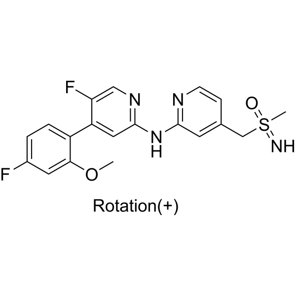
Enitociclib ((+)-BAY-1251152; (+)-VIP152) is a selective CDK9 inhibitor (IC50=3 nM) that inhibits transcriptional elongation by blocking Ser2/Ser5 phosphorylation of RNA polymerase II. Enitociclib specifically depletes key short-lived proteins such as c-MYC, MCL-1 and induces tumor cell apoptosis. Enitociclib also interferes with the production of enhancer RNAs (eRNA) and enhancer-promoter interactions, and downregulates oncogene expression at the epigenetic level. Enitociclib exerts synergistic effects with agents including Bortezomib (HY-10227), Lenalidomide (HY-A0003), Pomalidomide (HY-10984), Venetoclax (HY-15531) and Paclitaxel (HY-B0015), and even reverses paclitaxel resistance. Enitociclib serves as a vital research tool for various malignancies such as double-hit diffuse large B-cell lymphoma, multiple myeloma and pancreatic ductal adenocarcinoma.
For research use only. We do not sell to patients.
- Purity : 99.89%
- CAS No.: 1610408-97-3
- Formula: C19H18F2N4O2S
- Molecular Weight:404.43
-
Storage:Powder -20°C, 3 years , 4°C, 2 years ; In solvent -80°C, 2 years , -20°C, 1 year

To place orders, for customer services and technical support, please contact: MedChemExpress USA
Tel: 609-228-6898 E-mail: [email protected] [email protected]


-
 Biological Activity
-
Chemical Information
-
Solvent & Solubility
- Protocols
- Purity & Documentation
- References
-
 Help & FAQs


-
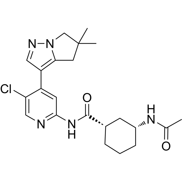
HY-112088
AZD4573CDK9 Inhibitor -
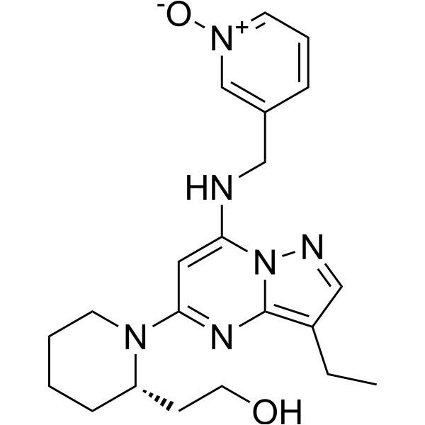
HY-10492
DinaciclibCDK2/5/9 Inhibitor -
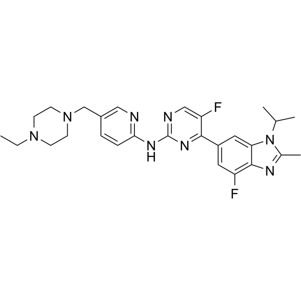
HY-16297A
AbemaciclibCDK4/6 Inhibitor -
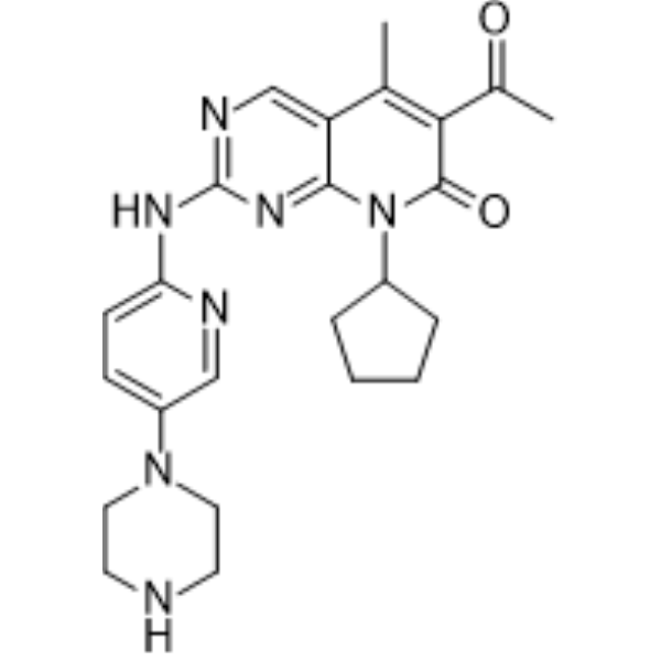
HY-50767
PalbociclibCDK4/6 Inhibitor -
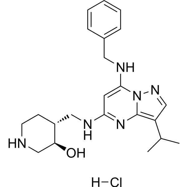
HY-103712A
Samuraciclib hydrochlorideCDK7 Inhibitor -
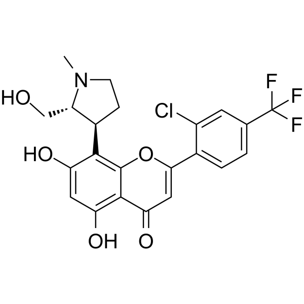
HY-12422
VoruciclibCDK Inhibitor -
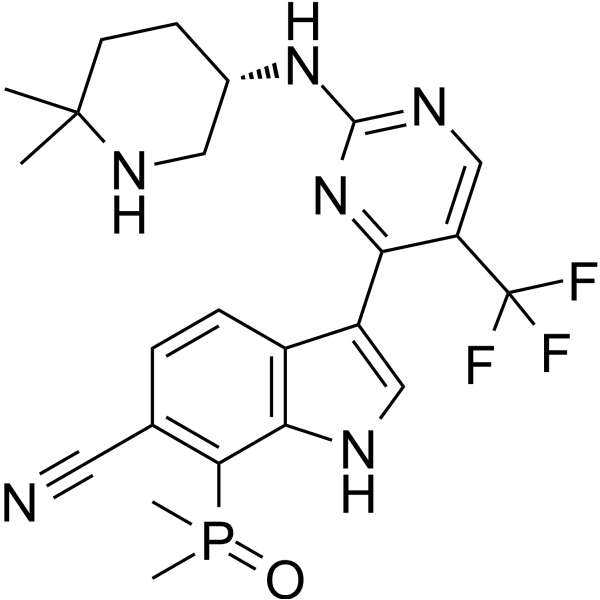
HY-138293
SY-5609CDK7 Inhibitor -
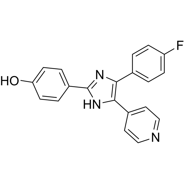
HY-10295
SB 202190p38 MAPK Inhibitor -
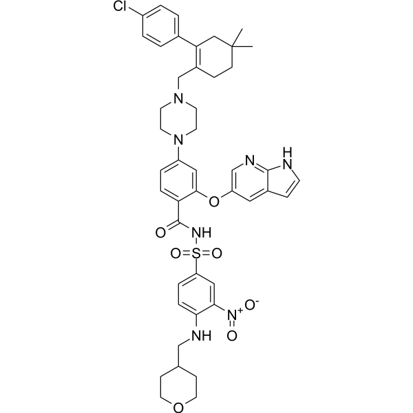
HY-15531
VenetoclaxBCL-2 Inhibitor -

HY-14648
DexamethasoneGR Agonist -
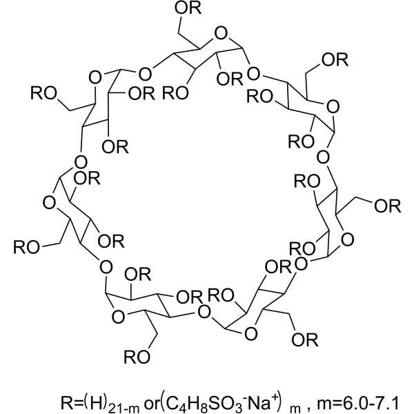
HY-17031
SBE-β-CDExcipient -
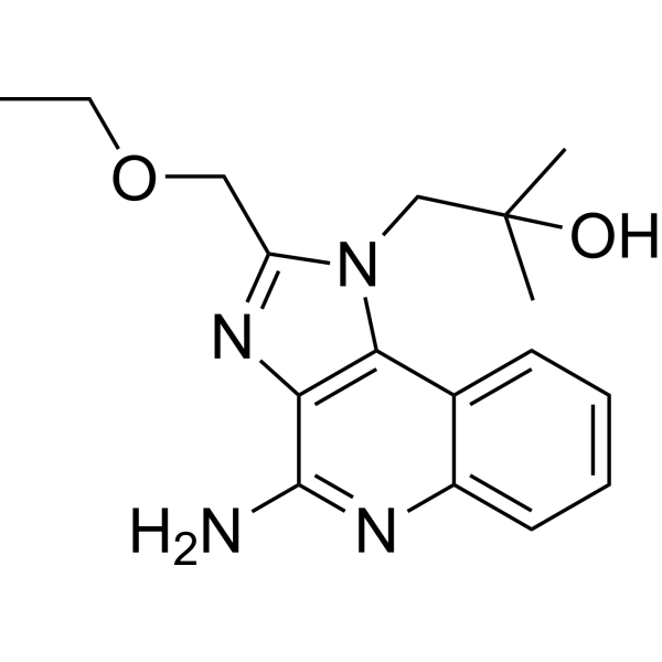
HY-13740
ResiquimodTLR7/8 Agonist
Publications Citing Use of MedChemExpress (MCE) Enitociclib
More Customer Validation & Images

-
Cell Proliferation/Viability Assay

-
WB

-
RT-PCR

-
In Vivo Efficacy Study

-
Cell Proliferation/Viability Assay
All DNA/RNA Synthesis Isoforms
More

Biological Activity
Description
IC50 & Target
|
RNA Polymerase |
CDK9/CycT1 3 nM (IC50) |
Cellular Effect
|
Cell Line

|
Type | Value | Description | References |
|---|---|---|---|---|
| A2780 | IC50 |
52 nM
Compound: 32
|
Antiproliferative activity against human A2780 cells assessed as inhibition of cell growth in presence of fetal calf serum incubated for 96 hrs measured by celltiter-glo luminescent cell viability assay
Antiproliferative activity against human A2780 cells assessed as inhibition of cell growth in presence of fetal calf serum incubated for 96 hrs measured by celltiter-glo luminescent cell viability assay
|
[PMID: 34264057] |
| MOLM-13 | IC50 |
29 nM
Compound: 13; VIP152
|
Antiproliferative activity against human MOLM-13 cells assessed as reduction in cell viability incubated for 72 hrs by MTT assay
Antiproliferative activity against human MOLM-13 cells assessed as reduction in cell viability incubated for 72 hrs by MTT assay
|
[PMID: 38564299] |
| MOLM-13 | IC50 |
29 nM
Compound: 8; BAY-1251152
|
Antiproliferative activity against human MOLM-13 cells
Antiproliferative activity against human MOLM-13 cells
|
[PMID: 32866383] |
| MOLM-13 | IC50 |
42 nM
Compound: 32
|
Antiproliferative activity against human MOLM-13 cells assessed as inhibition of cell growth in presence of fetal calf serum incubated for 96 hrs measured by celltiter-glo luminescent cell viability assay
Antiproliferative activity against human MOLM-13 cells assessed as inhibition of cell growth in presence of fetal calf serum incubated for 96 hrs measured by celltiter-glo luminescent cell viability assay
|
[PMID: 34264057] |
| T-cell | IC50 |
3 nM
Compound: 53; BAY-1251152
|
Inhibition of CDK9/Cyclin T (unknown origin)
Inhibition of CDK9/Cyclin T (unknown origin)
|
[PMID: 35485642] |
In Vitro
Enitociclib effectively reduces the viability of a panel of human lymphoma cell lines, including the DLBCL cell lines SU-DHL-4 (IC50=43 nmol/L) and SU-DHL-10 (IC50=74 nmol/L)[1].
Enitociclib (0.25-1 μM; 4 h) induces transcriptomic changes dominated by significant downregulation in SU-DHL-4 and SU-DHL-10 DLBCL cell lines, including marked downregulation of novel targets PHF23 and TP53RK, as well as enrichment of the RNA polymerase II-mediated transcription pathway[1].
Enitociclib (0.6-800 nM; 6-48 h) potently inhibits the viability of primary mantle cell lymphoma (MCL) cells, MCL cell lines, and diffuse large B-cell lymphoma (DLBCL) cell lines, and induces caspase-3-dependent apoptosis in these cells, with an IC50 of 11.5-172.3 nM[2].
Enitociclib (12.5-200 nM; 96 h) significantly reduces the cell viability of multiple myeloma cell lines MM1.S, NCI-H929, OPM-2 and U266B1, with an IC50 ranging from 36 to 78 nM after 96 hours of treatment[3].
Enitociclib (0.5-1 μM; 1-24 h) inhibits phosphorylation of RNA Pol II CTD at Ser2/Ser5 sites in a dose- and time-dependent manner, induces apoptosis, and suppresses oncoprotein expression in NCI-H929 and OPM-2 multiple myeloma cell lines[3].
Enitociclib (25-500 nM; combined with paclitaxel) synergistically inhibits the growth of paclitaxel-resistant L3.6pl pancreatic cancer cells[4].
MedChemExpress (MCE) has not independently confirmed the accuracy of these methods. They are for reference only. Further protocols information, click here.
-
Cell Line:SU-DHL-4, SU-DHL-10 (DLBCL) cell lines
-
Concentration:0.25 μmol/L; 1 μmol/L
-
Incubation Time:4 h (treatment); up to 48 h post-washout (SU-DHL-4); up to 24 h post-washout (SU-DHL-10)
-
Result:Decreased p-Ser2 levels by 50% for up to 48 hours post-washout in SU-DHL-4 cells treated with 0.25 μmol/L.
Caused a greater extent of p-Ser2 inhibition that sustained for 48 hours post-washout in SU-DHL-4 cells treated with 1 μmol/L.
Achieved a 50% reduction of p-Ser2 for up to 24 hours post-washout in SU-DHL-10 cells treated with 1 μmol/L.
-
Cell Line:SU-DHL-4, SU-DHL-10 (DLBCL) cell lines
-
Concentration:0.25 μmol/L; 1 μmol/L
-
Incubation Time:4 h (treatment); up to 48 h post-washout
-
Result:Inhibited MYC and MCL1 mRNA levels by approximately 75% for 16 to 48 hours post-washout in SU-DHL-10 cells.
Maintained durable depletion of MYC and MCL1 mRNA levels over the 48-hour time course in SU-DHL-4 cells treated with 1 μmol/L.
-
Cell Line:MM1.S, NCI-H929, OPM-2, U266B1 multiple myeloma cell lines
-
Concentration:12.5-50 nM (in combination with other agents)
-
Incubation Time:96 h
-
Result:Exhibited synergistic effects (ZIP synergy score >10) with bortezomib in OPM-2 cells at 50 nM enitociclib and 1 nM bortezomib.
Showed synergistic effects with lenalidomide in MM1.S, NCI-H929, and U266B1 cells at 12.5 to 25 nM enitociclib and 313 nM to 5 μM lenalidomide.
Demonstrated synergistic effects with pomalidomide in NCI-H929 and U266B1 cells at 12.5 nM enitociclib and 12.5 to 50 nM pomalidomide.
In Vivo
Enitociclib (10 mg/kg; i.v.; twice a week) potently inhibits tumor growth and prolongs survival in JeKo-1 MCL cell line-derived xenografts in NSG mice (p < 0.0001 for both endpoints) without significant toxicity[2].
Enitociclib (15 mg/kg; i.v.; once per week) significantly reduces JJN-3 multiple myeloma tumor volumes in SCID/Beige mice, and a single dose (15 mg/kg; i.v.) transiently suppresses oncogene transcription and induces tumor apoptosis[3].
Enitociclib (15 mg/kg; i.v.; once per week) significantly reduces OPM-2 multiple myeloma tumor volumes in NOD/SCID mice, and enhances efficacy when combined with lenalidomide or bortezomib[3].
Enitociclib (10 mg/kg; i.v.; once weekly; 4 weeks) does not reduce tumor size as a single agent, but in combination with paclitaxel, it induces significant tumor shrinkage in a paclitaxel-resistant PDAC patient-derived xenograft model[4].
MedChemExpress (MCE) has not independently confirmed the accuracy of these methods. They are for reference only.
-
Animal Model:C.B-17 SCID (female, 10-12 weeks old at study start)[1]
-
Dosage:5 mg/kg (mechanistic single dose); 10 mg/kg (i.v. once weekly for 3 weeks; mechanistic single dose); 15 mg/kg (i.v. once weekly for 3 weeks; mechanistic single dose)
-
Administration:i.v.; single dose (mechanistic studies); once weekly; 3 weeks (antitumor efficacy)
-
Result:Achieved a tumor growth inhibition (T/C) value of 0.19 on day 16/20, with sustained tumor growth control at 10 mg/kg once weekly.
Induced complete tumor regression during the 3-week treatment period, with a T/C value of 0.005 on day 16/20 at 15 mg/kg once weekly; tumors regrew after treatment cessation, but a second cycle of 15 mg/kg once weekly dosing induced tumor regression again.
Reduced RNA polymerase II Ser2 phosphorylation (p-Ser2) by >50% for 8 hours postdose; depleted MYC and MCL1 mRNA levels by 4 hours postdose, with depletion sustained for 8 hours at 5 mg/kg single dose.
Depleted p-Ser2 levels within 8 hours postdose; depleted MYC and MCL1 mRNA levels by 4 hours postdose, with depletion sustained for 8 hours; reduced Myc protein levels by 50% and Mcl-1 protein levels robustly within 4-8 hours postdose; increased cleaved PARP (apoptosis marker) levels postdose at 10 mg/kg and 15 mg/kg single dose.
-
Animal Model:NSG (NOD.Cg-Prkdcscid Il2rgtm1Wjl/SzJ) (immunodeficient)[2]
-
Dosage:10 mg/kg
-
Administration:i.v.; twice a week
-
Result:Markedly inhibited tumor growth.
Significantly prolonged mouse survival.
Caused no significant body weight loss or other apparent adverse effects.\nEfficaciously inhibited PDX-1 tumor growth.
Caused no significant body weight loss.\nEfficaciously inhibited PDX-2 tumor growth.
Caused no significant body weight loss.\nEfficaciously inhibited PDX-3 tumor growth.
Caused no significant body weight loss.
-
Animal Model:NOD/SCID (gender not specified; injected subcutaneously with 5.0×10⁶ OPM-2 MM cells)[3]
-
Dosage:15 mg/kg (monotherapy); 15 mg/kg (in combination with lenalidomide or bortezomib)
-
Administration:i.v.; once per week
-
Result:Significantly reduced OPM-2 tumor volumes compared with vehicle-treated mice as monotherapy.
Further enhanced antitumor efficacy compared with lenalidomide or bortezomib alone when combined with 50 mg/kg Lenalidomide (p.o.
daily) or 0.8 mg/kg Bortezomib (i.p.
twice weekly).


Chemical Information
-
CAS No. 1610408-97-3
-
Appearance Solid
-
Molecular Weight 404.43
-
Formula C19H18F2N4O2S
-
Color Off-white to yellow
-
SMILES
COC1=C(C2=C(F)C=NC(NC3=CC(C[S@@](=O)(C)=N)=CC=N3)=C2)C=CC(F)=C1
-
Synonyms
(+)-BAY-1251152; (+)-VIP152; (S)-Enitociclib
-
Shipping
Room temperature in continental US; may vary elsewhere.
-
Storage
Powder -20°C 3 years 4°C 2 years In solvent -80°C 2 years -20°C 1 year


Publications (2)
-
Journal Impact Factor
-
Most Recent
-
Acta Pharm Sin B
2023 Sep;13(9):3694-3707. PMID: 37719386
Enitociclib purchased from MedChemExpress. Usage Cited in: Acta Pharm Sin B. 2023 Sep;13(9):3694-3707. [Abstract]
The anti-proliferative activity of BAY1251152 (Enitociclib) against parental MOLM13 and MOLM13-BR cell lines was assessed using the CellTiter-Glo assay kit.
Enitociclib purchased from MedChemExpress. Usage Cited in: Acta Pharm Sin B. 2023 Sep;13(9):3694-3707. [Abstract]
After 3 hours of treatment with BAY1251152 (Enitociclib), changes in the phosphorylation levels of RNA Pol II and MYC/MCL were detected by Western blot.
Enitociclib purchased from MedChemExpress. Usage Cited in: Acta Pharm Sin B. 2023 Sep;13(9):3694-3707. [Abstract]
After treatment with 1 μM BAY1251152 (Enitociclib) for 6 hours, changes in MCL1 and MYC gene expression levels were measured in HeLa and HeLa CDK9 L156F cells.
Enitociclib purchased from MedChemExpress. Usage Cited in: Acta Pharm Sin B. 2023 Sep;13(9):3694-3707. [Abstract]
Relative tumor sizes in each group were measured following intravenous injection of IHMT-CDK9-36 or BAY1251152 (Enitociclib: 8 mg/kg, Q2D).
-
Oncotarget
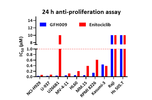
The pharmacodynamic and mechanistic foundation for the antineoplastic effects of GFH009, a potent and highly selective CDK9 inhibitor for the treatment of hematologic malignancies. [Abstract]2023 Dec 20:14:997-1008. PMID: 38117531
Enitociclib purchased from MedChemExpress. Usage Cited in: Oncotarget. 2023 Dec 20:14:997-1008. [Abstract]
Cell viability IC50s for GFH009 on human hematological malignancy-derived cell lines after 24 hours of exposure to the agent were determined using CellTiter Glo.


Solvent & Solubility
In Vitro:
DMSO : 100 mg/mL (247.26 mM; Need ultrasonic; Hygroscopic DMSO has a significant impact on the solubility of product, please use newly opened DMSO)
Please refer to the solubility information to select the appropriate solvent. Once prepared, please aliquot and store the solution to prevent product inactivation from repeated freeze-thaw cycles.
Storage method and period of stock solution: -80°C, 2 years; -20°C, 1 year. When stored at -80°C, please use it within 2 years. When stored at -20°C, please use it within 1 year.
Please refer to the solubility information to select the appropriate solvent. Once prepared, please aliquot and store the solution to prevent product inactivation from repeated freeze-thaw cycles.
Storage method and period of stock solution: -80°C, 2 years; -20°C, 1 year. When stored at -80°C, please use it within 2 years. When stored at -20°C, please use it within 1 year.
Concentration (start) × Volume (start) = Concentration (final) × Volume (final)
In Vivo:
Select the appropriate dissolution method based on your experimental animal and administration route.
- For the following dissolution methods, please ensure to first prepare a clear stock solution using an In Vitro approach and then sequentially add co-solvents:
- To ensure reliable experimental results, the clarified stock solution can be appropriately stored based on storage conditions. As for the working solution for In Vivo experiments, it is recommended to prepare freshly and use it on the same day.
- The percentages shown for the solvents indicate their volumetric ratio in the final prepared solution. If precipitation or phase separation occurs during preparation, heat and/or sonication can be used to aid dissolution.
Add each solvent one by one: 10% DMSO 40% PEG300 5% Tween-80 45% Saline
Solubility: ≥ 2.5 mg/mL (6.18 mM); Clear solution
This protocol yields a clear solution of ≥ 2.5 mg/mL (saturation unknown).
Taking 1 mL working solution as an example, add 100 μL DMSO stock solution (25.0 mg/mL) to 400 μL PEG300, and mix evenly; then add 50 μL Tween-80 and mix evenly; then add 450 μL Saline to adjust the volume to 1 mL.
Preparation of Saline: Dissolve 0.9 g sodium chloride in ddH₂O and dilute to 100 mL to obtain a clear Saline solution.
Add each solvent one by one: 10% DMSO 90% (20% SBE-β-CD in Saline)
Solubility: ≥ 2.5 mg/mL (6.18 mM); Clear solution
This protocol yields a clear solution of ≥ 2.5 mg/mL (saturation unknown).
Taking 1 mL working solution as an example, add 100 μL DMSO stock solution (25.0 mg/mL) to 900 μL 20% SBE-β-CD in Saline, and mix evenly.
Preparation of 20% SBE-β-CD in Saline (4°C, storage for one week): 2 g SBE-β-CD powder is dissolved in 10 mL Saline, completely dissolve until clear.
In Vivo Dissolution Calculator
Please enter the basic information of animal experiments:
-
-
-
-
Recommended: Prepare an additional quantity of animals to account for potential losses during experiments.
Please enter your animal formula composition:
-
%DMSO +
Recommended: Keep the proportion of DMSO in working solution below 2% if your animal is weak.
-
%+
-
+%Tween-80 + +
-
%Saline +
The co-solvents required include: DMSO, . All of co-solvents are available by MedChemExpress (MCE). , Tween 80. All of co-solvents are available by MedChemExpress (MCE).
Working solution concentration: 0.22 mg/mL
Method for preparing stock solution: mg drug dissolved in μL DMSO. Stock solution concentration: mg/mL.
1. Take μL DMSO stock solution;
2. Add μL .
μL , mix evenly;
3. Then add μL Tween 80, mix evenly;
4. Then add μL
Please ensure that the stock solution in the first step is dissolved to a clear state, and add co-solvents in sequence. You can use ultrasonic heating (ultrasonic cleaner, recommended frequency 20-40 kHz), vortexing, etc. to assist dissolution.

Protocols
-
Kinase activity and phosphorylation assays
Kinase activity assays measure the ability of kinases to transfer phosphate groups from ATP to specific substrates, while phosphorylation assays detect the presence and levels of phosphorylated proteins. Common methods include radiolabeled ATP incorporation (e. g. ,), ADP release detection via bioluminescence (e. g. ,[3]), enzyme-linked immunosorbent assays (ELISA) for phospho-specific epitopes (e. g. ,[6]), and microtiter-based formats for high-throughput screening (e. g. ,[8]). The ADP-Glo assay quantifies kinase activity by measuring ADP produced during phosphorylation using a luciferase-based system. Radiometric assays involve autoradiography or scintillation counting after incorporation of 32P-labeled ATP into substrate proteins. ELISA-based approaches rely on phospho-specific antibodies to detect activated kinases in cell lysates or purified samples.
-
Western Blot
Western blotting (WB) is a commonly used experimental method in molecular biology, biochemistry, and immunogenetics for identifying and quantifying target proteins. It combines gel electrophoresis with immunoassay, enabling researchers to analyze protein expression, post-translational modifications, and molecular weight.
-
RNA extraction experimental
By lysing cells, releasing RNA, and removing impurities such as proteins and DNA, high-purity RNA products are finally obtained. The commonly used traditional method is the guanidine isothiocyanate/phenol/chloroform method (Trizol), which is suitable for a variety of animal materials including animal tissues, microorganisms, cultured cells, etc., and most plant materials.
-
Apoptosis
Apoptosis, also called programmed cell death, is generally characterized by distinct morphological characteristics.
-
TUNEL staining for apoptotic DNA fragmentation
TUNEL staining detects DNA strand breaks by using terminal deoxynucleotidyl transferase to add labeled nucleotides to exposed 3′-OH DNA termini, generating either microscopic staining in fixed cells or tissue sections, or fluorescence/cytometric signal in cell suspensions. TUNEL positivity reflects DNA fragmentation but should not be interpreted alone as definitive apoptosis, because TUNEL can also label necrotic, autolytic, mechanically damaged, or DNA-repair-associated DNA breaks.
-
Cell Cytotoxicity Assay
Cytotoxicity assays are usually based on the assessment of cell membrane damage, which can also be indirectly detected by measuring cell viability. Detection methods include MTT assay, CKK-8 assay, LDH assay and ATP assay, etc.
-
Annexin V plus membrane-impermeant dye apoptosis staining
Annexin V-based apoptosis assays rely on the detection of phosphatidylserine (PS) externalization from the inner leaflet of the plasma membrane to the outer leaflet, an early biochemical hallmark of apoptosis. Fluorescently labeled Annexin V binds PS in a calcium-dependent manner, enabling identification of early apoptotic cells by flow cytometry or fluorescence microscopy. When combined with a membrane-impermeant DNA-binding dye (e. g. , propidium iodide), this approach allows discrimination between viable (Annexin V−/dye−), early apoptotic (Annexin V+/dye−), and late apoptotic or necrotic (Annexin V+/dye+) cell populations by assessing membrane integrity and PS exposure.
-
Apoptosis Solutions
Apoptosis is a regulated, generally non-lytic cell-death pathway that removes unwanted, damaged, infected, or abnormal cells through coordinated morphological changes, caspase activation, DNA fragmentation, and membrane remodeling. The intrinsic apoptosis pathway is controlled mainly by mitochondrial outer membrane permeabilization, BCL-2 family proteins, cytochrome c release, apoptosome formation, caspase-9 activation, and downstream executioner caspase-3/7 activation. The extrinsic apoptosis pathway is initiated by death receptors such as Fas, TNFR, and TRAIL receptors, which recruit adaptor proteins and activate caspase-8 before engaging executioner caspases or mitochondrial amplification through BID cleavage. Apoptosis is linked to many phenotypes, including cancer cell killing, tissue homeostasis, immune regulation, neurodegeneration, infection response, and treatment-induced cytotoxicity; unresolved questions include how apoptosis interacts with necroptosis, pyroptosis, ferroptos
-
Protocol for Kinase activity and phosphorylation assays
Kinase activity assays measure transfer of phosphate from ATP to a protein or peptide substrate, generating phosphorylated substrate, ADP, or incorporated radiolabeled phosphate as the readout; phosphorylation assays measure site-specific phosphorylation in cells or tissues as a proxy for kinase-pathway activation, inhibition, or substrate regulation. Phosphorylation can be detected by phospho-specific Western blot, immunoprecipitation kinase assay, phospho-immunofluorescence, phospho-flow cytometry, luminescent ADP detection, radiolabeled ATP incorporation, or reporter-based pathway assays, and these readouts can be applied to cancer cells, primary neurons, mouse tumors, organoids, inflammatory macrophages, ferroptosis studies, and mitophagy studies when the kinase target is biologically relevant.


Purity & Documentation
-
Data Sheet (286 KB)
-
SDS (254 KB)
- English - EN (254 KB)
- Français - FR (254 KB)
- Deutsch - DE (254 KB)
- Norwegian - NO (254 KB)
- Español - ES (254 KB)
- Swedish - SV (254 KB)
- Italian - IT (254 KB)
- Korean - KR (254 KB)
- Portuguese - PT (254 KB)
-
Handling Instructions (2659 KB)
References
[1]. Frigault MM, et al. Enitociclib, a Selective CDK9 Inhibitor, Induces Complete Regression of MYC+ Lymphoma by Downregulation of RNA Polymerase II Mediated Transcription. Cancer Res Commun. 2023;3(11):2268-2279. [Content Brief]
[2]. Jiang V, et al. The CDK9 inhibitor enitociclib overcomes resistance to BTK inhibition and CAR-T therapy in mantle cell lymphoma. Biomark Res. 2024;12(1):62. Published 2024 Jun 18. [Content Brief]
[3]. Tran S, et al. Enitociclib, a selective CDK9 inhibitor: in vitro and in vivo preclinical studies in multiple myeloma. Blood Neoplasia. 2024;2(1):100050. Published 2024 Oct 24. [Content Brief]
[4]. Hamdan FH, et al. Interactive enhancer hubs (iHUBs) mediate transcriptional reprogramming and adaptive resistance in pancreatic cancer. Gut. 2023;72(6):1174-1185. [Content Brief]


Complete Stock Solution Preparation Table
Please refer to the solubility information to select the appropriate solvent. Once prepared, please aliquot and store the solution to prevent product inactivation from repeated freeze-thaw cycles.
Storage method and period of stock solution: -80°C, 2 years; -20°C, 1 year. When stored at -80°C, please use it within 2 years. When stored at -20°C, please use it within 1 year.
| Optional Solvent | Concentration Solvent Mass | 1 mg | 5 mg | 10 mg | 25 mg |
|---|---|---|---|---|---|
| DMSO | 1 mM | 2.4726 mL | 12.3631 mL | 24.7262 mL | 61.8154 mL |
| 5 mM | 0.4945 mL | 2.4726 mL | 4.9452 mL | 12.3631 mL | |
| 10 mM | 0.2473 mL | 1.2363 mL | 2.4726 mL | 6.1815 mL | |
| 15 mM | 0.1648 mL | 0.8242 mL | 1.6484 mL | 4.1210 mL | |
| 20 mM | 0.1236 mL | 0.6182 mL | 1.2363 mL | 3.0908 mL | |
| 25 mM | 0.0989 mL | 0.4945 mL | 0.9890 mL | 2.4726 mL | |
| 30 mM | 0.0824 mL | 0.4121 mL | 0.8242 mL | 2.0605 mL | |
| 40 mM | 0.0618 mL | 0.3091 mL | 0.6182 mL | 1.5454 mL | |
| 50 mM | 0.0495 mL | 0.2473 mL | 0.4945 mL | 1.2363 mL | |
| 60 mM | 0.0412 mL | 0.2061 mL | 0.4121 mL | 1.0303 mL | |
| 80 mM | 0.0309 mL | 0.1545 mL | 0.3091 mL | 0.7727 mL | |
| 100 mM | 0.0247 mL | 0.1236 mL | 0.2473 mL | 0.6182 mL |


Enitociclib Related Classifications
HY-103019 Related Classifications
 Powered by Bioz
Keywords
- Enitociclib
- 1610408-97-3
- (+)-BAY-1251152
- (+)-VIP152
- (S)-Enitociclib
- Drug Isomer
- CDK
- Apoptosis
- DNA/RNA Synthesis
- multiple myeloma
- mantle cell lymphoma
- MCL-1
- MYC-positive non-Hodgkin lymphoma
- caspase-3
- double-hit diffuse large B-cell lymphoma
- CDK9
- c-MYC
- RNA polymerase II
- cyclin-dependent kinase 9
- Inhibitor
- inhibitor
- inhibit