



Sodium propionate
Based on 2 publication(s) in Google Scholar
Sodium propionate is an orally active short-chain fatty acid. Sodium propionate can be produced by intestinal bacteria from the metabolism of dietary fiber. Sodium propionate increases PPAR-γ, inhibits NF-κB activation, and reduces COX-2 expression and NO production. Sodium propionate also induces Apoptosis and Autophagy. Sodium propionate reduces HSV-1-induced keratitis. Sodium propionate has anticancer effects against glioblastoma. Sodium propionate exhibits neuroprotective, antioxidant, and anti-inflammatory activities. Sodium propionate can be used in the research of spinal cord injury and Alzheimer's disease.
For research use only. We do not sell to patients.
- Assay : 99.55%
- CAS No.: 137-40-6
- Formula: C3H5NaO2
- Molecular Weight:96.06
-
Storage:
4°C, sealed storage, away from moisture
* In solvent : -80°C, 6 months; -20°C, 1 month (sealed storage, away from moisture)


To place orders, for customer services and technical support, please contact: MedChemExpress USA
Tel: 609-228-6898 E-mail: [email protected] [email protected]


-
 Biological Activity
-
Chemical Information
-
Solvent & Solubility
- Protocols
- Purity & Documentation
- References
-
 Help & FAQs


-
HY-B0350A
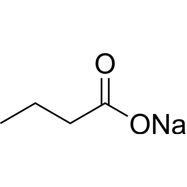
Sodium butyrateHDAC Inhibitor -
HY-Y1325H
Sodium acetate trihydrate, meets analytical specification of Ph. Eur. BP USP FCC E262, ≤0.00002% AlInorganic salt -

HY-Y1325I
Sodium acetate trihydrate, 99.5%Inorganic salt -
HY-W010452
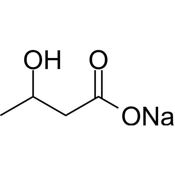
3-Hydroxybutyric acid sodium -
HY-10585
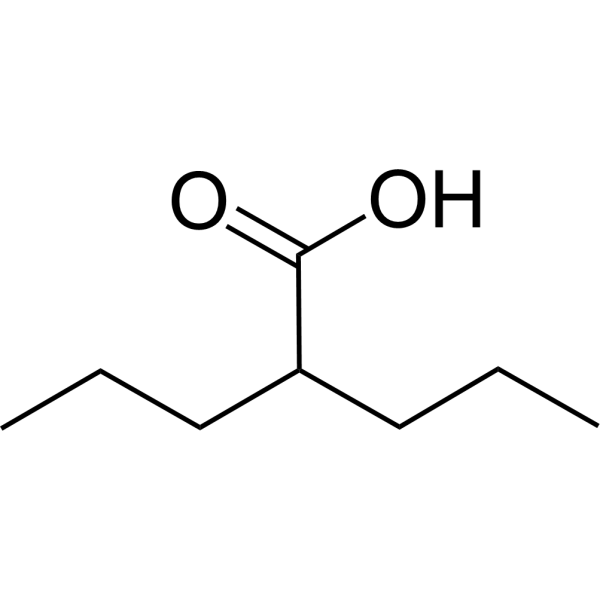
Valproic acidHDAC Inhibitor -
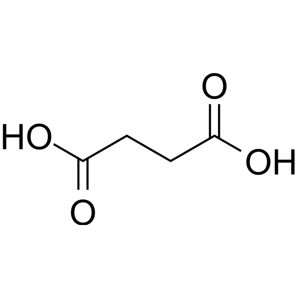
HY-N0420
Succinic acidFood Additive, Platform chemical -
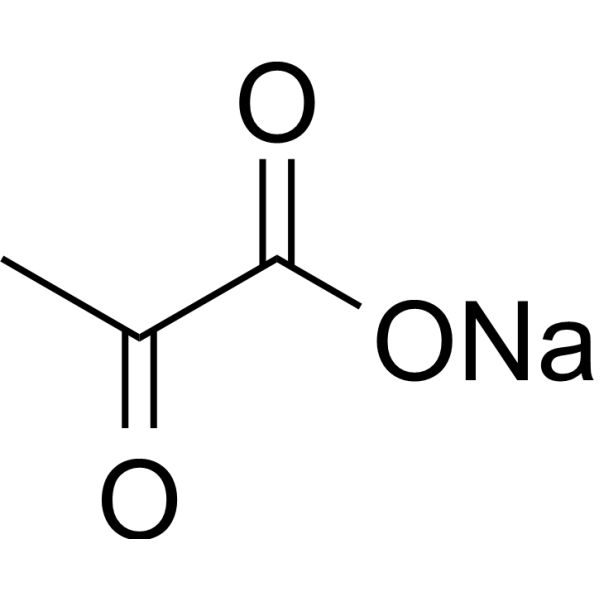
HY-W015913
Pyruvic acid sodiumGlycolysis Intermediate -
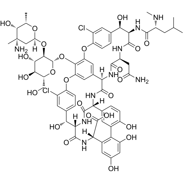
HY-B0671
VancomycinAntibiotic -
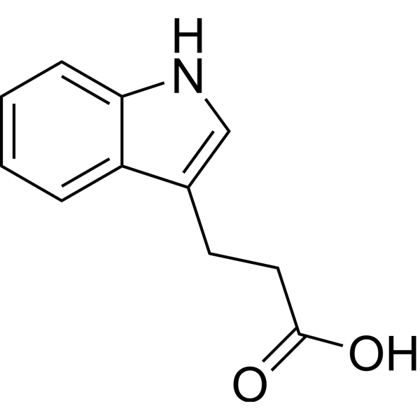
HY-W015229
3-Indolepropionic acid -
HY-113099
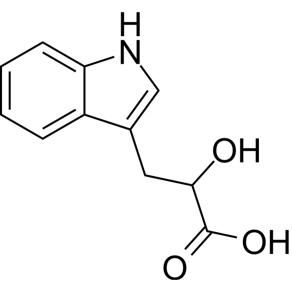
Indolelactic acidTryptophan Catabolite -

HY-15763
ErastinFerroptosis Inducer -

HY-B1773AR
Sodium propionate (Standard)Analytical Standard
Publications Citing Use of MedChemExpress (MCE) Sodium propionate
More Customer Validation & Images

-
WB
-
Cell Imaging/Staining

-
IHC
-
WB
-
In Vivo Efficacy Study
All Endogenous Metabolite Isoforms
More

Biological Activity
Description
|
PPAR-γ |
HSV-1 |
COX-2 |
In Vitro
Sodium propionate (10-100 mM; 24 h for viability test) inhibits viability of GBM cell lines (U-87, A-172, U-138) in a concentration-dependent manner, induces apoptosis and autophagy in U-87 cells[1].
Sodium propionate (1-10 μM) shows potential anti-oxidant effect in J774-A1 cell cultures stimulated with hydrogen peroxide[2].
Sodium propionate (0.1 μM-10 mM) protects SH-SY5Y neuroblastoma cells from Aβ1-42-induced neurotoxicity by inhibiting NF-κB activation, reducing iNOS/COX-2 expression and NO production[3].
Sodium propionate (100 μM-3.2 mM) inhibits differentiation of naive splenocytes into Th1/Th17 cells and promotes Treg differentiation[5].
MedChemExpress (MCE) has not independently confirmed the accuracy of these methods. They are for reference only. Further protocols information, click here.
-
Cell Line:U-87
-
Concentration:50 mM, 100 mM
-
Incubation Time:
-
Result:Induced apoptosis.
Upregulated p53, Bax, and caspase-3 protein levels.
Downregulated Bcl-2 protein level.
-
Cell Line:U-87
-
Concentration:50 mM, 100 mM
-
Incubation Time:
-
Result:Activated autophagy.
Upregulated LC3II/I, Beclin-1, and Atg5 protein levels.
Downregulated p62 protein level.
In Vivo
Sodium propionate (10-100 mg/kg; p.o.; 30 min before CAR subplantar injection) reduces Carrageenan (HY-125474)-induced paw edema and KO₂-induced pain/inflammation in Sprague-Dawley rats by inhibiting iNOS/COX-2 and enhancing antioxidant enzymes[2].
Sodium propionate (10-100 mg/kg; p.o.; 1 h and 6 h after injury) alleviates spinal cord trauma (SCI) in CD1 mice by reducing neuroinflammation, lipid peroxidation, and improving motor function [3].
Sodium propionate (500 mM; p.o. via drinking water; 3 weeks before infection to experiment end) reduces HSV-1-induced ocular stromal keratitis (SK) in C57BL/6 mice by regulating T cell balance (increasing Treg/Th1/Th17 ratio) and reducing inflammation[5].
MedChemExpress (MCE) has not independently confirmed the accuracy of these methods. They are for reference only.
-
Animal Model:BALB/c nude male mice (25-30 g; 6-8 weeks old) bearing U-87 GBM xenografts[1]
-
Dosage:10 mg/kg, 30 mg/kg, 100 mg/kg
-
Administration:oral gavage, 2 weeks
-
Result:Decreased tumor necrosis and neutrophil infiltration.
Reduced Ki-67 (proliferation marker) expression.
Induced apoptosis and autophagy.
Clinical Trial
| NCT Number | Sponsor | Condition | Start Date |
Phase

|
|---|---|---|---|---|
| NCT01329991 | Plexxikon| | 2011-05 | PHASE1 |


Chemical Information
-
CAS No. 137-40-6
-
Appearance Solid
-
Molecular Weight 96.06
-
Formula C3H5NaO2
-
Color White to off-white
-
SMILES
O=C(CC)O[Na]
-
Shipping
Room temperature in continental US; may vary elsewhere.
-
Storage
4°C, sealed storage, away from moisture
* In solvent : -80°C, 6 months; -20°C, 1 month (sealed storage, away from moisture)


Publications (2)
-
Journal Impact Factor
-
Most Recent
-
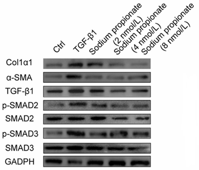
Sodium propionate purchased from MedChemExpress. Usage Cited in: Life Metab. 2025 Oct 8.
Sodium propionate (2-8 nM; 24 h). Fibrosis-related markers such as TIMP-1, α-SMA, and Col1α1 and TGF-β1 protein expression assayed by western blotting.
Sodium propionate purchased from MedChemExpress. Usage Cited in: Life Metab. 2025 Oct 8.
Sodium propionate (2-8 nM; 24 h). ROS visualized by microscopy using the 2’,7’-dichlorodihydrofluorescein diacetate (DCFH-DA) probe at a final concentration of 10 μmol/L.
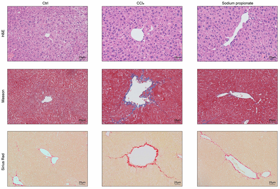
Sodium propionate purchased from MedChemExpress. Usage Cited in: Life Metab. 2025 Oct 8.
Sodium propionate (400 mM; drinking water). Representative images of H&E, Masson, and Sirius Red staining (Scale bar = 25 μm) in the liver.
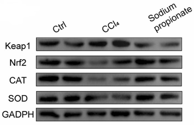
Sodium propionate purchased from MedChemExpress. Usage Cited in: Life Metab. 2025 Oct 8.
Sodium propionate (400 mM; drinking water). Oxidative stress-related Keap1/Nrf2 pathway protein expression assayed by western blotting.
-
iScience
Biglycan regulated colorectal cancer progress by modulating enteric neuron-derived IL-10 and abundance of Bacteroides thetaiotaomicron. [Abstract]2023 Aug 2;26(9):107515. PMID: 37664615
Sodium propionate purchased from MedChemExpress. Usage Cited in: iScience. 2023 Aug 2;26(9):107515. [Abstract]
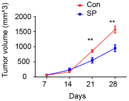
Sodium propionate (SP; 100 mM; 100 μL; intratumoral injection). Sodium propionate reduced the growth of MC38 allograft tumors.


Solvent & Solubility
In Vitro:
H2O : ≥ 200 mg/mL (2082.03 mM)
DMSO : 2 mg/mL (20.82 mM; ultrasonic and warming and heat to 60°C; Hygroscopic DMSO has a significant impact on the solubility of product, please use newly opened DMSO)
* "≥" means soluble, but saturation unknown.
Please refer to the solubility information to select the appropriate solvent. Once prepared, please aliquot and store the solution to prevent product inactivation from repeated freeze-thaw cycles.
Storage method and period of stock solution: -80°C, 6 months; -20°C, 1 month (sealed storage, away from moisture). When stored at -80°C, please use it within 6 months. When stored at -20°C, please use it within 1 month.
* Note: If you choose water as the stock solution, please dilute it to the working solution, then filter and sterilize it with a 0.22 μm filter before use.
Please refer to the solubility information to select the appropriate solvent. Once prepared, please aliquot and store the solution to prevent product inactivation from repeated freeze-thaw cycles.
Storage method and period of stock solution: -80°C, 6 months; -20°C, 1 month (sealed storage, away from moisture). When stored at -80°C, please use it within 6 months. When stored at -20°C, please use it within 1 month.
* Note: If you choose water as the stock solution, please dilute it to the working solution, then filter and sterilize it with a 0.22 μm filter before use.
Concentration (start) × Volume (start) = Concentration (final) × Volume (final)

Protocols
-
RNA extraction experimental
By lysing cells, releasing RNA, and removing impurities such as proteins and DNA, high-purity RNA products are finally obtained. The commonly used traditional method is the guanidine isothiocyanate/phenol/chloroform method (Trizol), which is suitable for a variety of animal materials including animal tissues, microorganisms, cultured cells, etc., and most plant materials.
-
Apoptosis
Apoptosis, also called programmed cell death, is generally characterized by distinct morphological characteristics.
-
TUNEL staining for apoptotic DNA fragmentation
TUNEL staining detects DNA strand breaks by using terminal deoxynucleotidyl transferase to add labeled nucleotides to exposed 3′-OH DNA termini, generating either microscopic staining in fixed cells or tissue sections, or fluorescence/cytometric signal in cell suspensions. TUNEL positivity reflects DNA fragmentation but should not be interpreted alone as definitive apoptosis, because TUNEL can also label necrotic, autolytic, mechanically damaged, or DNA-repair-associated DNA breaks.
-
Annexin V plus membrane-impermeant dye apoptosis staining
Annexin V-based apoptosis assays rely on the detection of phosphatidylserine (PS) externalization from the inner leaflet of the plasma membrane to the outer leaflet, an early biochemical hallmark of apoptosis. Fluorescently labeled Annexin V binds PS in a calcium-dependent manner, enabling identification of early apoptotic cells by flow cytometry or fluorescence microscopy. When combined with a membrane-impermeant DNA-binding dye (e. g. , propidium iodide), this approach allows discrimination between viable (Annexin V−/dye−), early apoptotic (Annexin V+/dye−), and late apoptotic or necrotic (Annexin V+/dye+) cell populations by assessing membrane integrity and PS exposure.
-
Autophagy
Autophagy is a process in which eukaryotic cells use lysosomes to degrade their own cytoplasmic proteins and damaged organelles under the regulation of autophagy related gene (Atg). Microtubule-associated proteins light chain 3 (LC3) is recognized as autophagy marker, which transfers from cytoplasmic LC3 (LC3-I) to membrane type (LC3-II). LC3-II/I ratio could be detected by Western Blot and fluorescence microscopy.
-
Apoptosis Solutions
Apoptosis is a regulated, generally non-lytic cell-death pathway that removes unwanted, damaged, infected, or abnormal cells through coordinated morphological changes, caspase activation, DNA fragmentation, and membrane remodeling. The intrinsic apoptosis pathway is controlled mainly by mitochondrial outer membrane permeabilization, BCL-2 family proteins, cytochrome c release, apoptosome formation, caspase-9 activation, and downstream executioner caspase-3/7 activation. The extrinsic apoptosis pathway is initiated by death receptors such as Fas, TNFR, and TRAIL receptors, which recruit adaptor proteins and activate caspase-8 before engaging executioner caspases or mitochondrial amplification through BID cleavage. Apoptosis is linked to many phenotypes, including cancer cell killing, tissue homeostasis, immune regulation, neurodegeneration, infection response, and treatment-induced cytotoxicity; unresolved questions include how apoptosis interacts with necroptosis, pyroptosis, ferroptos
-
Research Protocol for Inflammation-related Diseases
The NLRP3 inflammasome is a cytosolic innate immune signaling platform that integrates priming signals and danger-signal activation to promote caspase-1 activation, maturation of IL-1β and IL-18, and gasdermin D-mediated pyroptotic cell death. The core experimental logic is to determine whether inflammatory disease phenotypes are driven by increased NLRP3 expression, ASC-containing inflammasome assembly, caspase-1 cleavage, GSDMD cleavage, and extracellular release of IL-1β/IL-18 rather than by nonspecific cell injury alone. The pathway is strongly linked to inflammation-related disease phenotypes because monosodium urate crystals activate NALP3/NLRP3 inflammasome signaling in gout-like crystal inflammation, cholesterol crystals activate NLRP3 inflammasomes in atherogenesis models, and DSS-induced intestinal inflammation has been reported to involve NLRP3 inflammasome activity. However, experimental colitis studies also show context-dependent protective effects of NLRP3 inflammasome co
-
Lysosome and acidic-vesicle live-cell staining
Lysosome and acidic-vesicle live-cell staining detects acidic intracellular compartments by using membrane-permeant acidotropic probes that accumulate in low-pH vesicles, including lysosomes, late endosomes, autolysosomes, and acidic phagosomes. LysoTracker staining is commonly used as an intensity-based readout of acidic lysosomal compartment abundance or enlargement, while acridine orange produces green fluorescence in less concentrated compartments and red fluorescence after concentration-dependent accumulation in acidic vesicular organelles. Loss or reduction of acridine-orange red signal can be used as a readout of lysosomal membrane permeabilization or reduced acidic-vesicle integrity. This protocol is designed for live cultured cells and can be adapted for fluorescence microscopy, high-content imaging, plate-reader readout, or flow cytometry when the selected literature supports the readout. Because these dyes report acidotropic accumulation rather than lysosome identity alone,
-
Macroautophagy Solutions
Macroautophagy is a conserved lysosome-dependent degradation pathway in which cytoplasmic material is sequestered into double-membrane autophagosomes and delivered to lysosomes for degradation and recycling. The pathway supports cellular homeostasis during nutrient limitation, organelle stress, protein-aggregate accumulation, infection, differentiation, and tissue remodeling by coupling cargo sequestration, autophagosome maturation, lysosomal fusion, and degradation of cargo-derived macromolecules. The core molecular sequence includes initiation by nutrient- and stress-regulated autophagy machinery, autophagosome nucleation, LC3/ATG8-family conjugation to autophagosomal membranes, cargo selection through receptors such as SQSTM1/p62, autophagosome-lysosome fusion, and lysosomal degradation. LC3 was identified as a mammalian homolog of yeast Atg8 that localizes to autophagosomal membranes after processing, and p62/SQSTM1 was shown to connect ubiquitinated cargo with autophagic degradati
-
Alzheimer’s Disease Modeling
Alzheimer’s Disease (AD) is a neurodegenerative disorder characterized by a progressive decline in cognitive functions and loss of specific types of neurons and synapses. Alzheimer's symptoms can be simulated in mice by injecting drugs (such as Aβ) or genetically modified.


Purity & Documentation
-
Data Sheet (279 KB)
-
SDS (394 KB)
- English - EN (394 KB)
- Français - FR (394 KB)
- Deutsch - DE (394 KB)
- Norwegian - NO (394 KB)
- Español - ES (394 KB)
- Swedish - SV (394 KB)
- Italian - IT (394 KB)
- Korean - KR (394 KB)
- Portuguese - PT (394 KB)
-
Handling Instructions (2659 KB)
References
[1]. Filippone A, et al. Sodium Propionate Contributes to Tumor Cell Growth Inhibition through PPAR-γ Signaling. Cancers (Basel). 2022 Dec 29;15(1):217. [Content Brief]
[3]. Filippone A, et al. Protective effect of sodium propionate in Aβ1-42-induced neurotoxicity and spinal cord trauma. Neuropharmacology. 2020 Apr;166:107977. [Content Brief]
[4]. Park HS, et al. Sodium propionate exerts anticancer effect in mice bearing breast cancer cell xenograft by regulating JAK2/STAT3/ROS/p38 MAPK signaling. Acta Pharmacol Sin. 2021 Aug;42(8):1311-1323. [Content Brief]
[5]. Sumbria D, et al. Supplementing the Diet with Sodium Propionate Suppresses the Severity of Viral Immuno-inflammatory Lesions. J Virol. 2021 Jan 28;95(4):e02056-20. [Content Brief]
[6]. Lang W, et al. Sodium propionate improves cognitive and memory function in mouse models of Alzheimer's disease. Neurosci Lett. 2022 Nov 20;791:136887. [Content Brief]


Complete Stock Solution Preparation Table
Please refer to the solubility information to select the appropriate solvent. Once prepared, please aliquot and store the solution to prevent product inactivation from repeated freeze-thaw cycles.
Storage method and period of stock solution: -80°C, 6 months; -20°C, 1 month (sealed storage, away from moisture). When stored at -80°C, please use it within 6 months. When stored at -20°C, please use it within 1 month.
| Optional Solvent | Concentration Solvent Mass | 1 mg | 5 mg | 10 mg | 25 mg |
|---|---|---|---|---|---|
| DMSO / H2O | 1 mM | 10.4102 mL | 52.0508 mL | 104.1016 mL | 260.2540 mL |
| 5 mM | 2.0820 mL | 10.4102 mL | 20.8203 mL | 52.0508 mL | |
| 10 mM | 1.0410 mL | 5.2051 mL | 10.4102 mL | 26.0254 mL | |
| 15 mM | 0.6940 mL | 3.4701 mL | 6.9401 mL | 17.3503 mL | |
| 20 mM | 0.5205 mL | 2.6025 mL | 5.2051 mL | 13.0127 mL | |
| H2O | 25 mM | 0.4164 mL | 2.0820 mL | 4.1641 mL | 10.4102 mL |
| 30 mM | 0.3470 mL | 1.7350 mL | 3.4701 mL | 8.6751 mL | |
| 40 mM | 0.2603 mL | 1.3013 mL | 2.6025 mL | 6.5064 mL | |
| 50 mM | 0.2082 mL | 1.0410 mL | 2.0820 mL | 5.2051 mL | |
| 60 mM | 0.1735 mL | 0.8675 mL | 1.7350 mL | 4.3376 mL | |
| 80 mM | 0.1301 mL | 0.6506 mL | 1.3013 mL | 3.2532 mL | |
| 100 mM | 0.1041 mL | 0.5205 mL | 1.0410 mL | 2.6025 mL |
* Note: If you choose water as the stock solution, please dilute it to the working solution, then filter and sterilize it with a 0.22 μm filter before use.


Sodium propionate Related Classifications
HY-B1773A Related Classifications
 Powered by Bioz