



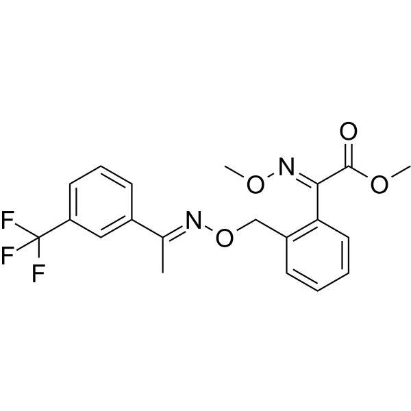
Trifloxystrobin

Based on 1 Customer Validation
Trifloxystrobin (CGA 279202) is a type of fungicide. Trifloxystrobin has toxicity, antiparasitic activity and induce apoptosis, oxidative stress and DNA damage. Trifloxystrobin can be used for the reaesrch of fungal diseases.
For research use only. We do not sell to patients.
- Purity : 99.49%
- CAS No.: 141517-21-7
- Formula: C20H19F3N2O4
- Molecular Weight:408.37
-
Storage:Powder -20°C, 3 years , 4°C, 2 years ; In solvent -80°C, 2 years , -20°C, 1 year

To place orders, for customer services and technical support, please contact: MedChemExpress USA
Tel: 609-228-6898 E-mail: [email protected] [email protected]


-
 Biological Activity
-
Chemical Information
-
Solvent & Solubility
- Protocols
- Purity & Documentation
- References
-
 Help & FAQs


-
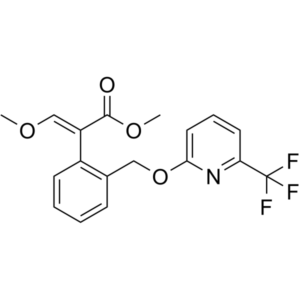
HY-136355
PicoxystrobinFungicide -
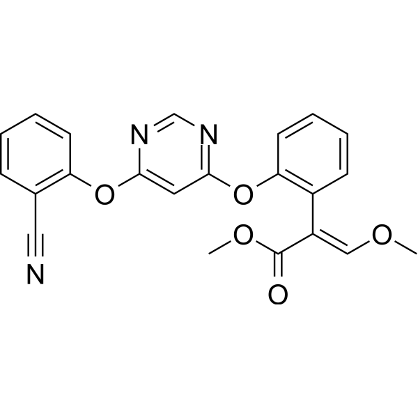
HY-B0849
AzoxystrobinFungicide -
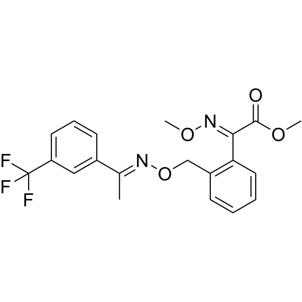
HY-123230R
Trifloxystrobin (Standard)Fungicide -
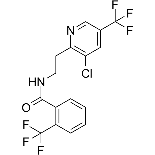
HY-119459
FluopyramSuccinate Dehydrogenase Inhibitor -
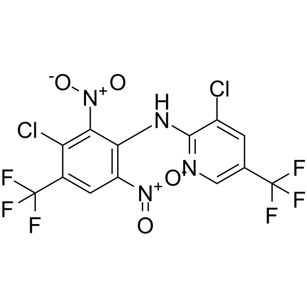
HY-B1839
FluazinamFungicide -
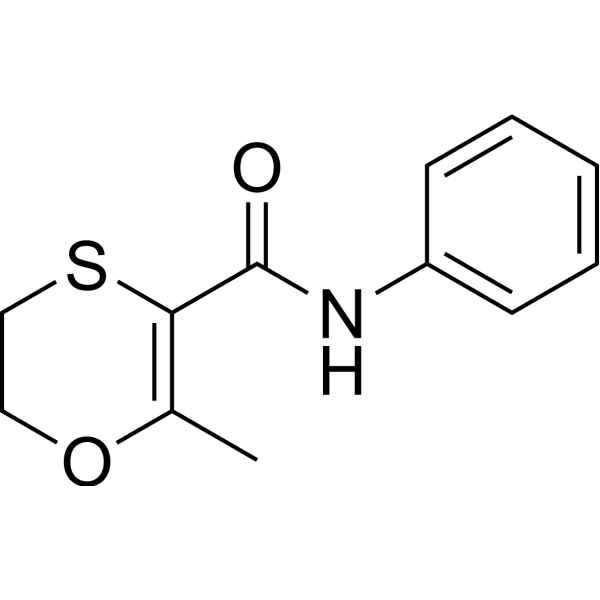
HY-B2064
CarboxinAntibiotic -
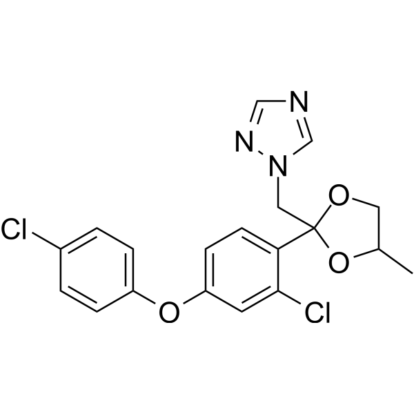
HY-B0850
DifenoconazoleTriazole Fungicide -

HY-B2064R
Carboxin (Standard)Antibiotic -

HY-B1839R
Fluazinam (Standard)Fungicide -
HY-B0850R
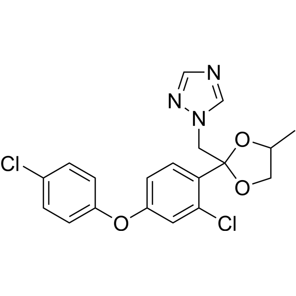
Difenoconazole (Standard)Triazole Fungicide -
HY-B2148
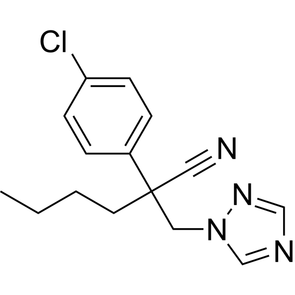
MyclobutanilFungal Inhibitor -
HY-B2004
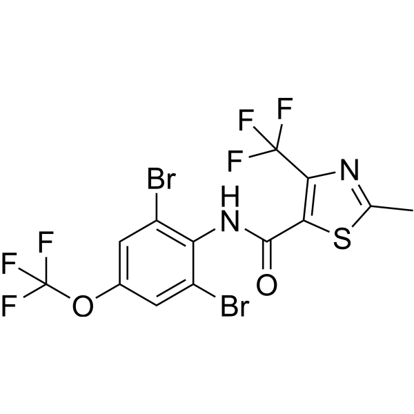
ThifluzamideAntifungal Agent
All Caspase Isoforms
More

Biological Activity
Description
IC50 & Target
|
Caspase 3 |
Caspase-8 |
Caspase-9 |
In Vitro
Trifloxystrobin (2-500 μM, 24-48 h) reduces the viability of HaCaT cells[1].
Trifloxystrobin (0.5 μM, 48 h) induces apoptosis via the tumor necrosis factor-related apoptosis-inducing ligand (TRAIL)-mediated pathway in HaCaT cells[1].
Trifloxystrobin (25-200 μM, 4-48 h) have neurotoxicity in differentiated human SH-SY5Y cells[2].
MedChemExpress (MCE) has not independently confirmed the accuracy of these methods. They are for reference only. Further protocols information, click here.
-
Cell Line:HaCaT cell
-
Concentration:2 μM, 3.91 μM, 7.81 μM, 14.63 μM, 31.25 μM, 62.50 μM, 125 μM, 250 μM, 500 μM
-
Incubation Time:24 h, 48 h
-
Result:Decreased the cell viability in a concentration-dependent manner.
Showed the IC50 value of cell viability at 24 h was calculated to be 22.9 μM, and that at 48 h was 5.14 μM.
-
Cell Line:HaCaT cell
-
Concentration:0.5 μM
-
Incubation Time:24 h, 48 h
-
Result:Induced the expression of pro-apoptotic proteins, apaf-1, procasp-9, procasp-8, and cyt c. Observed both only Annexin V-FITC (green)-positive cells and Annexin V-FITC (green)-/PI (red)-positive cells.
In Vivo
Trifloxystrobin (Soils were prepared by spiking 50 g of soil with 1mL of trifloxystrobin-acetonitrile solution. Following mixing and acetonitrile volatilization, spiked soil was mixed with additional soil to prepare 500 g aliquots containing 0.1, 1.0 and 2.5 mg/kg trifloxystrobin for 7-56 days) induces oxidative stress and DNA damage in two soils[4].
MedChemExpress (MCE) has not independently confirmed the accuracy of these methods. They are for reference only.
-
Animal Model:Zebrafish embryos[3]
-
Dosage:30.0 μg/L, 37.5 μg/L, 47.0 μg/L, 58.6 μg/L
-
Administration:Zebrafish embryos were exposed to trifloxystrobin
-
Result:Caused severe developmental abnormalities of zebrafish embryos.
Induced oxidative stress in embryos.
Changed the transcription of immune related genes of embryos.


Chemical Information
-
CAS No. 141517-21-7
-
Appearance Solid
-
Molecular Weight 408.37
-
Formula C20H19F3N2O4
-
Color White to off-white
-
SMILES
C/C(C1=CC(C(F)(F)F)=CC=C1)=N\OCC2=C(C=CC=C2)/C(C(OC)=O)=N\OC
-
Synonyms
CGA 279202
-
Shipping
Room temperature in continental US; may vary elsewhere.
-
Storage
Powder -20°C 3 years 4°C 2 years In solvent -80°C 2 years -20°C 1 year


Solvent & Solubility
In Vitro:
DMSO : 100 mg/mL (244.88 mM; Need ultrasonic; Hygroscopic DMSO has a significant impact on the solubility of product, please use newly opened DMSO)
Please refer to the solubility information to select the appropriate solvent. Once prepared, please aliquot and store the solution to prevent product inactivation from repeated freeze-thaw cycles.
Storage method and period of stock solution: -80°C, 2 years; -20°C, 1 year. When stored at -80°C, please use it within 2 years. When stored at -20°C, please use it within 1 year.
Please refer to the solubility information to select the appropriate solvent. Once prepared, please aliquot and store the solution to prevent product inactivation from repeated freeze-thaw cycles.
Storage method and period of stock solution: -80°C, 2 years; -20°C, 1 year. When stored at -80°C, please use it within 2 years. When stored at -20°C, please use it within 1 year.
Concentration (start) × Volume (start) = Concentration (final) × Volume (final)
In Vivo:
Select the appropriate dissolution method based on your experimental animal and administration route.
- For the following dissolution methods, please ensure to first prepare a clear stock solution using an In Vitro approach and then sequentially add co-solvents:
- To ensure reliable experimental results, the clarified stock solution can be appropriately stored based on storage conditions. As for the working solution for In Vivo experiments, it is recommended to prepare freshly and use it on the same day.
- The percentages shown for the solvents indicate their volumetric ratio in the final prepared solution. If precipitation or phase separation occurs during preparation, heat and/or sonication can be used to aid dissolution.
Add each solvent one by one: 10% DMSO 40% PEG300 5% Tween-80 45% Saline
Solubility: ≥ 2.17 mg/mL (5.31 mM); Clear solution
This protocol yields a clear solution of ≥ 2.17 mg/mL (saturation unknown).
Taking 1 mL working solution as an example, add 100 μL DMSO stock solution (21.7 mg/mL) to 400 μL PEG300, and mix evenly; then add 50 μL Tween-80 and mix evenly; then add 450 μL Saline to adjust the volume to 1 mL.
Preparation of Saline: Dissolve 0.9 g sodium chloride in ddH₂O and dilute to 100 mL to obtain a clear Saline solution.
In Vivo Dissolution Calculator
Please enter the basic information of animal experiments:
-
-
-
-
Recommended: Prepare an additional quantity of animals to account for potential losses during experiments.
Please enter your animal formula composition:
-
%DMSO +
Recommended: Keep the proportion of DMSO in working solution below 2% if your animal is weak.
-
%+
-
+%Tween-80 + +
-
%Saline +
The co-solvents required include: DMSO, . All of co-solvents are available by MedChemExpress (MCE). , Tween 80. All of co-solvents are available by MedChemExpress (MCE).
Working solution concentration: 0.22 mg/mL
Method for preparing stock solution: mg drug dissolved in μL DMSO. Stock solution concentration: mg/mL.
1. Take μL DMSO stock solution;
2. Add μL .
μL , mix evenly;
3. Then add μL Tween 80, mix evenly;
4. Then add μL
Please ensure that the stock solution in the first step is dissolved to a clear state, and add co-solvents in sequence. You can use ultrasonic heating (ultrasonic cleaner, recommended frequency 20-40 kHz), vortexing, etc. to assist dissolution.

Protocols
-
Apoptosis
Apoptosis, also called programmed cell death, is generally characterized by distinct morphological characteristics.
-
TUNEL staining for apoptotic DNA fragmentation
TUNEL staining detects DNA strand breaks by using terminal deoxynucleotidyl transferase to add labeled nucleotides to exposed 3′-OH DNA termini, generating either microscopic staining in fixed cells or tissue sections, or fluorescence/cytometric signal in cell suspensions. TUNEL positivity reflects DNA fragmentation but should not be interpreted alone as definitive apoptosis, because TUNEL can also label necrotic, autolytic, mechanically damaged, or DNA-repair-associated DNA breaks.
-
Annexin V plus membrane-impermeant dye apoptosis staining
Annexin V-based apoptosis assays rely on the detection of phosphatidylserine (PS) externalization from the inner leaflet of the plasma membrane to the outer leaflet, an early biochemical hallmark of apoptosis. Fluorescently labeled Annexin V binds PS in a calcium-dependent manner, enabling identification of early apoptotic cells by flow cytometry or fluorescence microscopy. When combined with a membrane-impermeant DNA-binding dye (e. g. , propidium iodide), this approach allows discrimination between viable (Annexin V−/dye−), early apoptotic (Annexin V+/dye−), and late apoptotic or necrotic (Annexin V+/dye+) cell populations by assessing membrane integrity and PS exposure.
-
ROS/oxidative-stress fluorescent staining
ROS/oxidative-stress fluorescent staining uses cell-permeant fluorogenic probes that become fluorescent after oxidation inside cells or tissues; commonly used examples include DCFH-DA/DCFDA for broad cellular oxidant detection, DHE for superoxide-related signal detection, MitoSOX for mitochondrial superoxide-related signal detection, and CellROX probes for oxidative-stress-associated fluorescence readouts. The assay detects probe oxidation rather than a single ROS species unless the probe and analysis method have been chemically validated for that species. DCFH-DA enters cells, is deacetylated by intracellular esterases to DCFH, and produces fluorescent DCF after oxidation, so the readout is used as an operational measure of total cellular oxidative stress rather than a species-specific ROS measurement. DHE and MitoSOX can report superoxide-related oxidation, but red fluorescence alone can include non-specific ethidium-like oxidation products; HPLC or optimized spectral approaches are
-
Apoptosis Solutions
Apoptosis is a regulated, generally non-lytic cell-death pathway that removes unwanted, damaged, infected, or abnormal cells through coordinated morphological changes, caspase activation, DNA fragmentation, and membrane remodeling. The intrinsic apoptosis pathway is controlled mainly by mitochondrial outer membrane permeabilization, BCL-2 family proteins, cytochrome c release, apoptosome formation, caspase-9 activation, and downstream executioner caspase-3/7 activation. The extrinsic apoptosis pathway is initiated by death receptors such as Fas, TNFR, and TRAIL receptors, which recruit adaptor proteins and activate caspase-8 before engaging executioner caspases or mitochondrial amplification through BID cleavage. Apoptosis is linked to many phenotypes, including cancer cell killing, tissue homeostasis, immune regulation, neurodegeneration, infection response, and treatment-induced cytotoxicity; unresolved questions include how apoptosis interacts with necroptosis, pyroptosis, ferroptos
-
Genotoxicity/Mutagenicity Study
The bacterial reverse mutation assay detects point mutations that restore amino-acid prototrophy in auxotrophic Salmonella typhimurium or Escherichia coli tester strains; after exposure to a test article, mutagenic activity is read out as an increased number of revertant colonies on minimal agar compared with the vehicle control. The assay uses tester strains with different mutation targets so that base-substitution and frameshift mutagens can be detected, and testing is performed with and without exogenous mammalian metabolic activation because some chemicals require biotransformation to become mutagenic.


Purity & Documentation
-
Data Sheet (277 KB)
-
SDS (597 KB)
- English - EN (597 KB)
- Français - FR (597 KB)
- Deutsch - DE (597 KB)
- Norwegian - NO (597 KB)
- Español - ES (597 KB)
- Swedish - SV (597 KB)
- Italian - IT (597 KB)
- Korean - KR (597 KB)
- Portuguese - PT (597 KB)
-
Handling Instructions (2659 KB)
References
[1]. Jang Y, et al. Trifloxystrobin induces tumor necrosis factor-related apoptosis-inducing ligand (TRAIL)-mediated apoptosis in HaCaT, human keratinocyte cells [J]. Drug and Chemical Toxicology, 2017, 40(1): 67-73. [Content Brief]
[2]. Nguyen K, et al. Neurotoxicity assessment of QoI strobilurin fungicides azoxystrobin and trifloxystrobin in human SH-SY5Y neuroblastoma cells: Insights from lipidomics and mitochondrial bioenergetics[J]. Neurotoxicology, 2022, 91: 290-304. [Content Brief]
[3]. Li H, et al. Developmental toxicity, oxidative stress and immunotoxicity induced by three strobilurins (pyraclostrobin, trifloxystrobin and picoxystrobin) in zebrafish embryos [J]. Chemosphere, 2018, 207: 781-790. [Content Brief]
[4]. Wu R, et al. Oxidative stress and DNA damage induced by trifloxystrobin on earthworms (Eisenia fetida) in two soils [J]. Science of The Total Environment, 2021, 797: 149004. [Content Brief]
[5]. Villares M, et al. Trifloxystrobin blocks the growth of Theileria parasites and is a promising drug to treat Buparvaquone resistance [J]. Communications Biology, 2022, 5(1): 1253. [Content Brief]


Complete Stock Solution Preparation Table
Please refer to the solubility information to select the appropriate solvent. Once prepared, please aliquot and store the solution to prevent product inactivation from repeated freeze-thaw cycles.
Storage method and period of stock solution: -80°C, 2 years; -20°C, 1 year. When stored at -80°C, please use it within 2 years. When stored at -20°C, please use it within 1 year.
| Optional Solvent | Concentration Solvent Mass | 1 mg | 5 mg | 10 mg | 25 mg |
|---|---|---|---|---|---|
| DMSO | 1 mM | 2.4488 mL | 12.2438 mL | 24.4876 mL | 61.2190 mL |
| 5 mM | 0.4898 mL | 2.4488 mL | 4.8975 mL | 12.2438 mL | |
| 10 mM | 0.2449 mL | 1.2244 mL | 2.4488 mL | 6.1219 mL | |
| 15 mM | 0.1633 mL | 0.8163 mL | 1.6325 mL | 4.0813 mL | |
| 20 mM | 0.1224 mL | 0.6122 mL | 1.2244 mL | 3.0609 mL | |
| 25 mM | 0.0980 mL | 0.4898 mL | 0.9795 mL | 2.4488 mL | |
| 30 mM | 0.0816 mL | 0.4081 mL | 0.8163 mL | 2.0406 mL | |
| 40 mM | 0.0612 mL | 0.3061 mL | 0.6122 mL | 1.5305 mL | |
| 50 mM | 0.0490 mL | 0.2449 mL | 0.4898 mL | 1.2244 mL | |
| 60 mM | 0.0408 mL | 0.2041 mL | 0.4081 mL | 1.0203 mL | |
| 80 mM | 0.0306 mL | 0.1530 mL | 0.3061 mL | 0.7652 mL | |
| 100 mM | 0.0245 mL | 0.1224 mL | 0.2449 mL | 0.6122 mL |


Trifloxystrobin Related Classifications
HY-123230 Related Classifications
 Powered by Bioz