



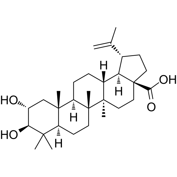
Alphitolic acid
Based on 1 publication(s) in Google Scholar
Alphitolic acid (Aophitolic acid), an anti-inflammatory triterpenoid compound, is a selective HMGCS2 inhibitor (IC50 17.17 μM, Kd 5.6 μM). Alphitolic acid inhibits ORAI1/STIM1-mediated CRAC currents, store-operated calcium entry, the Akt-mTOR-NF-κB signaling pathway, p70S6K/S6/IκBα phosphorylation, NF-κB/Bcl-2 expression, and NO/TNF-α production, while upregulating p53 and downregulating MDM2. Alphitolic acid induces apoptosis (apoptosis) and autophagy (autophagy) through Beclin 1, Atg7, LC3B-II, and autophagosomes, inhibits oral squamous cell carcinoma proliferation/viability, and exhibits antibacterial/antimalarial/antioxidant activities. Alphitolic acid can be used in research on inflammatory diseases, allergic diseases, Tamoxifen (HY-13757A)-resistant breast cancer, oral squamous cell carcinoma, and inflammation.
For research use only. We do not sell to patients.
- CAS No.: 19533-92-7
- Formula: C30H48O4
- Molecular Weight:472.70
-
Storage:
4°C, protect from light
* In solvent : -80°C, 6 months; -20°C, 1 month (protect from light)

To place orders, for customer services and technical support, please contact: MedChemExpress USA
Tel: 609-228-6898 E-mail: [email protected] [email protected]


-
 Biological Activity
-
Chemical Information
-
Solvent & Solubility
- Protocols
- Purity & Documentation
- References
-
 Help & FAQs



Publications Citing Use of MedChemExpress (MCE) Alphitolic acid
MoreAll Parasite Isoforms
More

Biological Activity
Description
IC50 & Target
[2]|
HMGCS2 17.17 μM (IC50) |
HMGCS2 5.6 μM (Kd) |
Cellular Effect
|
Cell Line

|
Type | Value | Description | References |
|---|---|---|---|---|
| HEK-293T | IC50 |
17.52 μM
|
Inhibition of ICRAC in ORAI1/STIM1-transfected HEK293T cells by patch-clamp electrophysiology.
Inhibition of ICRAC in ORAI1/STIM1-transfected HEK293T cells by patch-clamp electrophysiology.
|
38139137 |
| SCC-4 | IC50 |
12 μM
|
Antiproliferative activity against human SCC4 oral squamous cell carcinoma cells assessed as viability reduction by MTT assay.
Antiproliferative activity against human SCC4 oral squamous cell carcinoma cells assessed as viability reduction by MTT assay.
|
j.jff.2015.07.017 |
| RAW264.7 | IC50 |
17.6 μM
|
Inhibition of nitric oxide production in LPS and IFN-γ activated RAW 264.7 macrophages by Griess assay with 1 hr pre-incubation and 24 hrs activation.
Inhibition of nitric oxide production in LPS and IFN-γ activated RAW 264.7 macrophages by Griess assay with 1 hr pre-incubation and 24 hrs activation.
|
27845729 |
| RAW264.7 | IC50 |
22.7 μM
|
Inhibition of TNF-α production in LPS and IFN-γ activated RAW 264.7 macrophages by ELISA with 1 hr pre-incubation and 24 hrs activation.
Inhibition of TNF-α production in LPS and IFN-γ activated RAW 264.7 macrophages by ELISA with 1 hr pre-incubation and 24 hrs activation.
|
27845729 |
| RAW264.7 | LC50 |
18.6 μM
|
Cytotoxicity against LPS and IFN-γ activated RAW 264.7 macrophages assessed as viability reduction by Alamar Blue assay after 24 hrs activation and 2 hrs incubation.
Cytotoxicity against LPS and IFN-γ activated RAW 264.7 macrophages assessed as viability reduction by Alamar Blue assay after 24 hrs activation and 2 hrs incubation.
|
27845729 |
In Vitro
Alphitolic acid (30 µM) suppresses Thapsigargin (HY-13433)-induced calcium influx in Jurkat T cells[1].
Alphitolic acid (3-30 µM; three-day culture before flow cytometry) inhibits anti-CD3/anti-CD28-stimulated human CD4+ T cell proliferation in a concentration-dependent manner, with 80.48% inhibition at 30 µM[1].
Alphitolic acid suppresses proliferation of SCC4 and SCC2095 OSCC cells with IC50 values of 12 and 15 μM, respectively, whereas p53-null HSC-3 cells are resistant[3].
Alphitolic acid (10-25 μM) upregulates p53 phosphorylation/expression and downregulates MDM2 phosphorylation/expression in SCC4 cells[3].
Alphitolic acid (25 μM; 24 h)-mediated cytotoxicity in SCC4 cells is partially reduced by p53 knockdown[3].
Alphitolic acid (10-30 μM; 72 h) induces apoptosis in SCC4 OSCC cells, producing 25.1% apoptotic cells at 30 μM after 72 h[3].
Alphitolic acid (10-20 μM; 72 h) induces apoptotic chromatin condensation in SCC4 cells at 10 and 20 μM after 72 h[3].
Alphitolic acid (10-25 μM; 72 h) blocks Akt-NF-κB signaling in SCC4 cells, decreasing p-Akt, p-p70S6K, p-S6, p-IκBα, NF-κB, and Bcl-2[3].
Alphitolic acid (20 μM; 24 h) induces autophagosome formation in SCC4 cells[3].
Alphitolic acid (10-25 μM; 72 h [concentration response]; 20 μM; 3-72 h [time course]) induces autophagy markers LC3B-II, Beclin-1, and Atg7 in SCC4 cells in concentration- and time-dependent manners[3].
Alphitolic acid (20-25 μM; 48 h) induces GFP-LC3 puncta accumulation in SCC4 cells[3].
Alphitolic acid exhibits moderate antibacterial activity against Staphylococcus aureus, Enterococcus faecalis and Escherichia coli (MICs 32, 64 and 128 μg/mL, respectively)[5].
Alphitolic acid (2) (0-36 µg/mL; 1 h pre-incubation; 24 h after activation; 18 h plating; 2 h Alamar Blue) inhibits NO production with an IC50 of 17.6 µM and TNF-α with an IC50 of 22.7 µM, while showing cytotoxicity with an LC50 of 18.6 µM in LPS + IFN-γ activated RAW 264.7 macrophages[4].
Alphitolic acid is a selective HMGCS2 inhibitor with a Kd of 5.6 μM and an IC50 of 17.17 μM, sparing HMGCS1[2].
Alphitolic acid (10 μM; 24 h) lowers intracellular β-OHB in T47D/TR-2 cells in an HMGCS2-dependent manner without altering cholesterol[2].
Alphitolic acid (0-20 μM; 24 h-14 days) selectively increases mitochondrial ROS, reduces viability and clonogenicity, and triggers caspase-dependent apoptosis in T47D/TR-2 cells via HMGCS2[2].
Alphitolic acid (1.0-100 µM) inhibits ICRAC in ORAI1/STIM1-transfected HEK293T cells with an IC50 of 17.52 µM[1].
MedChemExpress (MCE) has not independently confirmed the accuracy of these methods. They are for reference only.
-
Cell Line:SCC4 human OSCC cells
-
Concentration:10, 20, 30 μM
-
Incubation Time:72 h
-
Result:Led to a dose-dependent increase in apoptosis: 10 μM, 11.8%; 20 μM, 16.1%; 30 μM, 25.1%.
-
Cell Line:SCC4 human OSCC cells
-
Concentration:10, 20 μM
-
Incubation Time:72 h
-
Result:Induced apoptosis in SCC4 cells, which showed increased chromatin condensation, as evidenced by intense bluish-white fluorescence in pyknotic nuclei at 10 and 20 μM.
-
Cell Line:SCC4 human OSCC cells
-
Concentration:10, 15, 20, 25 μM
-
Incubation Time:72 h
-
Result:Blocked Akt signalling, decreasing phosphorylation of Akt and its downstream substrates p70S6K, S6, and IκBα, and downregulating NF-κB and Bcl-2 expression.
-
Cell Line:SCC4 human OSCC cells
-
Concentration:20 μM
-
Incubation Time:24 h
-
Result:Transmission electron microscopy demonstrated formation of autophagosomes as membranous vacuoles with double-membrane structure.
-
Cell Line:SCC4 human OSCC cells
-
Concentration:10, 15, 20, 25 μM (Concentration response); 20 μM (Time course)
-
Incubation Time:72 h (Concentration response); 3, 6, 12, 24, 48, 72 h (Time course)
-
Result:Induced autophagy, which was confirmed by cellular accumulations of LC3B-II in concentration- and time-dependent manners, accompanied by parallel increases in Atg7 and Beclin-1 expression.
-
Cell Line:SCC4 human OSCC cells
-
Concentration:25 μM
-
Incubation Time:24 h (after transfection)
-
Result:p53 knockdown resulted in a higher degree of resistance to the antiproliferative activity.
Western blotting showed reduced p53 and p-p53 expression after p53 shRNA transfection.
In Vivo
MedChemExpress (MCE) has not independently confirmed the accuracy of these methods. They are for reference only.
-
Animal Model:BALB/c (female, 5-week-old, athymic nu/nu)[2]
-
Dosage:100 mg/kg
-
Administration:i.p.; every 3-4 days; ten injections
-
Result:At 100 mg/kg, significantly retarded tumor growth after two weeks of administration, and the tumor growth-inhibiting effect was retained throughout the entire administration period.
Cleavage of caspases was observed.
β-OHB and acetoacetate levels were significantly lower, whereas cholesterol levels remained unchanged .
- Western Blot
- RNA extraction experimental
- Research Protocol for Infectious Diseases
- Annexin V plus membrane-impermeant dye apoptosis staining
- Research Protocol for Inflammation-related Diseases
- Breast Cancer Modeling
- Cell Viability Determination by MTT Colorimetric Assay
- Kinase activity and phosphorylation assays
- Detection of Apoptosis
- TUNEL staining for apoptotic DNA fragmentation
- Autophagy
- Apoptosis Solutions
- Protocol for Kinase activity and phosphorylation assays
- Lysosome and acidic-vesicle live-cell staining
- Macroautophagy Solutions


Chemical Information
-
CAS No. 19533-92-7
-
Appearance Solid
-
Molecular Weight 472.70
-
Formula C30H48O4
-
Color White to off-white
-
SMILES
OC([C@]12[C@@]([C@@H](CC2)C(C)=C)([H])[C@]3([H])[C@@](CC1)([C@]4([C@]([C@@]5([C@@](C(C)([C@H]([C@@H](C5)O)O)C)([H])CC4)C)([H])CC3)C)C)=O
-
Synonyms
Aophitolic acid
-
Structure Classification
-
Initial Source
-
Shipping
Room temperature in continental US; may vary elsewhere.
-
Storage
4°C, protect from light
* In solvent : -80°C, 6 months; -20°C, 1 month (protect from light)


Publications (1)
-
Journal Impact Factor
-
Most Recent
-
Mol Immunol
Autotaxin promotes the degradation of the mucus layer by inhibiting autophagy in mouse colitis. [Abstract]2023 Aug:160:44-54. PMID: 37356325


Solvent & Solubility
In Vitro:
DMSO : 50 mg/mL (105.78 mM; Need ultrasonic; Hygroscopic DMSO has a significant impact on the solubility of product, please use newly opened DMSO)
Please refer to the solubility information to select the appropriate solvent. Once prepared, please aliquot and store the solution to prevent product inactivation from repeated freeze-thaw cycles.
Storage method and period of stock solution: -80°C, 6 months; -20°C, 1 month (protect from light). When stored at -80°C, please use it within 6 months. When stored at -20°C, please use it within 1 month.
Please refer to the solubility information to select the appropriate solvent. Once prepared, please aliquot and store the solution to prevent product inactivation from repeated freeze-thaw cycles.
Storage method and period of stock solution: -80°C, 6 months; -20°C, 1 month (protect from light). When stored at -80°C, please use it within 6 months. When stored at -20°C, please use it within 1 month.
Concentration (start) × Volume (start) = Concentration (final) × Volume (final)
In Vivo:
Select the appropriate dissolution method based on your experimental animal and administration route.
- For the following dissolution methods, please ensure to first prepare a clear stock solution using an In Vitro approach and then sequentially add co-solvents:
- To ensure reliable experimental results, the clarified stock solution can be appropriately stored based on storage conditions. As for the working solution for In Vivo experiments, it is recommended to prepare freshly and use it on the same day.
- The percentages shown for the solvents indicate their volumetric ratio in the final prepared solution. If precipitation or phase separation occurs during preparation, heat and/or sonication can be used to aid dissolution.
In Vivo Dissolution Calculator
Please enter the basic information of animal experiments:
-
-
-
-
Recommended: Prepare an additional quantity of animals to account for potential losses during experiments.
Please enter your animal formula composition:
-
%DMSO +
Recommended: Keep the proportion of DMSO in working solution below 2% if your animal is weak.
-
%+
-
+%Tween-80 + +
-
%Saline +
The co-solvents required include: DMSO, . All of co-solvents are available by MedChemExpress (MCE). , Tween 80. All of co-solvents are available by MedChemExpress (MCE).
Working solution concentration: 0.22 mg/mL
Method for preparing stock solution: mg drug dissolved in μL DMSO. Stock solution concentration: mg/mL. * In solvent : -80°C, 6 months; -20°C, 1 month (protect from light)
1. Take μL DMSO stock solution;
2. Add μL .
μL , mix evenly;
3. Then add μL Tween 80, mix evenly;
4. Then add μL
Please ensure that the stock solution in the first step is dissolved to a clear state, and add co-solvents in sequence. You can use ultrasonic heating (ultrasonic cleaner, recommended frequency 20-40 kHz), vortexing, etc. to assist dissolution.


Purity & Documentation
-
Data Sheet (303 KB)
-
SDS (252 KB)
- English - EN (252 KB)
- Français - FR (252 KB)
- Deutsch - DE (252 KB)
- Norwegian - NO (252 KB)
- Español - ES (252 KB)
- Swedish - SV (252 KB)
- Italian - IT (252 KB)
- Korean - KR (252 KB)
- Portuguese - PT (252 KB)
-
Handling Instructions (2659 KB)
References


Complete Stock Solution Preparation Table
Please refer to the solubility information to select the appropriate solvent. Once prepared, please aliquot and store the solution to prevent product inactivation from repeated freeze-thaw cycles.
Storage method and period of stock solution: -80°C, 6 months; -20°C, 1 month (protect from light). When stored at -80°C, please use it within 6 months. When stored at -20°C, please use it within 1 month.
| Optional Solvent | Concentration Solvent Mass | 1 mg | 5 mg | 10 mg | 25 mg |
|---|---|---|---|---|---|
| DMSO | 1 mM | 2.1155 mL | 10.5775 mL | 21.1551 mL | 52.8877 mL |
| 5 mM | 0.4231 mL | 2.1155 mL | 4.2310 mL | 10.5775 mL | |
| 10 mM | 0.2116 mL | 1.0578 mL | 2.1155 mL | 5.2888 mL | |
| 15 mM | 0.1410 mL | 0.7052 mL | 1.4103 mL | 3.5258 mL | |
| 20 mM | 0.1058 mL | 0.5289 mL | 1.0578 mL | 2.6444 mL | |
| 25 mM | 0.0846 mL | 0.4231 mL | 0.8462 mL | 2.1155 mL | |
| 30 mM | 0.0705 mL | 0.3526 mL | 0.7052 mL | 1.7629 mL | |
| 40 mM | 0.0529 mL | 0.2644 mL | 0.5289 mL | 1.3222 mL | |
| 50 mM | 0.0423 mL | 0.2116 mL | 0.4231 mL | 1.0578 mL | |
| 60 mM | 0.0353 mL | 0.1763 mL | 0.3526 mL | 0.8815 mL | |
| 80 mM | 0.0264 mL | 0.1322 mL | 0.2644 mL | 0.6611 mL | |
| 100 mM | 0.0212 mL | 0.1058 mL | 0.2116 mL | 0.5289 mL |


Alphitolic acid Related Classifications
HY-N2855 Related Classifications
 Powered by Bioz