



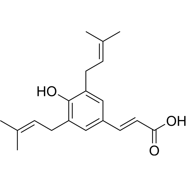
Artepillin C

Based on 1 Customer Validation
Artepillin C is an orally active CREB/CRTC2 inhibitor and TRPA1 covalent agonist (EC50=1.8 μM). Artepillin C inhibits CREB/CRTC2-mediated gene transcription and downregulates BMAL1 expression to regulate glucose and lipid metabolism. Artepillin C can also activate TRPA1 channels to induce spicy taste signals. Artepillin C can inhibit tumor cell proliferation, induce necroptosis, improve insulin resistance and inhibit liver lipid synthesis. Artepillin C can be used in the study of metabolic syndrome, tumor prevention and treatment, and inflammation.
Nos produits utilisent uniquement pour la recherche. Nous ne vendons pas aux patients.
- Pureté : 99.07%
- CAS No.: 72944-19-5
- Formule: C19H24O3
- Masse moléculaire:300.39
-
Stockage:Powder -20°C, 3 years ; In solvent -80°C, 6 months , -20°C, 1 month

To place orders, for customer services and technical support, please contact: MedChemExpress USA
Tel: 609-228-6898 E-mail: [email protected] [email protected]


-
 Activité biologique
-
Chemical Information
-
Solvant et solubilité
- Protocole
- Pureté et documentation
- Références
-
 Help & FAQs




Activité biologique
Description
IC50 & Target
[3]|
CREB/CRTC2 |
TRPA1 1.8 μM () |
Cellular Effect
|
Cell Line

|
Type | Value | Description | References |
|---|---|---|---|---|
| A549 | IC50 |
25 μM
Compound: ARC
|
Growth inhibition of human A549 cells
Growth inhibition of human A549 cells
|
[PMID: 28814374] |
| DU-145 | IC50 |
179 μM
Compound: 1
|
Antiproliferative activity against human DU-145 cells assessed as cell viability for 72 hrs by MTT assay
Antiproliferative activity against human DU-145 cells assessed as cell viability for 72 hrs by MTT assay
|
[PMID: 34454129] |
| HeLa | IC50 |
7 μg/mL
Compound: 1
|
Antiallergic activity in Ca(2+)-stimulated differentiated human HeLa cells assessed as inhibition of cys-leukotriene release after 6 days by ELISA
Antiallergic activity in Ca(2+)-stimulated differentiated human HeLa cells assessed as inhibition of cys-leukotriene release after 6 days by ELISA
|
[PMID: 19942440] |
| HT-1080 | ED50 |
45.47 μg/mL
Compound: 5
|
Cytotoxicity against human HT1080 cells after 24 hrs by MTT assay
Cytotoxicity against human HT1080 cells after 24 hrs by MTT assay
|
[PMID: 9677271] |
| MCF-10A | CC50 |
>200 μM
Compound: 1
|
Cytotoxicity against human MCF-10A cells assessed as cell viability for 72 hrs by MTT assay
Cytotoxicity against human MCF-10A cells assessed as cell viability for 72 hrs by MTT assay
|
[PMID: 34454129] |
| MCF7 | IC50 |
162.3 μM
Compound: 1
|
Antiproliferative activity against human MCF7 cells assessed as cell viability for 72 hrs by MTT assay
Antiproliferative activity against human MCF7 cells assessed as cell viability for 72 hrs by MTT assay
|
[PMID: 34454129] |
| MDA-MB-231 | IC50 |
172.7 μM
Compound: 1
|
Antiproliferative activity against human MDA-MB-231 cells assessed as cell viability for 72 hrs by MTT assay
Antiproliferative activity against human MDA-MB-231 cells assessed as cell viability for 72 hrs by MTT assay
|
[PMID: 34454129] |
| PC-3 | IC50 |
178.9 μM
Compound: 1
|
Antiproliferative activity against human PC-3 cells assessed as cell viability for 72 hrs by MTT assay
Antiproliferative activity against human PC-3 cells assessed as cell viability for 72 hrs by MTT assay
|
[PMID: 34454129] |
| PNT2 | CC50 |
187.6 μM
Compound: 1
|
Cytotoxicity against human PNT2 cells assessed as cell viability for 72 hrs by MTT assay
Cytotoxicity against human PNT2 cells assessed as cell viability for 72 hrs by MTT assay
|
[PMID: 34454129] |
In Vitro
Artepillin C (1 μM-1 mM; 12-48 h) inhibits the viability of HEp-2 tumor cells in a concentration- and time-dependent manner, with a CC50 of 4.07 μM-0.9 μM[1].
Artepillin C (1-0.5 μM; 24 h) induces HEp-2 cell necrosis and significantly increases cell membrane permeability[1].
Artepillin C (1 μM; 30 min; pH 3.2) enhances the membrane permeability of DPPS-containing macropinosomes (GUVs) and significantly accelerates the efflux of fluorescent probes[1].
Artepillin C (3.13-50 μg/mL; 36 h) inhibits HUVEC cell tube formation in a concentration-dependent manner, and completely inhibits it at 50 μg/mL[2].
Artepillin C (3.13-50 μg/mL; 3 d) inhibits HUVEC cell proliferation with an IC50 of approximately 37.2 μg/mL[2].
MedChemExpress (MCE) has not independently confirmed the accuracy of these methods. They are for reference only.
-
Cell Line:HEp-2 cells
-
Concentration:0.01 mM, 0.05 mM, 0.1 mM, 0.5 mM
-
Incubation Time:24 h
-
Result:Increased propidium iodide (PI)-positive cells, indicating membrane permeabilization and necrosis.
Morphological changes included cell swelling and granular nuclei, characteristic of necrotic cell death, without significant apoptosis induction.
In Vivo
Artepillin C (20 mg/kg; intraperitoneal injection; once a day; 1-3 weeks) significantly improves glucose homeostasis, reduces fasting blood glucose and insulin resistance, inhibits hepatic BMAL1 expression and lipid synthesis-related genes, and reduces hepatic lipid accumulation in db/db obese mice[4].
Artepillin C (20 mg/kg; intraperitoneal injection; once a day; 3 weeks) improves insulin sensitivity, reduces hepatic gluconeogenesis and lipid synthesis, and regulates the expression of the circadian rhythm-related gene Bmal1 in DIO mice fed a high-fat diet[4].
MedChemExpress (MCE) has not independently confirmed the accuracy of these methods. They are for reference only.
-
Animal Model:ICR mice (female, 30±5 g, 7-week-old), dorsal air sac (DAS) tumor angiogenesis model[2]
-
Dosage:0.125%, 0.25%, 0.5% (w/w in diet)
-
Administration:Oral administration via diet, once daily, 6 days starting from chamber implantation
-
Result:Significantly reduced tumor-induced angiogenesis in a dose-dependent manner.
No signs of toxicity were observed, with stable body weight throughout the treatment period.
Histological analysis confirmed reduced vascular density in the treated groups, indicating anti-angiogenic activity without systemic toxicity.
- RNA extraction experimental
- BrdU Incorporation Assay
- Somatic Cell Culture
- CFSE Dye Dilution Proliferation Assay
- Dye-dilution cell tracking and proliferation staining
- CCK-8/WST-8 Cell Proliferation Assay
- Protocol for Cell Cycle
- Cell Counting-Based Growth Curve Assay
- Research Protocol for Inflammation-related Diseases
- Colony Formation (Clonogenic) Assay
- Research Protocol for Metabolic Diseases
- Cell Viability Determination by MTT Colorimetric Assay
- EdU Incorporation Assay (Click Chemistry-Based DNA Synthesis Measurement)
- Ki-67 Immunostaining Proliferation Assay
- PCNA Immunodetection Proliferation Assay
- Protocol for Cell Counting and Cell Density Analysis
- Research Protocol for Endocrine Diseases
- MTT Cell Proliferation Assay


Chemical Information
-
CAS No. 72944-19-5
-
Appearance Solid
-
Masse moléculaire 300.39
-
Formule C19H24O3
-
Color Light yellow to yellow
-
SMILES
C/C(C)=C\CC1=C(C(C/C=C(C)\C)=CC(/C=C/C(O)=O)=C1)O
-
Structure Classification
-
Livraison
Room temperature in continental US; may vary elsewhere.
-
Stockage
Powder -20°C 3 years In solvent -80°C 6 months -20°C 1 month


Solvant et solubilité
In Vitro:
DMSO : 100 mg/mL (332.90 mM; Need ultrasonic; Hygroscopic DMSO has a significant impact on the solubility of product, please use newly opened DMSO)
Please refer to the solubility information to select the appropriate solvent. Once prepared, please aliquot and store the solution to prevent product inactivation from repeated freeze-thaw cycles.
Storage method and period of stock solution: -80°C, 6 months; -20°C, 1 month. When stored at -80°C, please use it within 6 months. When stored at -20°C, please use it within 1 month.
Please refer to the solubility information to select the appropriate solvent. Once prepared, please aliquot and store the solution to prevent product inactivation from repeated freeze-thaw cycles.
Storage method and period of stock solution: -80°C, 6 months; -20°C, 1 month. When stored at -80°C, please use it within 6 months. When stored at -20°C, please use it within 1 month.
Concentration (start) × Volume (start) = Concentration (final) × Volume (final)
In Vivo:
Select the appropriate dissolution method based on your experimental animal and administration route.
- For the following dissolution methods, please ensure to first prepare a clear stock solution using an In Vitro approach and then sequentially add co-solvents:
- To ensure reliable experimental results, the clarified stock solution can be appropriately stored based on storage conditions. As for the working solution for In Vivo experiments, it is recommended to prepare freshly and use it on the same day.
- The percentages shown for the solvents indicate their volumetric ratio in the final prepared solution. If precipitation or phase separation occurs during preparation, heat and/or sonication can be used to aid dissolution.
Add each solvent one by one: 10% DMSO 40% PEG300 5% Tween-80 45% Saline
Solubility: ≥ 2.5 mg/mL (8.32 mM); Clear solution
This protocol yields a clear solution of ≥ 2.5 mg/mL (saturation unknown).
Taking 1 mL working solution as an example, add 100 μL DMSO stock solution (25.0 mg/mL) to 400 μL PEG300, and mix evenly; then add 50 μL Tween-80 and mix evenly; then add 450 μL Saline to adjust the volume to 1 mL.
Preparation of Saline: Dissolve 0.9 g sodium chloride in ddH₂O and dilute to 100 mL to obtain a clear Saline solution.
In Vivo Dissolution Calculator
Please enter the basic information of animal experiments:
-
-
-
-
Recommended: Prepare an additional quantity of animals to account for potential losses during experiments.
Please enter your animal formula composition:
-
%DMSO +
Recommended: Keep the proportion of DMSO in working solution below 2% if your animal is weak.
-
%+
-
+%Tween-80 + +
-
%Saline +
The co-solvents required include: DMSO, . All of co-solvents are available by MedChemExpress (MCE). , Tween 80. All of co-solvents are available by MedChemExpress (MCE).
Working solution concentration: 0.22 mg/mL
Method for preparing stock solution: mg drug dissolved in μL DMSO. Stock solution concentration: mg/mL.
1. Take μL DMSO stock solution;
2. Add μL .
μL , mix evenly;
3. Then add μL Tween 80, mix evenly;
4. Then add μL
Please ensure that the stock solution in the first step is dissolved to a clear state, and add co-solvents in sequence. You can use ultrasonic heating (ultrasonic cleaner, recommended frequency 20-40 kHz), vortexing, etc. to assist dissolution.


Pureté et documentation
-
Fiche technique (277 KB)
-
SDS (393 KB)
- English - EN (393 KB)
- Français - FR (393 KB)
- Deutsch - DE (393 KB)
- Norwegian - NO (393 KB)
- Español - ES (393 KB)
- Swedish - SV (393 KB)
- Italian - IT (393 KB)
- Korean - KR (393 KB)
- Portuguese - PT (393 KB)
-
Instruction de manipulation (2659 KB)
Références
[1]. Kobal MB,et al. Correlating Artepillin C cytotoxic activity on HEp-2 cells with bioinspired systems of plasma membranes. Mater Sci Eng C Mater Biol Appl. 2020 Jul;112:110943. [Content Brief]
[2]. Ahn MR, et al. Suppression of tumor-induced angiogenesis by Brazilian propolis: major component artepillin C inhibits in vitro tube formation and endothelial cell proliferation. Cancer Lett. 2007 Jul 18;252(2):235-43. [Content Brief]
[3]. Hata T, et al. Artepillin C, a major ingredient of Brazilian propolis, induces a pungent taste by activating TRPA1 channels. PLoS One. 2012;7(11):e48072. [Content Brief]
[4]. Wang L, et al. Artepillin C Time-Dependently Alleviates Metabolic Syndrome in Obese Mice by Regulating CREB/CRTC2-BMAL1 Signaling. Nutrients. 2023 Mar 28;15(7):1644. [Content Brief]


Complete Stock Solution Preparation Table
Please refer to the solubility information to select the appropriate solvent. Once prepared, please aliquot and store the solution to prevent product inactivation from repeated freeze-thaw cycles.
Storage method and period of stock solution: -80°C, 6 months; -20°C, 1 month. When stored at -80°C, please use it within 6 months. When stored at -20°C, please use it within 1 month.
| Optional Solvent | Concentration Solvent Mass | 1 mg | 5 mg | 10 mg | 25 mg |
|---|---|---|---|---|---|
| DMSO | 1 mM | 3.3290 mL | 16.6450 mL | 33.2901 mL | 83.2251 mL |
| 5 mM | 0.6658 mL | 3.3290 mL | 6.6580 mL | 16.6450 mL | |
| 10 mM | 0.3329 mL | 1.6645 mL | 3.3290 mL | 8.3225 mL | |
| 15 mM | 0.2219 mL | 1.1097 mL | 2.2193 mL | 5.5483 mL | |
| 20 mM | 0.1665 mL | 0.8323 mL | 1.6645 mL | 4.1613 mL | |
| 25 mM | 0.1332 mL | 0.6658 mL | 1.3316 mL | 3.3290 mL | |
| 30 mM | 0.1110 mL | 0.5548 mL | 1.1097 mL | 2.7742 mL | |
| 40 mM | 0.0832 mL | 0.4161 mL | 0.8323 mL | 2.0806 mL | |
| 50 mM | 0.0666 mL | 0.3329 mL | 0.6658 mL | 1.6645 mL | |
| 60 mM | 0.0555 mL | 0.2774 mL | 0.5548 mL | 1.3871 mL | |
| 80 mM | 0.0416 mL | 0.2081 mL | 0.4161 mL | 1.0403 mL | |
| 100 mM | 0.0333 mL | 0.1665 mL | 0.3329 mL | 0.8323 mL |


Artepillin C Related Classifications
HY-N10319 Related Classifications
 Powered by Bioz