



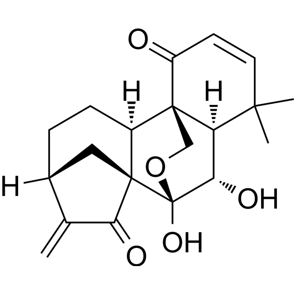
Eriocalyxin B
Based on 3 publication(s) in Google Scholar
Eriocalyxin B is a diterpenoid compound that can be isolated from Chinese herb Isodon eriocalyx. Eriocalyxin B exhibits multiple activities, such as anti-cancer, anti-inflammatory, and inhibition of adipogenesis. Eriocalyxin B is capable of inducing apoptosis and autophagy in tumor cells. Eriocalyxin B can be used in the research of cancers, autoimmune diseases, and other conditions.
Nur für Forschungszwecke. Wir verkaufen nicht an Patienten.
- Reinheit : 99.93%
- CAS. Nr.: 84745-95-9
- Formel: C20H24O5
- Molecular Weight:344.40
-
Speicherung:
4°C, protect from light
* In solvent : -80°C, 6 months; -20°C, 1 month (protect from light)

To place orders, for customer services and technical support, please contact: MedChemExpress USA
Tel: 609-228-6898 E-mail: [email protected] [email protected]


-
 Biologische Aktivität
-
Chemical Information
-
Lösungsmittel & Löslichkeit
- Protokoll
- Reinheit & Dokumentation
- Verweise
-
 Help & FAQs


Publications Citing Use of MedChemExpress (MCE) Eriocalyxin B
More

Biologische Aktivität
Beschreibung
Cellular Effect
|
Cell Line

|
Type | Value | Description | References |
|---|---|---|---|---|
| A549 | IC50 |
3.1 μM
Compound: 8
|
Cytotoxicity against human A549 cells after 48 hrs by MTT assay
Cytotoxicity against human A549 cells after 48 hrs by MTT assay
|
[PMID: 22624550] |
| HL-60 | IC50 |
0.3 μM
Compound: 8
|
Cytotoxicity against human HL60 cells after 48 hrs by MTT assay
Cytotoxicity against human HL60 cells after 48 hrs by MTT assay
|
[PMID: 22624550] |
| MCF7 | IC50 |
0.6 μM
Compound: 8
|
Cytotoxicity against human MCF7 cells after 48 hrs by MTT assay
Cytotoxicity against human MCF7 cells after 48 hrs by MTT assay
|
[PMID: 22624550] |
| SMMC-7721 | IC50 |
0.8 μM
Compound: 8
|
Cytotoxicity against human SMMC7721 cells after 48 hrs by MTT assay
Cytotoxicity against human SMMC7721 cells after 48 hrs by MTT assay
|
[PMID: 22624550] |
| SW480 | IC50 |
0.5 μM
Compound: 8
|
Cytotoxicity against human SW480 cells after 48 hrs by MTT assay
Cytotoxicity against human SW480 cells after 48 hrs by MTT assay
|
[PMID: 22624550] |
In Vitro
Eriocalyxin B (0-3.5 μM; 7 days) inhibits adipogenesis (IC50: 2.745 μM) and suppresses the expression of key regulators of adipocyte differentiation, including C/EBPα, C/EBPβ, PPARγ, and FABP4 in 3T3-L1 preadipocytes[1].
Eriocalyxin B (1.25-3.5 μM; 16-48 h) induces G2/M phase arrest and inhibits the expression levels of cell cycle regulators in differentiated 3T3-L1 cells[1].
Eriocalyxin B (48 h) is cytotoxic to tumor cells such as A-549, MCF-7, SMMC-7721, SW-480, and HL-60, with IC50 values ranging from 0.3-3.1 μM[2].
Eriocalyxin B (0.35-2.25 μM; 24-72 h) inhibits cell viability, increases intracellular ROS production, and induces apoptosis and autophagy in MCF-7 and MDA-MB-231. The mechanism involves the inhibition of the Akt/mTOR/p70S6K signaling pathway[3].
MedChemExpress (MCE) has not independently confirmed the accuracy of these methods. They are for reference only.
-
Cell Line:3T3-L1 cells
-
Concentration:1.25, 2.5, 3 and 3.5 μM
-
Incubation Time:24 h
-
Result:Inhibited the levels of CDK1, CDK2, Cyclin A and Cyclin B1.
In Vivo
Eriocalyxin B (10 mg/kg; intraperitoneal injection; 18-28 days) has an ameliorating effect in a mouse model of experimental autoimmune encephalomyelitis (EAE)[4].
MedChemExpress (MCE) has not independently confirmed the accuracy of these methods. They are for reference only.
-
Animal Model:Female NOD/SCID mice aged 6-8 weeks old treated MDA-MB-231 cells[3]
-
Dosage:10 mg/kg
-
Administration:Intraperitoneal injection (i.p.); 27 days
-
Result:Led to a slower tumor growth rate and reduced final tumor weight.
Caused no significant change in body weight and liver enzyme levels in plasma (ALT, AST and LDH) after treatment.
Activated autophagy and apoptosis in vivo.
-
Animal Model:Female C57BL/6 mice aged 6-8 weeks old with EAE model[4]
-
Dosage:10 mg/kg
-
Administration:Intraperitoneal injection (i.p.); 11 days after or on the day of immunization for the treatment, continued until 28 days
-
Result:Led to amelioration of EAE, which correlated with reduced spinal cord inflammation and demyelination.
Abolished encephalitogenic T-cell responses to myelin oligodendrocyte glycoprotein in an adoptive transfer EAE model. The underlying mechanism of EriB-induced effects involved inhibition of T helper (Th) 1 and Th17 cell differentiation through Janus Kinase/Signal Transducer and Activator Of Transcription and Nuclear factor-κB signaling pathways as well as elevation of reactive oxygen species.
- 3T3-L1 preadipocyte-to-adipocyte differentiation
- Detection of Apoptosis
- TUNEL staining for apoptotic DNA fragmentation
- Annexin V plus membrane-impermeant dye apoptosis staining
- Autophagy
- Apoptosis Solutions
- Research Protocol for Inflammation-related Diseases
- Lysosome and acidic-vesicle live-cell staining
- Macroautophagy Solutions
- Lipid Droplets: Oil Red O/Sudan Dye Lipid Staining


Chemical Information
-
CAS. Nr. 84745-95-9
-
Appearance Solid
-
Molecular Weight 344.40
-
Formel C20H24O5
-
Color White to off-white
-
SMILES
O[C@]1(OC2)[C@](C[C@]3([H])C4=C)(C4=O)[C@](CC3)([H])[C@]2(C(C=C5)=O)[C@](C5(C)C)([H])[C@@H]1O
-
Structure Classification
-
Initial Source
-
Versand
Room temperature in continental US; may vary elsewhere.
-
Speicherung
4°C, protect from light
* In solvent : -80°C, 6 months; -20°C, 1 month (protect from light)


Publications (3)
-
Journal Impact Factor
-
Most Recent
-
Phytomedicine
Eriocalyxin B induces ferroptosis through SIRT3 inhibition in triple-negative breast cancer. [Abstract]2025 Sep 11:148:157257. PMID: 40976126 -
Eur J Pharmacol
Eriocalyxin B ameliorated Crohn's disease-like colitis by restricting M1 macrophage polarization through JAK2/STAT1 signalling. [Abstract]2023 Sep 5:954:175876. PMID: 37391008 -


Lösungsmittel & Löslichkeit
In Vitro:
DMSO : 20 mg/mL (58.07 mM; Need ultrasonic and warming; Hygroscopic DMSO has a significant impact on the solubility of product, please use newly opened DMSO)
Please refer to the solubility information to select the appropriate solvent. Once prepared, please aliquot and store the solution to prevent product inactivation from repeated freeze-thaw cycles.
Storage method and period of stock solution: -80°C, 6 months; -20°C, 1 month (protect from light). When stored at -80°C, please use it within 6 months. When stored at -20°C, please use it within 1 month.
Please refer to the solubility information to select the appropriate solvent. Once prepared, please aliquot and store the solution to prevent product inactivation from repeated freeze-thaw cycles.
Storage method and period of stock solution: -80°C, 6 months; -20°C, 1 month (protect from light). When stored at -80°C, please use it within 6 months. When stored at -20°C, please use it within 1 month.
Konzentration (Stammlösung) × Volumen (Stammlösung) = Konzentration (Ziellösung) × Volumen (Ziellösung)


Reinheit & Dokumentation
-
Data Sheet (289 KB)
-
SDS (252 KB)
- English - EN (252 KB)
- Français - FR (252 KB)
- Deutsch - DE (252 KB)
- Norwegian - NO (252 KB)
- Español - ES (252 KB)
- Swedish - SV (252 KB)
- Italian - IT (252 KB)
- Korean - KR (252 KB)
- Portuguese - PT (252 KB)
-
Handling Instructions (2659 KB)
Verweise
[1]. Rong-Fang Mu, et al. Eriocalyxin B Inhibits Adipogenesis in 3T3-L1 Adipocytes by Cell Cycle Arrest. Nat Prod Bioprospect. 2020 Jun;10(3):131-140. [Content Brief]
[2]. Wang WG, et al. Laxiflorolides A and B, epimeric bishomoditerpene lactones from Isodon eriocalyx. J Nat Prod. 2012 Jun 22;75(6):1102-7. [Content Brief]
[3]. Zhou X, et al. Eriocalyxin B, a novel autophagy inducer, exerts anti-tumor activity through the suppression of Akt/mTOR/p70S6K signaling pathway in breast cancer. Biochem Pharmacol. 2017 Oct 15;142:58-70. [Content Brief]
[4]. Lu Y, et al. Eriocalyxin B ameliorates experimental autoimmune encephalomyelitis by suppressing Th1 and Th17 cells. Proc Natl Acad Sci U S A. 2013 Feb 5;110(6):2258-63. [Content Brief]
[5]. Leung CH, et al. Eriocalyxin B inhibits nuclear factor-kappaB activation by interfering with the binding of both p65 and p50 to the response element in a noncompetitive manner. Mol Pharmacol. 2006 Dec;70(6):1946-55. [Content Brief]


Complete Stock Solution Preparation Table
Please refer to the solubility information to select the appropriate solvent. Once prepared, please aliquot and store the solution to prevent product inactivation from repeated freeze-thaw cycles.
Storage method and period of stock solution: -80°C, 6 months; -20°C, 1 month (protect from light). When stored at -80°C, please use it within 6 months. When stored at -20°C, please use it within 1 month.
| Optional Solvent | Concentration Solvent Mass | 1 mg | 5 mg | 10 mg | 25 mg |
|---|---|---|---|---|---|
| DMSO | 1 mM | 2.9036 mL | 14.5180 mL | 29.0360 mL | 72.5900 mL |
| 5 mM | 0.5807 mL | 2.9036 mL | 5.8072 mL | 14.5180 mL | |
| 10 mM | 0.2904 mL | 1.4518 mL | 2.9036 mL | 7.2590 mL | |
| 15 mM | 0.1936 mL | 0.9679 mL | 1.9357 mL | 4.8393 mL | |
| 20 mM | 0.1452 mL | 0.7259 mL | 1.4518 mL | 3.6295 mL | |
| 25 mM | 0.1161 mL | 0.5807 mL | 1.1614 mL | 2.9036 mL | |
| 30 mM | 0.0968 mL | 0.4839 mL | 0.9679 mL | 2.4197 mL | |
| 40 mM | 0.0726 mL | 0.3630 mL | 0.7259 mL | 1.8148 mL | |
| 50 mM | 0.0581 mL | 0.2904 mL | 0.5807 mL | 1.4518 mL |


Eriocalyxin B Related Classifications
HY-N2303 Related Classifications
 Powered by Bioz